Week 1 HW: Final Project Ideas
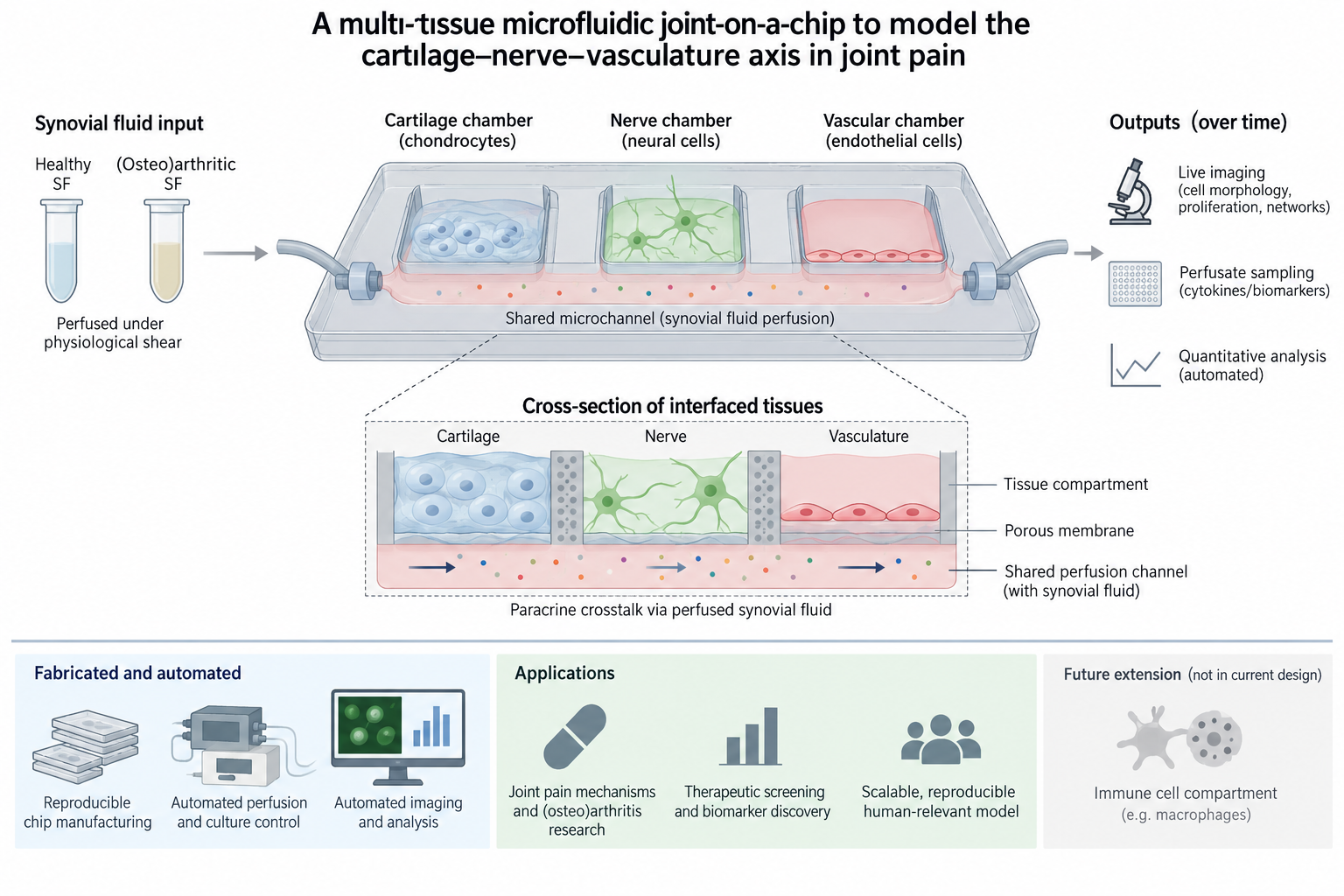
(1) What I would like to biomanufacturer and why Target: A multi-chamber microfluidic “joint-on-a-chip” device that co-cultures cartilage tissue (chondrocytes), vascular tissue (endothelial cells), and peripheral nerve tissue (neural cells) in adjacent chambers, connected by a shared microchannel perfused with human synovial fluid under physiological shear. Both the chip itself and the chip-co-culture platform are the manufactured products. This configuration is designed specifically to model the cartilage–nerve–vasculature axis underlying joint pain, as distinct from a purely inflammatory/immune model; adding an immune cell compartment (e.g., macrophages) is noted here as a possible future extension rather than part of the current design.
Why: Arthritis and pain research is currently constrained by two major gaps. First, animal and other in vivo models raise ethical concerns, differ physiologically and pathologically from human disease, are expensive, and are highly variable. Second, human patient studies are confounded by patient-to-patient variability and comorbidities that can obscure (osteo)arthritis-specific markers, progression, and pain mechanisms. A reproducible, biomanufactured joint-in-a-chip that recreates crosstalk across the cartilage–nerve–vasculature axis and synovial inflammation. Using this device at scale (i.e., many chips and conditions in parallel) could let researchers study joint pain pathways and screen therapeutics far more systematically than current models allow.
Who it’s for: researchers and pharmaceutical developers studying joint pain mechanisms, and eventually those seeking a therapeutics-screening or biomarker-discovery platform. The first focus would be for (osteo)arthritis research, but the platform would be designed in a way to allow the research of other joint diseases/conditions. This is infrastructure meant to accelerate other people’s research, not a single therapeutic in itself.
(2) What living system would I use? Chassis: A co-culture of three human cell types, seeded into a purpose-designed multi-chamber microfluidic chip: • Chondrocytes: forming a cartilage-like tissue chamber. • Endothelial cells: forming a vascular chamber. • Neural cells: representing peripheral nerve input to the joint. Starting with commercially available human cell lines, then progressing to primary patient-derived cells or, potentially, iPSC-derived cells as the system matures.
Process (high level): • In: expanded chondrocytes, endothelial cells, and neural cells; a fabricated multi-chamber chip; culture medium; and human synovial fluid (SF) collected from both healthy and (osteo)arthritic donors, perfused through the shared channel. • Biological process: each cell type is cultured in its own chamber, connected via a common perfusion channel carrying SF under physiological shear, so that the three tissue types are exposed to the same fluid environment and can signal to one another (paracrine cross-talk) the way they would across the joint capsule. Introducing SF from (osteo)arthritic donors, versus healthy donors, is intended to recreate a disease-relevant inflammatory/degenerative signal and reveal how it affects each tissue compartment. • Out: a living, multi-tissue chip that can be imaged and sampled over time, plus collected perfusate for cytokine/biomarker analysis.
Assumptions: commercial cell lines will adequately approximate primary-cell behaviour during early development; SF collected from donors can be stored/diluted into media without losing bioactivity; and a shared perfusion channel provides sufficient, physiologically realistic crosstalk between three separate chambers. Synovial fluid sourcing and quality control: The plan targets a minimum of n≥3 healthy donors and n≥3 osteoarthritic donors for SF collection, consistent with the biological-replicate structure described below. Each SF batch will be aliquoted and frozen at the time of collection under activity-preserving conditions (e.g., single-use aliquots to avoid freeze-thaw degradation), and baseline cytokine profiling of each batch will be performed before use, as part of the automated readout pipeline (Section 3), to characterize batch-to-batch variability rather than treat it purely as an unknown. Upstream cell supply: Based on the seeding densities and ratios described in Section 4(a), meeting the full combinatorial screen (360–1,200 chips) would require expanding on the order of 10⁷–10⁸ total cells per cell type (chondrocytes, endothelial cells, neural cells), assuming ~10⁵–10⁶ cells seeded per chip depending on density condition. This would be achieved through standard 2D flask expansion of each cell line, assuming realistic fold-expansion (~10–20× over 1–2 weeks) and staying within typical passage-number limits before phenotype drift becomes a concern. This upstream expansion step, rather than chip fabrication itself, may be the practical bottleneck feeding the full-scale screen.
(3) How I would use automation • Chip fabrication: a programmable microfluidics platform or automated soft-lithography/injection-moulding line, to produce chips with consistent channel geometry and dimensions. This is essential for reproducibility across the thousands of chips a later large-scale version of this platform would need. • Perfusion and co-culture control: automated syringe-pump/perfusion systems to systematically vary: (1) SF source and concentration (healthy vs. arthritic, 0–100% dilution in media), (2) Seeding density per cell line (3) Ratios between cell lines, and (4) Flow regime (static, pulsing, continuous) This would enable standardised control vs experimental comparisons and multiple chip screens to run in parallel rather than one at a time. This also allows testing multiple media protocols to identify the best composition (and any timepoint-specific changes) for maintaining the three-cell co-culture. • Readouts: automated (including live) imaging and cytokine assays to capture how each tissue compartment responds to different SF conditions over time, including the baseline SF batch characterization described above. • Image analysis: automated quantification of cell morphology, proliferation, and other endpoints from the imaging data, replacing manual scoring. Prototype vs. scale: early prototyping would likely use manual/benchtop fabrication (e.g., PDMS soft lithography) and a small number of syringe pumps for a single cell line and one SF condition. At larger scale, this would shift to injection-molded chips for cost and reproducibility, run on a multiplexed perfusion manifold with automated imaging and immunoassay readout. This would move the platform closer to an integrated automated screening line. Production-scale automation: if the platform is deployed as a commercial or shared research screening tool, chip fabrication would shift from manual PDMS casting to injection-molded chips for cost and reproducibility at volume; perfusion control would shift from individual benchtop syringe pumps to multiplexed perfusion manifolds capable of running hundreds of chips simultaneously under independently addressable conditions; and readout would shift from manual/small-batch imaging to an integrated automated imaging and immunoassay line, closer to a CloudLab-style screening facility than a benchtop setup. This is a substantively different automation stack from the prototype phase, not simply a scaled-up version of the same equipment.
(4) How much would you need to make, and how long would it take? (4a) How much? The platform optimisation and validation would include: • 10 SF concentrations (0–100%, including control) • 3–4 cell seeding densities per cell line • 2+ cell ratios (chondrocyte : neural : endothelial) • 2–5 chip designs • 3+ replicates per condition Even taking the low end of each range (10 × 3 × 2 × 2 × 3), a full combinatorial exploration is 360 chips; the high end (10 × 4 × 2 × 5 × 3) is 1,200 chips. Replicate structure: replicates are defined to include both technical replicates (same chip design, same cell/SF donor batch, run in parallel to assess device and assay reproducibility) and biological replicates (different donor batches of cells and/or SF, run to assess donor-to-donor variability). The 3+ replicates figure is intended as a minimum split across both types — for example, 3 technical replicates repeated across each of ≥3 SF donor batches per health status — directly operationalizing the “how much variability can automation remove” question from Section 5. Phasing: this full 360–1,200 chip combinatorial screen represents the eventual full-scale use of a validated platform, not the scope of the initial 8-month development phase described below. The 8-month phase uses a reduced, representative subset of these conditions (e.g., 2 chip designs × 2 cell ratios × 1 seeding density × a smaller set of SF concentrations) sufficient to validate that the platform works, before committing resources to the complete parameter sweep.
(4b) How long? Phase 1: Platform development and validation (~8 months), using the reduced condition set described above: • Chip design, fabrication, and prototype testing (single cell line, single SF condition): 1 month • Cell seeding optimization, run separately per cell line at ~3 weeks each (with some potential to parallelize across cell lines): ~2–2.5 months • Co-culture optimization: validating seeding density, cell ratios, and media protocols together: 3 months • Disease induction: comparing healthy vs. osteoarthritic SF effects: 1–2 months • Total estimated Phase 1 timeline: ~8 months Success/validation criteria for Phase 1: the platform will be considered validated if it shows a statistically significant, reproducible difference in a defined readout (e.g., a specific cytokine level or a quantified morphological/proliferation metric) between healthy and osteoarthritic SF conditions, consistent across at least 3 donor pairs. Meeting this criterion is the trigger for proceeding to Phase 2. Phase 2 Full-scale combinatorial screening (subsequent to Phase 1): once validated, the platform would be used to run the complete 360–1,200 chip parameter space described in Section 4(a). Using production-scale automation (Section 3), and assuming multiplexed perfusion manifolds capable of running on the order of 100+ chips in parallel with a 2–3 week culture/exposure window per batch, the full screen could plausibly be completed in an estimated 2–4 months of screening time, run as a separate, subsequent stage rather than within the original 8-month figure.
(5) What I don’t know yet • Whether the chip can detect a real disease signal: it’s unknown whether measurable differences in cell morphology, proliferation, or cytokine output actually emerge when comparing healthy vs. osteoarthritic synovial fluid, and if so, how large and reproducible that signal is. • How much of the variability is biological vs. technical: donor-to-donor differences in synovial fluid (and potentially donor age, sex, and injury/procedure type. For example, total vs. partial knee replacement) may significantly affect results, and it’s unclear how much of this variability automation can actually control for, versus needing to be addressed through donor selection/stratification. • Whether the platform can scale beyond commercial cell lines: it remains to be determined whether iPSC-derived cells could replace primary patient-derived cells for scalability, and whether matching cell donor and SF donor (i.e., autologous chip components) would meaningfully change results compared to mismatched combinations.
(6) Use of AI delcared: Claude.ai Prompts used: Please complete the following tasks, marked with upper case letters in parentheses: (A) Please read the following notes for a project proposal. Notes: What to biomanufacturer? Microfluidic synovial joint-on-a-chip device. This small device would enable co-culture of cartilage tissue, synovial fluid, nerve and vascular cells in adjacent microfluidic chambers connected by a shared perfused channel with synovial fluid, which is under physiological shear. Manufactured product: both the microfluidic chip itself and the microfluidic platform (chip + co-culture of cartilage-nerve-vasculature). Application: (1) model inflammatory joint diseases (e.g., osteoarthritis) in vitro; (2) platform for testing/screening therapeutics or, potentially, biomarker discovery. Gap/Need: (Osteo)arthritis research, including therapeutics development/treatments, are limited by: (1) ethical concerns, (patho)physiological differences, cost, variability with the use of in vivo models; (2) patient-to-patient differences, existence of co-morbidities that might obscure (osteo)arthritis markers/progression/consequences. There is a need for a biomanufactured, reproducible joint-in-a-chip device that recreates the cross-talk between the cartilage-nerve-vascular axis and synovial inflammation at a scale (i.e., many conditions in parallel, many chips). This could allow researchers to study pain pathway(s) or screen therapeutics. Key point: Platform to accelerate the research of other researchers on (osteo)arthritis and pain, including therapeutics screening. Device: Multi-chamber microfluidic device, to be designed as part of this project. Use human cell lines. Start with commercially available cells and then move onto primary cells obtained from patients or possibly iPSC-derived cells: • Chondrocytes – forming a cartilage-like tissue • Endothelial cells – vasculature • Neural cells – peripheral nerves • Synovial fluid – from healthy patients and from those with (osteo)arthritis Automation for: • chip fabrication (automate fabrication of chips through programmable microfluidics platform or automated soft-lithography/injection-moulding line) to produce chips that have consistent channel geometry/dimensions. This is so the device is reproducible across thousands of units (key for large scale manufacturing at a later stage). • Perfusion & co-culture: automated syringe-pump or perfusion control to run variations in (1) synovial fluid (e.g., healthy vs arthritic patient); (2) cell seeding number for each cell line; (3) cell ratios between cell lines; (4) dilution of synovial fluid variations; and (5) model different media protocols (to determine which is the best media composition for the co-culture and changes, if required, to the media used at specific timepoints). This would enable running standardised control vs. experimental parameters (flow rate, static vs. pulsing vs. continuous, etc.) across many chips in parallel; as well as running multiple chip screenings instead of one at a time. • Readouts: automated imaging (incl. live imaging) and cytokine assays to evaluate response to variations in synovial fluid, for example. • Image analysis: quantify changes in cell morphology, proliferation, etc. How much: • 10 concentrations of synovial fluid, including a control: 0-100% synovial fluid in media • 3-4 different cell densities per cell line. • 2+ different cell ratios (chondrocytes : neural cells : endothelial cells) • 2-5 different chip designs. • 3+ replicates of each device+co-culture mix. How long? • Chip design and fabrication, including prototype testing (using one single cell line and synovial fluid): 1 month • Cell seeding optimisation: run separately for each cell line, 3 weeks per cell line. *Could potentially run some things in parallel. • Co-culture optimisation: validation of the cell seeding and ratio between cell lines, as well as media protocols – 3 months • Disease induction: compare impact of using synovial fluid from healthy patient vs from osteoarthritic patient – 1-2 months • Total estimated time: ~8 months What I don’t know yet: • Can the joint-on-a-chip show differences in cell morphology/number or cytokines when using synovial fluid from healthy patient vs osteoarthritic patient? • Impact of using synovial fluid from different healthy patients / (osteo)arthritic patients? (so comparing 3+ samples of healthy patients). What about age? And sex? • Does the type of injury impact the synovial fluid’s impact on the cells? For example, would SF from a patient that has undergone a total knee replacement act differently than one that has undergone a partial knee replacement? • How much chip-to-chip and synovial fluid batch-to-batch variability automation can remove? • Could iPSC-derived cells replace primary cells to make this scalable? • Would using cells from the same patient as the synovial fluid patient make a difference?
(B) Now, please review the tasks assigned for the initial proposal. [Questions copied from the homework here]
(C) Please draft the proposal addressing each of the questions using the notes I provided in point (A). (D) Identify any gaps or inconsistencies and flag them at the end. Give me this output as a numbered list, with details. (E) Finally, propose ideas to address these gaps and inconsistencies.
After this, edits were made directly into the text produced by Claude.