Final Project
I. Abstract
Prions are misfolded proteins that exhibit ‘infectious’-like behaviour and are the root cause for multiple prion protein associated neurodiseases: Creutzfeldt–Jakob Disease, Kuru (not exclusively in humans) and Scrapie and Bovine spongiform encephalopathy (BSE, mad-cow disease) characteristically affecting ovines and bovines respectively.
In humans, the gene PrPC encodes for the premature version of the cellular prion protein (253-a.a. long) found within neuroglia and neurons, its function and role remain ambiguous and have not been described fully (or so it is believed). After post-translational modification in the endoplasmic reticulum, the precursor protein loses its N-terminal signal peptide (1-22) and C-terminal signal peptide (232-253) and carries on as a mature, functional PrPC surface glycoprotein that is 208-209 residues long. Faulty folding or interaction with other midfolded prion proteins can lead the native cellular variant of the protein to display a different structure and, thus, behaviour: resulting isoform is called PrPSc (scrapie-associated prion protein).
Present study explores how the native variant of the prion protein in humans behaves under different parameters set in-silico. Furthermore, a proposal for assessing prion diseases using binding peptides is described and showcased on PrPC.
II. Introduction
III. Structure
*EXPLAIN ‘GLYCO-’ PART OF THE PROTEIN: glycosylphosphatidylinositol; function within cell;
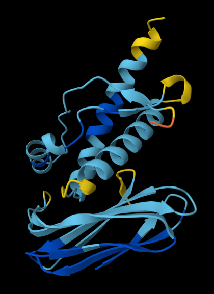
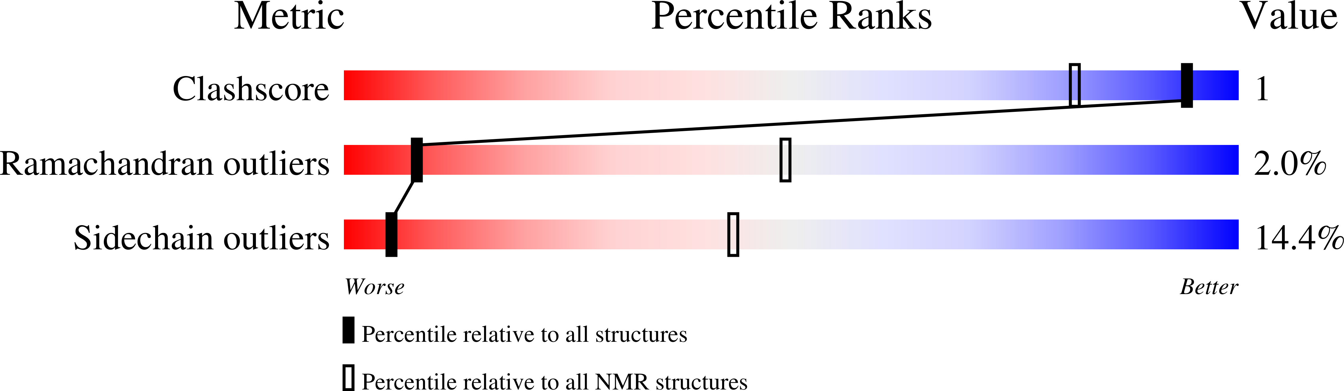
For the means of this in-silico workthrough, the chosen structure for the PrPC protein is the RCSB deposit under the name of 1QLX, obtained via NMR.
 The human PrP protein, including 2 C-terminal fragements NMR screeing resulted in the deposit 1QLX.
Important aspects of the globular domain (125-228):
three α-helices (144-154; 173-194; 200-228)
one short anti-parallel β-sheet (128-131/ 161-164)
three disordered peptides (167-171 - a loop-like structure; 187-194; 219-228)
Between the Cys179 and Cys214 residues, a stronger intramolecular force (disulfide bond) bridges helix 2 and helix 3; stabilising the native globular architecture, mechanical integrity and hydrophobic core of the monomeric protein.
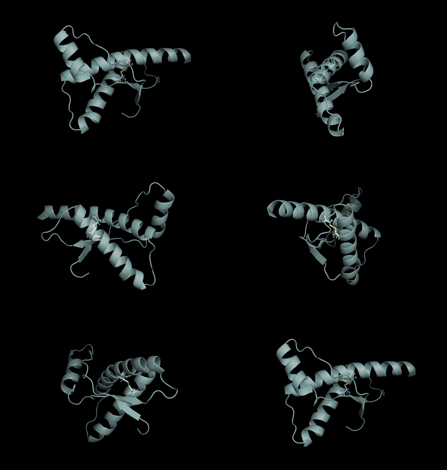
Amino acid asparagine on position 181 is documented to carry a sugar chain in real cells, thus the binder would be unable to contact it.
GROMACS analysis
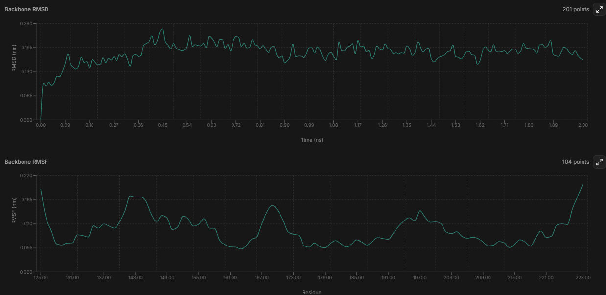
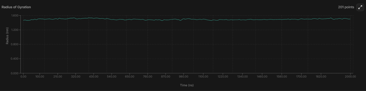
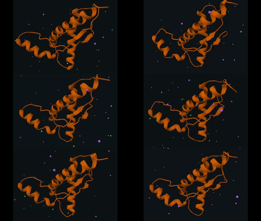
- with the small and flat RMSD and Radius of gyration graphics, the structure can be interpreted as compact and not really seeming to “drift away” from the starting shape;
- the parts that wiggle the most: the loop around 167-172 matches what the original 1QLX paper reported;
Adaptive Poisson-Boltzmann Solver in PyMol
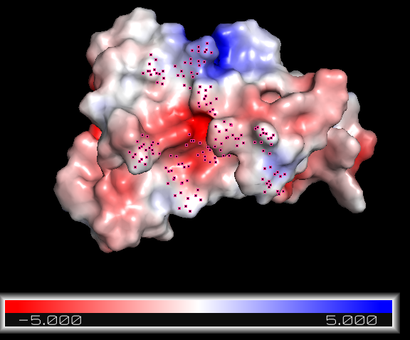
A good binder should be able to complementary attach to electronegative and/or electropositive regions of the protein.
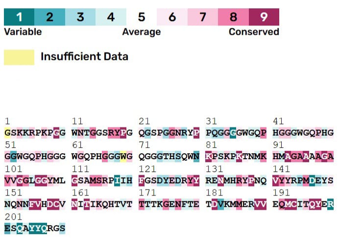
ConSurf analysis
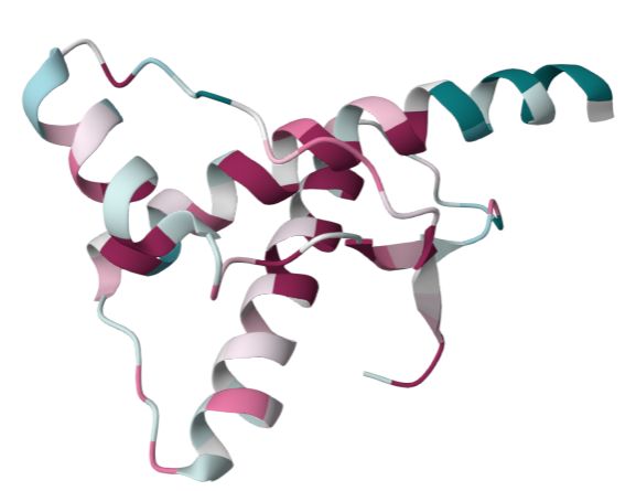
IV. Binding peptide approach
V. Binder generation
Asp178 and Gln212 are both positions of disease-linked mutations.
Batch #1 - BoltzGen
1. Try one
Target residues: all from helix 2 (173-194)
Best ipTM (.443), best min PAE: spec_19_4. RMSD = 2.01
95 residues: SVTVVASGGGLVQAGGTLRVSCAASGAGVVAVSAGVRPPGSDRLLGGGVVVGKASGTVEIKDLEPADSGVVQCEAAVAGSDTLARGDGPQVTVLP
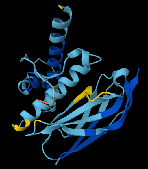
2. Try two
Target residues: solvent exposed parts from helix 2 plus some from helix 3 Filter RMSD stood within 1.2-1.9A for every speculative design, all structures should hypothetically fold accordingly.
Best ipTM (.362), best min PAE: spec_00_0. RMSD = 1.372, 2 H bonds, 0% β-sheet
49 residues: EEAKRYAIKLFVKKIEEGKPLTKRTLEELIKLAEELGDKEALEVFKKAL
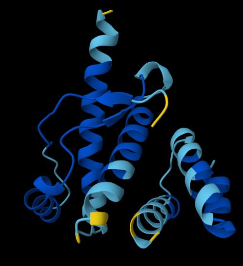
3. Try three
Target residues: none choosen; let BoltzGen choose Filter RMSD stood within 1.2-1.9A for every speculative design, all structures should hypothetically fold accordingly.
Best ipTM (.460), best min PAE: spec_49_2. RMSD = 14.111, 5 H bonds, 0% α-helix
91 residues: ATASNPTITATGDKATASGTCDAPGAARTVVSLLNAETGECLGIGESDGTTASAEGTGAKAGDTVAAGVSALDAEGNILASGGGTATATPA
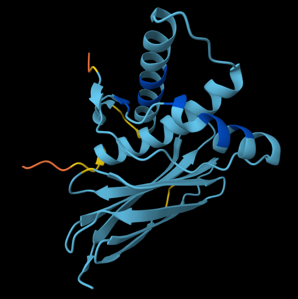
ipTM (.372), second best min PAE: spec_49_3. RMSD = , 12 H bonds
91 residues: ASRTCTTKSGDKLTCTASGTATDVTVTVTGEIAETSTSPSRPYTSPATGQTGSAATAVAKSKKSGEIVVCTGIVYADGTITVSCYLNGEAV