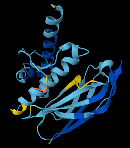
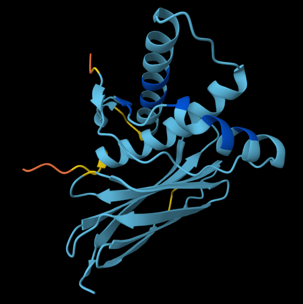
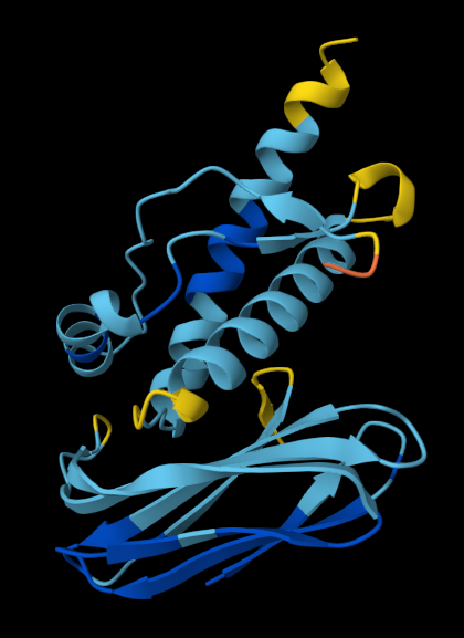
I really love biology and the sciences that make it up, be it molecular dynamics, organic chemistry, systems biology or even evolution. At the moment, neurobiology, endocrinology and bioinformatics are my focus.
✦ I love art.. probably listening to music right now
What do you want to make? → Custom designed protein; post translational modifications/ usage of nonstandard amino acids (e.g. selenocysteine) not clear.
For the purpose of this homework, may the custom protein be named Cuop. Coup is a peptidic macromolecule I plan to design, analyse and test using various computational tools. Its intended goal is to address prion activity in-vivo; work like an inhibitory drug against prion PrPSc “infectious” folding mechanism. The explanation for a protein’s ability to self-propagate a motif is intermolecular interactions between strongly bound-together amino acids (PrPSc beta sheets) and a less rigid structure (PrP alpha helices). Therefore a molecule able to prevent the interactions between the two proteins may be capable of preventing the spread of disease in high risk patients. The way I aim to achieve this is molecular docking simulations to assess if the custom protein can actually help.
Protein expression system in Asimov Kernel 1. Defining expression target What molecule are you expressing? A protein, an RNA, or an enzyme (or small set of enzymes) for a pathway. If your HW1 product is a material, food, or chemical, identify the protein(s) that produce it and design for those. Fortunately, the only molecule desribed for my final project is a single short protein (i.e. prion-binding ‘peptide’). The design logic stays simple throughout production: DNA -> mRNA -> protein of interest.
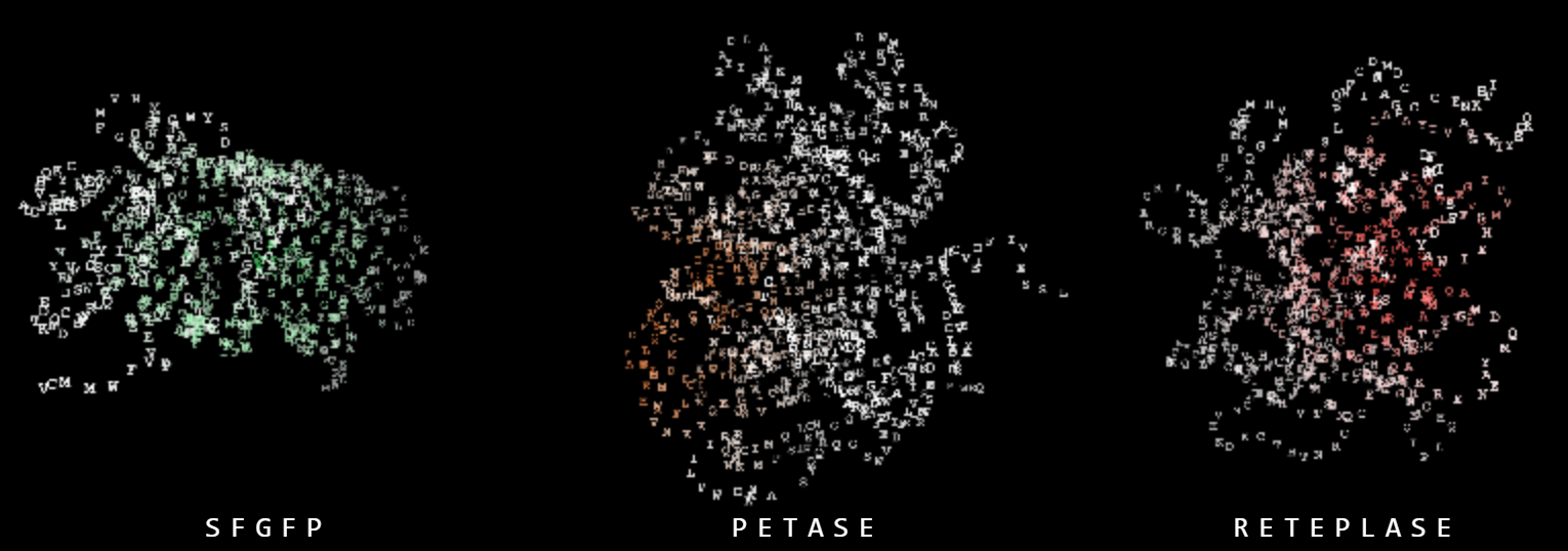
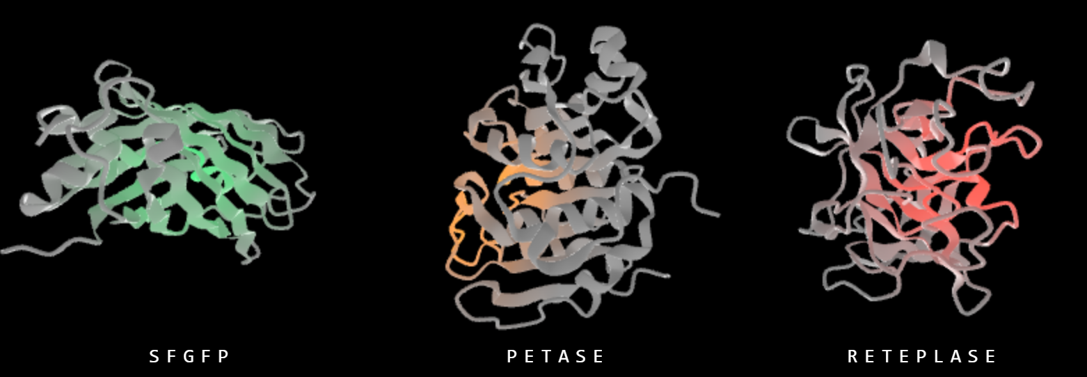
WEEK 3 ASSIGNMENT: CFPS REACTION DESIGN FOR SFGFP, PETASE, RETEPLASE A. COMPARE CELL-FREE REACTION COMPOSITIONS (20 minutes) Why does the concentration of potassium glutamate and magnesium glutamate differ between compositions? Which is more sensitive? Referance concentration: K-glutamate (279.84 mM), Mg-glutamate (8.22 mM) are to mimic and maintain osmotic parameters of lysate in CFPS, assure ribosome, energy-replenishing enzymes, polymerase function, stabilise DNA/RNA structure etc.; both glutamic acid salts are usually found in high concentrations.
→ Custom designed protein; post translational modifications/ usage of nonstandard amino acids (e.g. selenocysteine) not clear.
For the purpose of this homework, may the custom protein be named Cuop. Coup is a peptidic macromolecule I plan to design, analyse and test using various computational tools. Its intended goal is to address prion activity in-vivo; work like an inhibitory drug against prion PrPSc “infectious” folding mechanism.
The explanation for a protein’s ability to self-propagate a motif is intermolecular interactions between strongly bound-together amino acids (PrPSc beta sheets) and a less rigid structure (PrP alpha helices). Therefore a molecule able to prevent the interactions between the two proteins may be capable of preventing the spread of disease in high risk patients. The way I aim to achieve this is molecular docking simulations to assess if the custom protein can actually help.
The synthesis of Coup is contextualised for industry-scale (working with software like Kernel and interpreting the data accordingly), though the proposed activity validation route for the drug may revolve around the ideas of batches and limited portions for quality-assessment bottlenecks.
What living system could make it?
→ Assuming a rather simple peptidic structure with no ions attached, E. coli can be used as a model organism as in-silico workflow parameters usually are determined by Escherichia systems. After clarifying the proposed amino acid sequence, BLAST tools could help in seeking homologous sequences, thus an organism already specialised in synthesizing similar enough structures.
*I am noting the fact that the host living system is a really important choice I need to settle down on later in the pipeline:
It should properly replicate the computationally validated lucrative structure and refrain from potentially altering it via unintended integration of the product in own metabolic chains or via its intracellular medium;
It should express the gene cost and time efficient;
The protein’s toxicity to the organism should be minimised;
*
Week 2 HW: DNA design
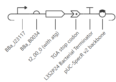
Protein expression system in Asimov Kernel
1. Defining expression target
What molecule are you expressing? A protein, an RNA, or an enzyme (or small set of enzymes) for a pathway. If your HW1 product is a material, food, or chemical, identify the protein(s) that produce it and design for those.
Fortunately, the only molecule desribed for my final project is a single short protein (i.e. prion-binding ‘peptide’). The design logic stays simple throughout production: DNA -> mRNA -> protein of interest.
What is the sequence? Give the protein or RNA sequence, or the specific gene, you will encode, and say where it comes from (a database accession such as UniProt or NCBI, a known part, or de novo). Cite it.
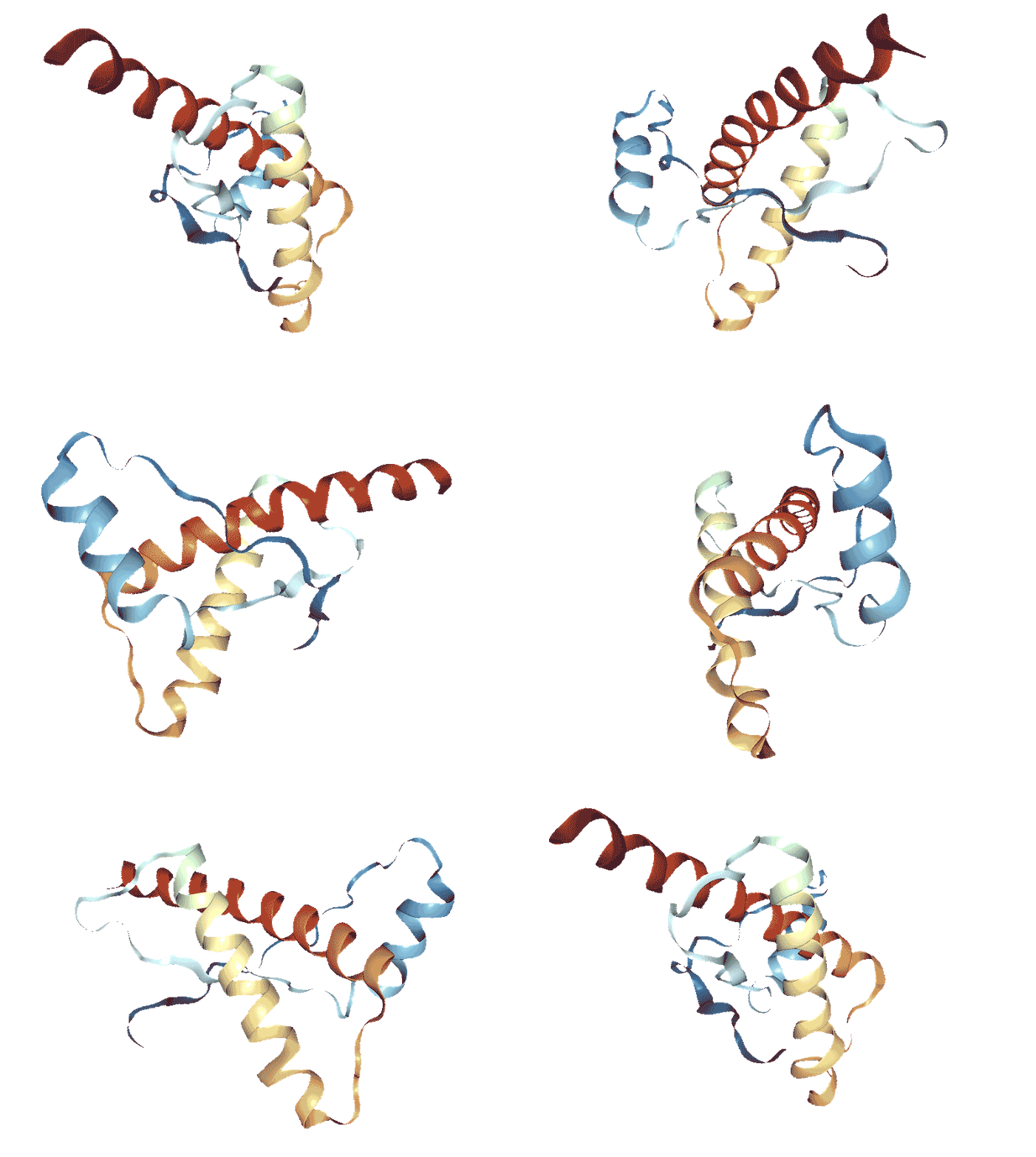
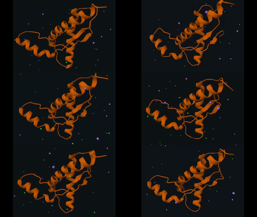
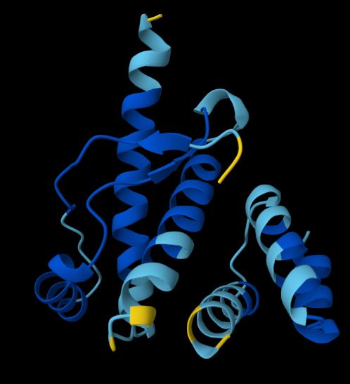
For this assignment I made the second binder peptide from the original batch the expression target. The sequence is 49 amino acids long (the shortest from its batch) and encodes for a trouple of α-helices, supposedly having a high-affinity-for-PrP S surface.
Unsurprisingly, reverse translating it into a 147 nt sequence outputed nothing when BLAST-ed.
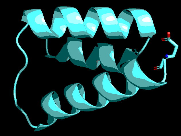
the residue shown as sticks is the N-terminus of the peptide
What host or chassis? The living system from HW1, for example E. coli, S. cerevisiae, CHO or another mammalian line, or a cell-free system. Your host drives almost every part choice that follows.
E.coli is the best pick for expressing a short simple singular polypeptide unit with no disulfide bridges or other tertiary structure requirements that would make synthesis increasigly complicated.
Where does the product need to go? Cytoplasm, secreted, membrane, or a compartment? This decides whether you need a signal peptide or targeting sequence.
I have not tought of this yet, upcoming updates will come as I progress my final project research and engineering work.*
2. Genetic design
Layer
Prokaryote host
Justification
Data
Transcription start
Promoter
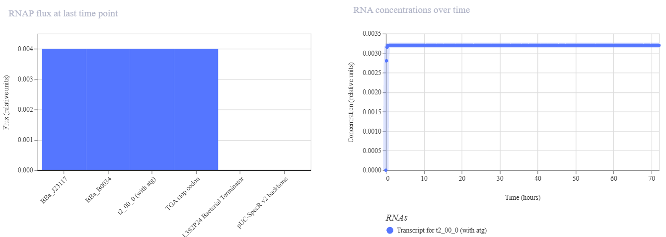
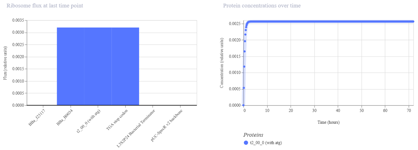
Noticeably weak Anderson promoter, further investigating the binder will reveal if its overexpression could pose a hazard to host-cell. J23106 is an alternative
-> no selection marker was attributed to the plasmid in Kernel as the chosen vector already had one: SpecR, streptomycin with its own native promoter ~430 bp upstream;
3. Construct & assembling
The design should pass as Golden-Gate clean: no BsaI, BsmBI/Esp3I, BbsI, SapI, PaqCI sites are found within the construct, risking later assembly.
Week 3 HW: CFPS for sfGFP, PETase, Reteplase
WEEK 3 ASSIGNMENT: CFPS REACTION DESIGN FOR SFGFP, PETASE, RETEPLASE
A. COMPARE CELL-FREE REACTION COMPOSITIONS (20 minutes)
Why does the concentration of potassium glutamate and magnesium glutamate differ between compositions? Which is more sensitive?
Referance concentration: K-glutamate (279.84 mM), Mg-glutamate (8.22 mM) are to mimic and maintain osmotic parameters of lysate in CFPS, assure ribosome, energy-replenishing enzymes, polymerase function, stabilise DNA/RNA structure etc.; both glutamic acid salts are usually found in high concentrations.
Mg2+ is needed by ribosomes, T7 polymerase, and ADP phosphorylating enzymes; nucleotides, phosphate groups and PEP all bind it => optimum Mg level shifts with the rest of the recipe.
K+ is a broader background that tolerates tens of mM shifts (Mg is the more sensitive one).
Explain how a PEP+NTP reaction formulation is different from a Ribose+NMP reaction formulation.
PEP+NTP: PEP regenerates ATP directly (+the four triphosphates) - quick, expensive, risky (PEP degradation releases phosphate that can tie up Mg);
Ribose+NMP: cheap sugar + monophosphates that will be metabolised by the lysate and supposedly turned into ATP (i.e. GMP can be partly replaced by guanine + ribose, like one paper specifies) - cheaper, harder to nail (reactions dependent on phosphate availablity, cofactors, pH etc)
“Surprisingly, we found that GMP could be replaced with a combination of guanine and ribose without sacrificing protein yield and that UMP could be replaced with its corresponding base and ribose, albeit with reduced yields.”
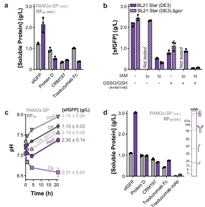
Choose two cell-free reaction compositions from the literature, compare and contrast the choice of reagents and the concentration of reagents.
PANOx-SP s.
Cytomin s.
Comment
Mg acetate (mM)a
3.3
3.3
Mg glutamate (mM)
20
8
NH4 acetate (mM)
0
0
NH4 glutamate (mM)
10
10
K acetate (mM)a
14.4
14.4
K glutamate (mM)
175
130
phosphoenolpyruvate (mM)b
33
0
Na pyruvate (mM)
0
33
Spermidine (mM)
1.5
1.5
Putrescine (mM)
1
1
Na oxalate (mM)
2.7
4
Tris (mM)
2.4
2.4
pH = 8.2
Note: A recommended starting point would be to review a least one published formulation, such as PANOx-SP (Jewett & Swartz 2004), a cost-optimized formulation from Olsen et al. (2026), and/or an AI-designed formulation from Smith et al. (2026).
B. DESIGN ONE REACTION COMPOSITION FOR EACH OF THE THREE PROTEINS (40 minutes)
Sign in to coopetition.htgaa.org and publish your first unique 20 µL CFPS reaction composition for each of the following (don’t worry, you can resubmit later):
sfGFP
PETase
Reteplase
Note: if you’re unable to login with your HTGAA keycloak account, please post in Zulip so that you can participate in the coopetition!
For each composition, specify the reagents you would adjust from the relevant categories, including salts, energy sources, building blocks, cofactors, and redox components.
C. ASIMOV KERNAL
Build T7 compatible plasmids for cell-free expression using the amino acid coding sequences below:
Bonus question: design a T7 helper plasmid that you could add to the cell-free reaction to improve the yield of functional reteplase (general chaperones, disulfide-bond machinery, etc).
Week 4 HW: CFPS in batches
Labs
Lab writeups:
Final Project
I. Abstract
Prions are misfolded proteins that exhibit ‘infectious’-like behaviour and are the root cause for multiple prion protein associated neurodiseases: Creutzfeldt–Jakob Disease, Kuru (not exclusively in humans) and Scrapie and Bovine spongiform encephalopathy (BSE, mad-cow disease) characteristically affecting ovines and bovines respectively.
In humans, the gene PrPC encodes for the premature version of the cellular prion protein (253-a.a. long) found within neuroglia and neurons, its function and role remain ambiguous and have not been described fully (or so it is believed). After post-translational modification in the endoplasmic reticulum, the precursor protein loses its N-terminal signal peptide (1-22) and C-terminal signal peptide (232-253) and carries on as a mature, functional PrPC surface glycoprotein that is 208-209 residues long. Faulty folding or interaction with other midfolded prion proteins can lead the native cellular variant of the protein to display a different structure and, thus, behaviour: resulting isoform is called PrPSc (scrapie-associated prion protein).
Present study explores how the native variant of the prion protein in humans behaves under different parameters set in-silico. Furthermore, a proposal for assessing prion diseases using binding peptides is described and showcased on PrPC.
II. Introduction
III. Structure
*EXPLAIN ‘GLYCO-’ PART OF THE PROTEIN: glycosylphosphatidylinositol; function within cell;
For the means of this in-silico workthrough, the chosen structure for the PrPC protein is the RCSB deposit under the name of 1QLX, obtained via NMR.
1QLX publication abstract
The NMR structures of the recombinant human prion protein,
hPrP(23–230), and two C-terminal fragments, hPrP(90 –230) and
hPrP(121–230), include a globular domain extending from residues
125–228, for which a detailed structure was obtained, and an
N-terminal flexibly disordered ‘‘tail.’’ The globular domain contains
three a-helices comprising the residues 144–154, 173–194, and
200 –228 and a short anti-parallelb-sheet comprising the residues
128 –131 and 161–164. Within the globular domain, three polypeptide segments show increased structural disorder: i.e., a loop of
residues 167–171, the residues 187–194 at the end of helix 2, and
the residues 219 –228 in the C-terminal part of helix 3. The local
conformational state of the polypeptide segments 187–193 in helix
2 and 219 –226 in helix 3 is measurably influenced by the length of
the N-terminal tail, with the helical states being most highly
populated in hPrP(23–230). When compared with the previously
reported structures of the murine and Syrian hamster prion proteins, the length of helix 3 coincides more closely with that in the
Syrian hamster protein whereas the disordered loop 167–171 is
shared with murine PrP. These species variations of local structure
are in a surface area of the cellular form of PrP that has previously
been implicated in intermolecular interactions related both to the
species barrier for infectious transmission of prion disease and to
immune reactions
The human PrP protein, including 2 C-terminal fragements NMR screeing resulted in the deposit 1QLX.
Important aspects of the globular domain (125-228):
three α-helices (144-154; 173-194; 200-228)
one short anti-parallel β-sheet (128-131/ 161-164)
three disordered peptides (167-171 - a loop-like structure; 187-194; 219-228)
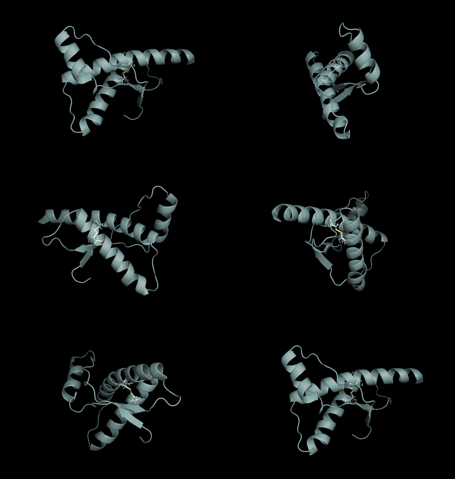
Between the Cys179 and Cys214 residues, a stronger intramolecular force (disulfide bond) bridges helix 2 and helix 3; stabilising the native globular architecture, mechanical integrity and hydrophobic core of the monomeric protein.
Amino acid asparagine on position 181 is documented to carry a sugar chain in real cells, thus the binder would be unable to contact it.
GROMACS analysis
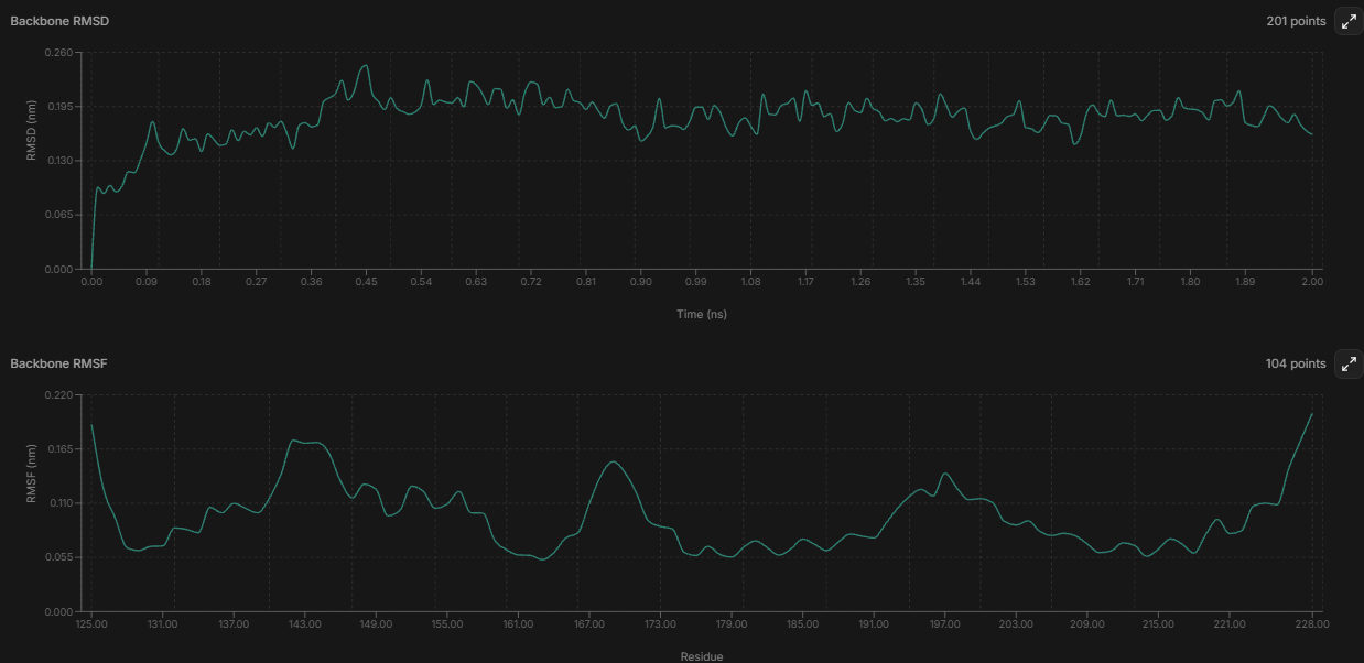
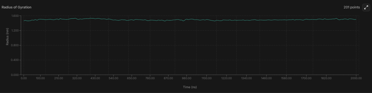
with the small and flat RMSD and Radius of gyration graphics, the structure can be interpreted as compact and not really seeming to “drift away” from the starting shape;
the parts that wiggle the most: the loop around 167-172 matches what the original 1QLX paper reported;
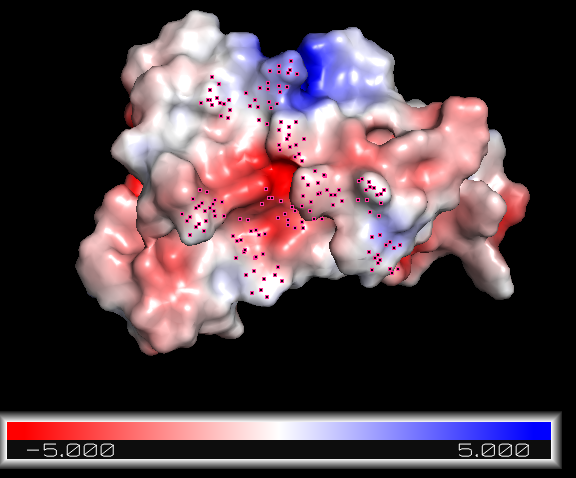
Adaptive Poisson-Boltzmann Solver in PyMol
A good binder should be able to complementary attach to electronegative and/or electropositive regions of the protein.
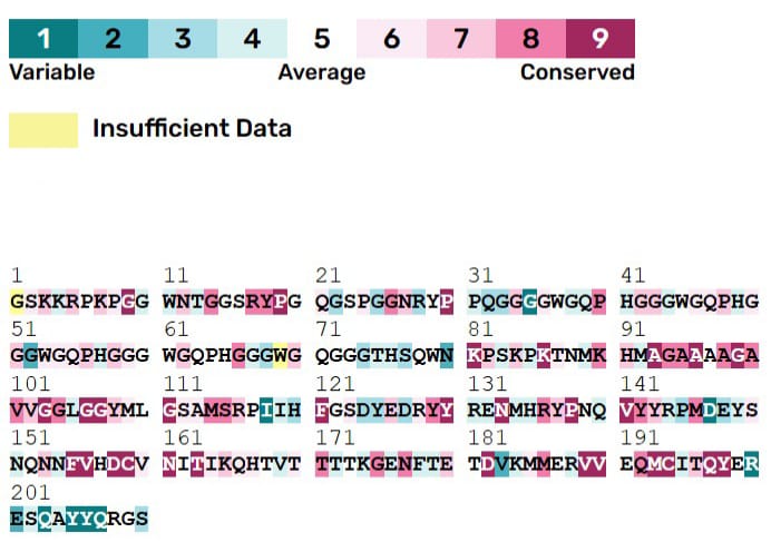
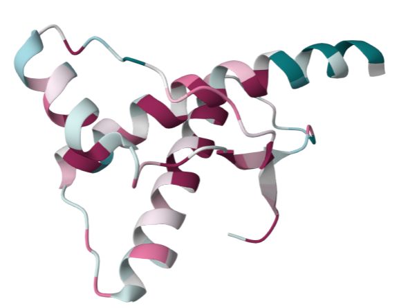
ConSurf analysis
IV. Binding peptide approach
V. Binder generation
Asp178 and Gln212 are both positions of disease-linked mutations.
Batch #1 - BoltzGen
1. Try one
Target residues: all from helix 2 (173-194)
Best ipTM (.443), best min PAE: spec_19_4. RMSD = 2.01
Target residues: solvent exposed parts from helix 2 plus some from helix 3
Filter RMSD stood within 1.2-1.9A for every speculative design, all structures should hypothetically fold accordingly.
Best ipTM (.362), best min PAE: spec_00_0. RMSD = 1.372, 2 H bonds, 0% β-sheet
Target residues: none choosen; let BoltzGen choose
Filter RMSD stood within 1.2-1.9A for every speculative design, all structures should hypothetically fold accordingly.
Best ipTM (.460), best min PAE: spec_49_2. RMSD = 14.111, 5 H bonds, 0% α-helix