Millions of probes are launched from Earth on a Starship, destined to be deployed across the universe, each probe contains a cassette, cell free sensors destined to prove human habitability. Their data will improve our understanding of habitable planets in the universe and help humans plan future colonisation missions to them
01
Stage 01 · payload
It 's not an organism, not alive, wont pollute
A cell free cassette, DNA templates to sense the environment. No mutation risks, fewer contamination risks, more radiation resistance than a live cell culture, hurtling through space, hunting, reporting back to human colonies.
02
Stage 02 · interlock
It stays inert, only responding if the planet says yes
Temperature, solvent and chemical triggers activate the logic gates and next actions (see the mindmap below for more info)
03
Stage 03 · sensing
It reads the local chemistry
A controlled panel turns a raw environment into inputs, the logic acts on these.
04
Stage 04 · inference
Molecular Inference
Cell-free logic gates weigh the inputs and perform inference against in-built classifiers, each weight is assessed against earth biocompatibility, allowing the inference to prove biocompatible planets instead of physical visitations
05
Stage 05 · signal
Logic becomes light
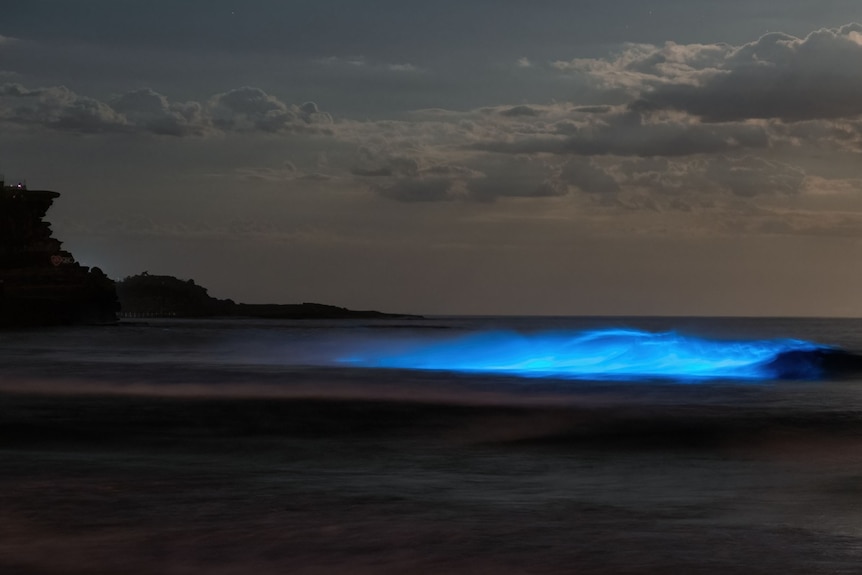
Only the inferenced reporters fire, different schema lighting their set spectral peak and a band in the encoded barcode. This will allow observers to map the spectral signals that the cassette has provided allowing for future colonisation targets
My current mindmap of the factors that each reporter will target.
mindmap
  root((mindmap))
    Physical
      Temperature
      Pressure
      ::icon(fa fa-book)
    Solvent
      Water
      Other solvent available
      No solvent found
    Chemical
      pH
      Redox
      Organics
    Biological
      Candidate biosignatures
    Toxicological
      compounds incompatible with humans
    Local environment
      Atmospheric composition/disequilibrium
    Geological	mineral/elemental environment
    Information anomalous artificial signatures
And the inference map of the planet
Temperature ████████░░
Water activity █████████░
Organic chemistry ███████░░░
Redox energy ████████░░
Toxicity ██░░░░░░░░
Biosignature █████░░░░░
Contamination risk ███████░░░
which is encoded as a fingerprint, wavelength-accurate spectral barcode and sent back to earth
for eg. sfGFP (510 nm), mScarlet-I (594 nm) and mTagBFP2 (457 nm):
When thinking about colonisation of the universe by human consciousness and earth biology, the limitations of our current technological stack become apparent and the challenges ahead seem enormous. I hope with my final project to be able to solve a small part of the problem, ie the ability to autonomously select planets that are biocompatible and suitable for human habitation using biological space probes.
What this would be is a cell-free protein analysis suite, because suitably engineered cell-free molecular system may provide greater containment and reduced biological persistence than a viable-organism probe during inter/intra galactic transit (a biological probe/coloniser as suggested by George Church risks being damaged by environmental radiation associated with space travel with the added risks of biological contamination and providing false positives, dangerous for future human missions).
This probe will wake up on arrival at a targeted planet. Its logic gates sweeps through the local chemistry searching for bio signatures or other signs of civilisations life and releases hyperspectral proteins encoded within a spectral barcode triggered by the logic, as its a cell-free design, the local temperature of the planet will anneal the proteins tot he point where the spectral signature gives us even more information on bio compatibility. These hyperspectral signals will be captured and monitored via a space telescope (maybe using gravitational lensing) or a local probe dedicated to the analysis of the data in that system and the information is transmitted autonomously to the nearest human civilisation outpost.
The costs associated with the space economy is rapidly subsiding, with the age of space data centres, costs of launch are being heavily subsidised by the infrastructure providers to make business sense for launch. Therefore the costs associated with testing this project in space will be trivial in the near future as well as longer term.
#Q2. What living system could make it?
A cell free system
#Q3. How could automation help you make it?
We need a detailed analysis to select the best reporters for the slots in the mindmap, I believe in terms of automation this will be my first step among many
#Q4. What would it take to make it at a meaningful scale?
This is a whole of system architecture that I’ve had in my head and want to test out among my peers, I am unsure if it will be viable to join all the parts together but they work individually and I see no reason why they wont work together, however I’m unsure how I would scale it without having first assembled one myself, I see a pathway to scale and the financial case to scale it, however I’m unsure of the biomanufacturing aspect of it at this stage.
References: CHATGPT Prompt: Hello Mr GPT what do you think of my idea? do you think its viable? how would inference look like here (and then I copied Q1 answer) and it provided me with the inference graph and mindmap ideas to work off.
Week 2: DNA Design & Applications
HTBAA Homework 2 — DNA design
Part 0
My Kernel repository is Clinton Monteiro and the project folder inside it is Space Invaders I, in the HTBAA 2026 workspace.
Construct name: pCF-T7-ho1-BV.
Part 1 — Expression target
What molecule. I am expressing one enzyme, heme oxygenase 1 (HO1). The product I care about is the small molecule that enzyme makes: biliverdin IXα. Biliverdin is a hyperspectral reporter pigment. HO1 opens a heme ring and releases biliverdin IXα, Fe²⁺, and CO. I am encoding the enzyme, because the pigment is not a gene.
This is the first flight readout for the cell-free probe I proposed in Homework 1. That probe is a dried DNA cassette that reads a planetary sample and reports with a spectral barcode a telescope can see. Homework 1 sketched the barcode with fluorescent proteins (sfGFP, mScarlet-I, mTagBFP2). Those stay in the project as bench controls. The long-range channel is the one Chemla, Levin, and colleagues built for hyperspectral cameras: an enzyme whose product has an absorption signature that can be unmixed under ambient light, including from a distance (Chemla et al., Nature Biotechnology, 2025, DOI 10.1038/s41587-025-02622-y). They used two pigments. Biliverdin IXα is made by one enzyme. Bacteriochlorophyll a is a pathway. This assignment is one coding sequence, so the construct is ho1.
What sequence.Synechocystis sp. PCC 6803 heme oxygenase 1, gene ho1 (ordered locus sll1184, synonym pbsA1).
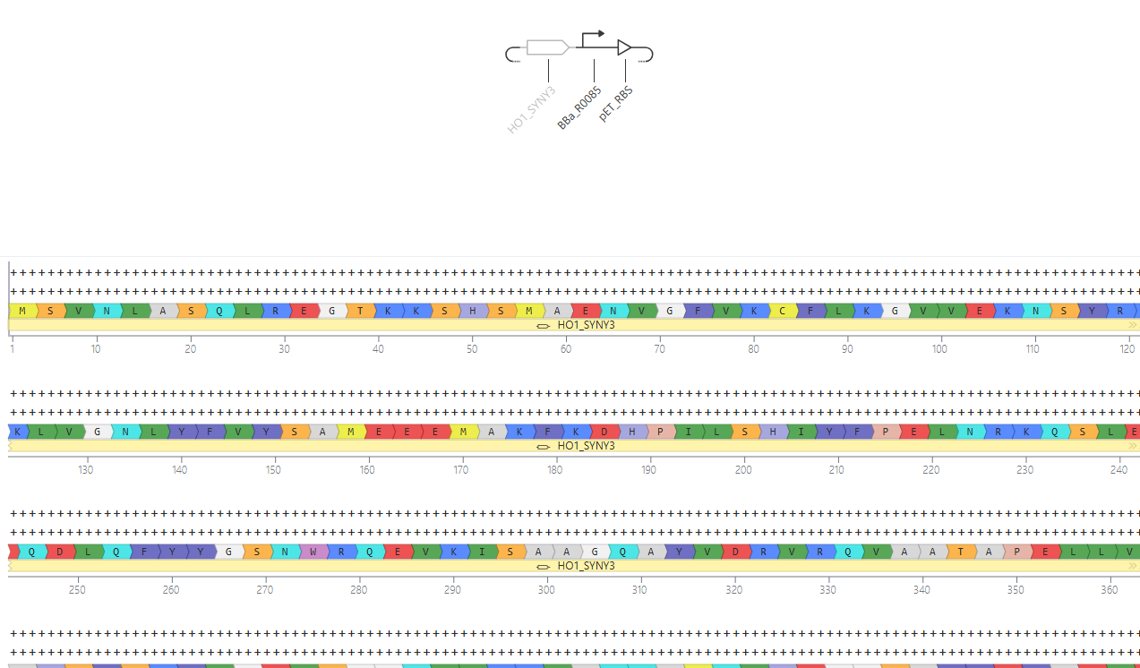
Protein: UniProt P72849 (HO1_SYNY3), 240 amino acids, reviewed, evidence at protein level. GenBank protein BAA16864.1, genome BA000022.
The prototype construct adds a C-terminal GSGSHHHHHH tag, so the translated product is 250 amino acids and then a stop. The tag is for a Western blot. It is not part of the natural enzyme, and the flight cassette can drop it once expression is confirmed.
Host. A T7 cell-free reaction (a PURExpress-style system, or a lysate supplemented with T7 RNA polymerase). Homework 1 committed to a cell-free payload so the probe is not a replicating organism at the destination. The ribosomes in that reaction are E. coli ribosomes, so the part list follows the prokaryotic column: promoter, ribosome binding site, coding sequence, terminator. Codon optimization is for E. coli for the same reason. The plasmid backbone is only how I copy the template on Earth.
Where the product goes. The enzyme is soluble and stays in the reaction volume. Biliverdin accumulates in that same volume. There is no periplasm and no compartment to target, so the construct has no signal peptide and no membrane anchor. The His tag does not change location. It is a detection handle.
One guess, stated as a guess: the dried reaction has to carry heme, oxygen, and a source of electrons. HO1 does not make pigment from amino acids alone. In Synechocystis the electron donor is reduced ferredoxin. I supply heme so that color means the cassette was expressed. Cornejo, Willows, and Beale showed that E. coli expressing this gene accumulates biliverdin and turns green, and that the extracted activity is ferredoxin-dependent (The Plant Journal 15:99–107, 1998). Whether a minimal cell-free mix will do the same is an experiment, not a result I already have. That is Part 6.
Part 2 — Parts
I am using the prokaryotic column because the cell-free ribosome is an E. coli ribosome and the polymerase I add is T7 RNA polymerase.
Name
Type
Sequence and source
Why this part, for this host, for this product
T7_promoter
Promoter
TAATACGACTCACTATAGGGAGA. T7 promoter including the GGGAGA transcribed start. Same element as iGEM BBa_R0085.
Cell-free kits supply T7 RNA polymerase. This promoter is what that polymerase binds. Transcription of the payload happens in the reaction, where the polymerase is added.
pET_RBS
RBS
AAGAAGGAGATATACAT. Ribosome binding site from the pET vector series. Shine–Dalgarno AAGGAG, then 8 bp, and the ATG follows immediately.
E. coli ribosomes initiate best with that spacing. A Kozak sequence would address a eukaryotic ribosome this chassis does not have.
ho1_P72849_His
CDS
Native sll1184 coding sequence from BA000022 / KEGG syn:sll1184, native stop removed, then linker, His6, and TAA. Full 753 bp sequence below. Protein identity is UniProt P72849 plus GSGSHHHHHH.
This is the enzyme that makes the pigment. I start from the natural gene and codon-optimize in Part 4, rather than editing the protein. The tag lets me see the polypeptide if the tube never changes color.
T7_terminator
Terminator
CTAGCATAACCCCTTGGGGCCTCTAAACGGGTCTTGAGGGGTTTTTTG. T7 terminator from pET vectors (iGEM BBa_K731721).
Ends the T7 transcript so the polymerase does not run into the backbone. The run of T’s is the terminator and stays.
Backbone
Origin + selection marker
Reused, not designed. [Name of the Kernel vector]. It provides a high-copy ColE1/pUC-type origin and an antibiotic marker (ampicillin, unless the vector I find uses another marker).
I need many copies of template DNA for cell-free reactions. The origin and the marker do that job in E. coli during template prep. They have no job inside the probe.
CDS to paste as ho1_P72849_His (753 bp). Frame: ATG, then P72849, then GSGSHHHHHH, then one TAA. No internal stop.
The cassette without the backbone is 841 bp: promoter 23, RBS 17, CDS 753, terminator 48. The ribosome binding site ends in ACAT and the coding sequence starts ATG, so the junction is ACATATG. That is one start codon, in frame, with 8 bp between the Shine–Dalgarno and the ATG. The natural ho1 stop is TAG. I removed it before the tag. Leaving it in would have ended the protein at residue 240 and deleted the His tag. The only in-frame stop in the coding sequence is the final TAA.
The backbone’s resistance gene is its own transcription unit. It does not have to point the same direction as the T7 cassette. The T7 cassette is the payload. The resistance gene is for selecting the manufacturing host on Earth.
After codon optimization in Part 4, the CDS DNA changes and this map should be rebuilt from the optimized part. The part order stays the same. The protein stays the same.
Part 4 — Optimize and quality-check
Codon optimization
I selected the coding sequence in pCF-T7-ho1-BV and ran Kernel’s codon optimizer with E. coli as the target. Constraints I set: avoid EcoRI (GAATTC), XbaI (TCTAGA), SpeI (ACTAGT), PstI (CTGCAG), and BsaI (GGTCTC / GAGACC), and keep GC in a synthesis-friendly range around 40–60%.
[Kernel report: CAI before → after, GC before → after. Screenshot.]
Codon usage matters here because the gene is from a cyanobacterium and the ribosome is from E. coli. Synonymous codons are not equally fast or equally available. The native 720 bp coding sequence uses several codons that are rare in E. coli: CCC (Pro, three times), CGG (Arg, four), CGA (Arg, once), and CTA (Leu, once), plus a less-preferred TTA leucine. It is also about 50.6% GC, which is already a comfortable synthesis number, but it contains AAAAA at position 39 and GGGGG at position 386. Those homopolymers are where chemical synthesis slips.
Optimization is allowed to change DNA and is not allowed to change protein. After the run I translate the new CDS and check that it is still P72849 plus GSGSHHHHHH, with one stop at the end. A higher codon adaptation index means the codons are the common E. coli ones. It does not mean the protein will fold, and it does not mean biliverdin will appear. Those are measured in Part 6.
A preview, calculated from the Kazusa E. coli K-12 codon table rather than from Kernel: CAI of the 240 native codons is about 0.69, and a synthesis-cleaned E. coli optimization of the same protein lands near 0.96, with the nine rare codons gone and GC still near 53%. If Kernel’s numbers differ, I report Kernel’s numbers. The optimized DNA in my notes is a sanity check, not a substitute for the run.
Sequence search
[BLAST: top hit, percent identity, E-value, on the native CDS and on the optimized CDS. Screenshot.]
I search the native coding sequence as nucleotides and the optimized coding sequence both as nucleotides and as a translation.
What I conclude from the pair of searches: before optimization, the DNA should be the natural Synechocystis gene, a full-length match to ho1 / sll1184 over 720 bp, with the His-tag tail unmatched because I added it. After optimization, nucleotide identity to that gene should fall, because the codons were rewritten, and a translated search should still hit UniProt P72849 at 100% over the 240 amino acids. The ten-residue tag is the only novel peptide, and it is a His tag. If the closest protein hit is some other enzyme, the construct is the wrong gene and I stop.
Safety screen
[SecureDNA result: clear, or the flagged coordinates. Screenshot.]
I ran Kernel’s SecureDNA screen on the construct. The screen compares a sequence to hazard lists synthesis companies use: toxin genes, pathogen sequences, virulence factors, and related sequences they are not supposed to print. It is a print-permission check.
HO1 is a cyanobacterial pigment enzyme. I expect a clear result. A clear result means a vendor has no biosecurity reason to refuse the DNA. It does not mean the probe is something to release; the design is a cell-free reaction I run in a tube. The reaction releases one CO per heme cleaved. That is catalytic chemistry in a reaction I supplied with substrate, and it is not a reason to mutate the enzyme.
If the screen flags a window, I read the coordinates before changing anything. A short accidental match gets recoded with synonymous codons if it is real, or ignored if it is a His-tag or terminator hit that the report itself explains. I would ask the TA before ordering a flagged gene. I would not edit the active site to chase a flag I have not read.
Part 5 — From design to build
How I would build one. I would order the 841 bp cassette as a single clonal synthetic gene, already cloned into the high-copy backbone, and I would read the vendor’s sequence alignment before I used it. At this length a commercial gene is one assembled fragment plus a sequenced colony. Gibson assembly is the wrong tool for the first build of one short cassette, and a chip oligo pool is the wrong tool for one sequence.
Gibson assembly is how this becomes the probe later. Gibson et al. (Nature Methods 6:343–345, 2009) chew back short overlaps and seal them in one isothermal reaction, and the method works up to hundreds of kilobases. I would use it to insert this cassette into a backbone I already have, and again when the barcode is several transcription units (this enzyme, a second pigment, a fluorescent bench control) on one plasmid.
What synthesis constrains. Chemical synthesis errs at a low rate per base, so a perfect 841-base oligonucleotide is the wrong object to order. Vendors synthesize short oligos, assemble them, and throw out the failures. Carr et al. (Nucleic Acids Research 32:e162, 2004) do that cleanup with MutS, which binds mismatches in a heteroduplex so error-containing strands can be removed. Tian et al. (Nature 432:1050–1054, 2004) do it by hybridization selection on oligos that came off a microarray. For this prototype I am buying the version of that pipeline in which the company sequences a clone and ships the one that matched. I still look at the trace.
The sequence-specific risks in my design are the native homopolymers, AAAAA and GGGGG, which codon optimization removes, and the six T’s in the T7 terminator, which are the terminator and stay. If a vendor’s complexity filter rejects that poly-T, I order promoter, RBS, and CDS only, and add the terminator from a stock plasmid. I do not shorten the poly-T.
Prototype versus scale. Testing the idea takes one correct plasmid and a few cell-free reactions. That is one gene order and a miniprep.
Putting the same cassette into many identical probes does not mean synthesizing it again. I transform E. coli, grow it, and prep plasmid, or I amplify the cassette by PCR. Scale of identical DNA is fermentation. Kong et al. (Nucleic Acids Research 35:e61, 2007) is the related move when I instead want many different assemblies in parallel at small volume.
A library is the point where chip synthesis earns its place. Different pigments, a ladder of ribosome binding sites, and the sensor gates that will eventually replace the always-on T7 promoter are many sequences. Kosuri et al. (Nature Biotechnology 28:1295–1299, 2010) amplify chosen subsets out of a microarray oligo pool so those variants can be built together. Elowitz and Leibler (Nature 403:335–338, 2000) are why the library has to include expression-strength variants: their three-repressor oscillator only oscillated inside a window of promoter and RBS strength. I should expect a barcode bit to be just as sensitive to dose. Stanton et al. (Nature Chemical Biology 10:99–105, 2014) are the part list for the gates themselves, mined orthogonal repressors, which is future work on top of this cassette rather than a change to ho1.
Part 6 — What I do not know yet
Will this promoter and RBS make enough enzyme in the kit I actually use? T7 plus the pET ribosome binding site is a strong default. Elowitz and Leibler had to tune dose before their circuit behaved, and I expect the same. I would run the cell-free reaction with and without the plasmid, Western-blot against the His tag, and read absorbance near 376 nm and near 660–670 nm, where biliverdin absorbs. The next design variant is a short RBS ladder in front of the same gene.
Will the enzyme turn over heme in a cell-free mix? A positive Western blot only shows the polypeptide. Cornejo et al. (1998) got green E. coli and showed the activity wants reduced ferredoxin. A minimal PURE reaction may make HO1 and make no pigment. I would run three tubes: complete reaction with heme, the same reaction without heme, and heme with no DNA or with boiled mix. If the blot is positive and the complete tube is colorless, I would move to a lysate that still has reducing metabolism, or add ferredoxin and a chemical reductant. I would not rewrite the gene before that result.
Is biliverdin a separate bit next to the fluorescent proteins from Homework 1? Chemla et al. showed it can be unmixed from outdoor backgrounds with a hyperspectral camera. I have not shown it can be unmixed from sfGFP, mScarlet-I, and a second pigment in one tube. I would scan an HO1 reaction and a fluorescent-protein reaction on the same instrument and see whether the peaks separate. That result decides whether bacteriochlorophyll is worth designing as bit two, and whether to replace this always-on T7 promoter with a Stanton-style sensor so the bit turns on only when the sample meets the condition.
The tag, the Earth-only backbone, and the always-on promoter are prototype choices. I know they are bets about manufacturing and measurement. The flight version drops the tag and puts logic in front of the same enzyme.
AI citation
Client: Grok 4.7 (xAI), 28 September 2026.
Prompt, in substance: using my Homework 1 cell-free planetary probe and Yonatan Chemla’s hyperspectral reporters, design the Homework 2 expression construct and draft the Kernel write-up. I asked for the part choices, the sequences, how to run codon optimization, BLAST, and the safety screen, and how the single cassette connects to the final project.
Kernel AI prompts I use while building:
“What is the difference between a part and a construct, and where do I codon-optimize a CDS?”
“Find a T7 promoter, a strong E. coli RBS, a T7 terminator, and a high-copy ampicillin backbone in this workspace.”
“I am expressing Synechocystis heme oxygenase in a T7 cell-free system. Is E. coli the right codon-optimization host? Does this protein need a signal peptide?”
Kernel’s codon-optimization report, BLAST hits, and SecureDNA result are the tool’s outputs, and the screenshots are mine. The Kazusa CAI numbers above are a preview, not a Kernel report.
References
Chemla, Y., Levin, I., Fan, Y., Johnson, A. A., Coley, C. W. & Voigt, C. A. Hyperspectral reporters for long-distance and wide-area detection of gene expression in living bacteria. Nature Biotechnology (2025). https://doi.org/10.1038/s41587-025-02622-y
Cornejo, J., Willows, R. D. & Beale, S. I. Phytobilin biosynthesis: cloning and expression of a gene encoding soluble ferredoxin-dependent heme oxygenase from Synechocystis sp. PCC 6803. The Plant Journal 15:99–107 (1998). https://doi.org/10.1046/j.1365-313x.1998.00186.x
Elowitz, M. B. & Leibler, S. A synthetic oscillatory network of transcriptional regulators. Nature 403:335–338 (2000). https://doi.org/10.1038/35002125
Stanton, B. C. et al. Genomic mining of prokaryotic repressors for orthogonal logic gates. Nature Chemical Biology 10:99–105 (2014). https://doi.org/10.1038/nchembio.1411
Carr, P. A. et al. Protein-mediated error correction for de novo DNA synthesis. Nucleic Acids Research 32:e162 (2004). https://doi.org/10.1093/nar/gnh160
Gibson, D. G. et al. Enzymatic assembly of DNA molecules up to several hundred kilobases. Nature Methods 6:343–345 (2009). https://doi.org/10.1038/nmeth.1318
Kosuri, S. et al. Scalable gene synthesis by selective amplification of DNA pools from high-fidelity microchips. Nature Biotechnology 28:1295–1299 (2010). https://doi.org/10.1038/nbt.1716
Kong, D. S. et al. Parallel gene synthesis in a microfluidic device. Nucleic Acids Research 35:e61 (2007). https://doi.org/10.1093/nar/gkm121
Tian, J. et al. Accurate multiplex gene synthesis from programmable DNA microchips. Nature 432:1050–1054 (2004). https://doi.org/10.1038/nature03151
Week 3: Cell-Free Systems, Cloud Labs, & Field trip to Ginkgo Bioworks
CFPS Arena → See how every coopetition design compares on $/g, explore the price map, and cost your own mix.
sfGFP (PDB 2B3P; UniProt P42212)
PETase (PDB 4CG1; UniProt Q6A0I4)
Reteplase (PDB 1RTF; tPA UniProt P00750)
Goal: using the collective intelligence of the HTBAA community and accessible AI systems, design novel CFPS reaction compositions for each of the three target proteins: sfGFP, PETase, and Reteplase to reduce the cost per gram of functional protein. Each design should be compatible with the experimental framework described above and should reflect the distinct expression and folding requirements of that protein. You can learn more about the details of the coopetition here.
A. COMPARE CELL-FREE REACTION COMPOSITIONS (20 minutes)
Why does the concentration of potassium glutamate and magnesium glutamate differ between compositions? Which is more sensitive?
Reasons for Differing Concentrations
The concentrations differ because these ions are essential for cell-free protein synthesis (CFPS) and the molecular interactions required for transcription and translation [5]. Specifically:
Optimization for specific strains: In some systems, the concentrations of these two ions are adjusted to account for the specific requirements of a given cell extract strain [5].
Mimicking the cytoplasmic environment: Research has shown that modifying salt concentrations to better recreate the E. coli cytoplasmic condition—such as using glutamate as the preferred anionic species—can influence protein expression [3].
Optimization goals: Different formulations aim for different outcomes, such as maximizing protein yield (e.g., the PANOx-SP system) or minimizing the reagent cost per gram of protein [1, 7].
Sensitivity
While the provided text does not explicitly state “magnesium is more sensitive than potassium” in a single direct sentence, it provides data regarding the sensitivity of different components:
Ammonium glutamate: Protein expression yields were noted to be “nearly insensitive” to ammonium glutamate concentrations between 0 and 100 mM [3].
Magnesium and Potassium Glutamate: One excerpt notes that the concentrations of these two ions are “essential” for CFPS [5]. In the context of optimization, researchers have performed sequential optimization of these ions (for example, optimizing magnesium glutamate between 1–6 mM while fixing potassium glutamate at a mean of 80 mM) to find the ideal balance for protein synthesis [5].
(Note: The specific comparison of which of the two—potassium or magnesium—is more sensitive is not explicitly answered in the provided excerpts, though they are both identified as key variables for optimization [5, 8].)
Sources consulted
[1]p.10
HTBAA Homework #3 Cell Free Reaction Design Google Docs
[2]p.5 [3]p.5 [4]p.4
Mimicking the Escherichia coli Cytoplasmic Environment Activates Long-Lived and Efficient Cell-Free Protein Synthesis (2004)
[5]p.33
GENETICALLY EXPANDED CELL FREE PROTEIN SYNTHESIS SYSTEMS, METHODS AND KITS (2017)
[6]p.16 [8]p.10
Using a GPT-5-driven autonomous lab to optimize the cost
[7]p.2
Olsen2026 low cost reagents NatCommun
Explain how a PEP+NTP reaction formulation is different from a Ribose+NMP reaction formulation.
Based on the provided excerpts, there is no information that explains the difference between a “PEP+NTP” reaction formulation and a “Ribose+NMP” reaction formulation.
The excerpts do discuss different energy substrates and their effects on cell-free protein synthesis (CFPS), but they do not mention “Ribose+NMP” formulations:
PEP (Phosphoenolpyruvate): The text notes that PEP is a high-energy phosphate compound used to regenerate ATP [3]. However, using PEP as an energy substrate can lead to a rapid and sustained increase in phosphate levels, which can eventually inhibit protein synthesis [5, 7].
Pyruvate: The “Cytomim” system uses pyruvate as an energy source, which results in more stable pH and much lower phosphate accumulation compared to PEP-based systems [3, 5].
Other substrates: The text mentions “sacrificial substrates” like kinases and polyphosphate systems used to maximize yield, as well as the use of natural catabolic pathways like glycolysis and oxidative phosphorylation [8].
Because the term “Ribose+NMP” does not appear in the provided library, I cannot provide a comparison between the two specific formulations requested.
Sources consulted
[1]p.10 [6]p.5
HTBAA Homework #3 Cell Free Reaction Design Google Docs
[2]p.3 [3]p.7 [5]p.6
Mimicking the Escherichia coli Cytoplasmic Environment Activates Long-Lived and Efficient Cell-Free Protein Synthesis (2004)
[4]p.5 [7]p.5
Olsen2026 low cost reagents NatCommun
[8]p.9
Rasor2021 sustainable cell free COBIOT OSTI
Choose two cell-free reaction compositions from the literature, compare and contrast the choice of reagents and the concentration of reagents.
observatory
Based on the provided literature, two cell-free reaction compositions that can be compared are the PANOx system and the Cytomim system [8].
Comparison of Reagents and Concentrations
The primary differences between these two systems lie in their energy substrates, the types of glutamate used, and their buffering strategies.
Energy Substrates and Metabolism
PANOx System: Utilizes Phosphoenolpyruvate (PEP) at a concentration of 33 mM as its energy substrate [8].
Cytomim System: Does not use PEP; instead, it uses Sodium pyruvate at a concentration of 33 mM [8].
2. Glutamate and Salt Composition
PANOx System: Relies heavily on Potassium glutamate at a high concentration of 200 mM, but contains 0 mM of both Magnesium glutamate and Ammonium glutamate [8]. It also uses 80 mM Ammonium acetate [8].
Cytomim System: Uses a more balanced approach to glutamate. It contains 8 mM Magnesium glutamate and 10 mM Ammonium glutamate [8]. Its Potassium glutamate concentration (130 mM) is significantly lower than that of the PANOx system [8].
3. Buffering and Other Additives
PANOx System: Uses HEPES KOH (57 mM) for pH buffering and includes Polyethylene-glycol (PEG) at 2% [8]. It also contains 2.7 mM Sodium oxalate [8].
Cytomim System: Uses Tris (2.4 mM) for buffering rather than HEPES [8]. It contains a slightly higher concentration of Sodium oxalate (4 mM) compared to the PANOx system [8].
Summary Table of Key Reagents
| Reagent | PANOx System | Cytomim System |
| :— | :— | :— |
| Energy Substrate | 33 mM PEP [8] | 33 mM Sodium pyruvate [8] |
| Potassium glutamate | 200 mM [8] | 130 mM [8] |
| Magnesium glutamate | 0 mM [8] | 8 mM [8] |
| Ammonium glutamate | 0 mM [8] | 10 mM [8] |
| Buffering Agent | 57 mM HEPES KOH [8] | 2.4 mM Tris [8] |
Sources consulted
[1]p.10 [3]p.10 [4]p.2
HTBAA Homework #3 Cell Free Reaction Design Google Docs
[2]p.33
GENETICALLY EXPANDED CELL FREE PROTEIN SYNTHESIS SYSTEMS, METHODS AND KITS (2017)
[5]p.1 [6]p.10 [7]p.2
Olsen2026 low cost reagents NatCommun
[8]p.2
Mimicking the Escherichia coli Cytoplasmic Environment Activates Long-Lived and Efficient Cell-Free Protein Synthesis (2004)
Note: A recommended starting point would be to review a least one published formulation, such as PANOx-SP (Jewett & Swartz 2004), a cost-optimized formulation from Olsen et al. (2026), and/or an AI-designed formulation from Smith et al. (2026).
B. DESIGN ONE REACTION COMPOSITION FOR EACH OF THE THREE PROTEINS (40 minutes)
Sign in to coopetition.htgaa.org and publish your first unique 20 µL CFPS reaction composition for each of the following (don’t worry, you can resubmit later):
sfGFP
PETase
Reteplase
Note: if you’re unable to login with your HTGAA keycloak account, please post in Zulip so that you can participate in the coopetition!
For each composition, specify the reagents you would adjust from the relevant categories, including salts, energy sources, building blocks, cofactors, and redox components.
As a HTBAA research team, we will have the opportunity to suggest new reagents to add to the cell-free reagent inventory. Contribute (or upvote) at least one reagent to the “Community Reagents” section of the website and provide a short rationale. Include in your homework documentation how you decided on that specific reagent and provide links to any relevant research.
Visit the cfps-coopetition forum page to ensure you will be able to take part in the discussion. Bonus: send a note in the channel about a CFPS optimization idea that you have! If you would like clarification on aspects of the project, please message in this dedicated channel.
C. ASIMOV KERNAL (OPTIONAL)
Build T7 compatible plasmids for cell-free expression using the amino acid coding sequences below:
Bonus question: design a T7 helper plasmid that you could add to the cell-free reaction to improve the yield of functional reteplase (general chaperones, disulfide-bond machinery, etc).
I like the idea of using light as a transmission mechanism, light by itself is faster than all other methods used for interstellar communication and encoding information using spectral differentiation makes sense. maybe one day we might be able to tune gravitational waves for higher accuracy in data transmission, but until that day light will provide the accuracy needed for our inference needs