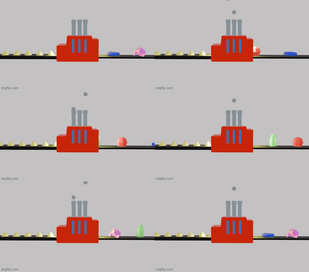
(Fun fact! This gif was made entirely by yours truely. You may find many images and gifs like this here. They’re not too impressive, but enjoy!)
About me
I am a lover of art, pholosophy, and their connection with biology.
Contact info
Homework, Labs, and Projects
See the nav bar on the left to look through my work for How To
Biomanufacture Almost Anything (Fall 2026)!
Subsections of Daryoush Afshinjoo — HTBAA Fall 2026
Week 1 HW: Final Project Ideas
A Factory of Accidents
One input, one process, a variety of products.
I am interested in creating a process, in which the varying of the output is used as a form of bioart.
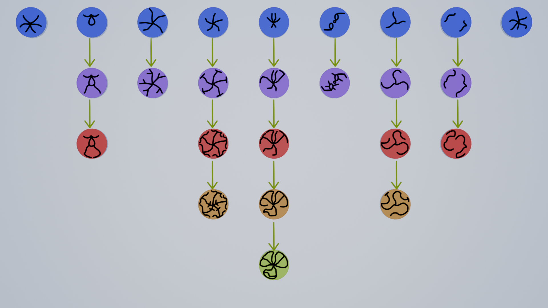
The basic idea is to create a biomanufacturing process in which a living system repeatedly produces different visual patterns. Each production cycle would generate several possible “artworks”. Rather than manufacturing one predetermined object, the process would produce a population of variations. Several human viewers will then select a number of results they see as desirable. The two least chosen designs are not continued, and the conditions to create the desirable artworks are then repeated with small variations. This process is repeated until one artwork remains, being statistically the most desirable of all results.
The catch to this kind of art is that the final peice is not simply the most chosen artwork but the entire tree that lead to the creation of its design.
(This example is purely to show the final displayed piece and its artworks are not accurate nor related to possible designs.)
This art is simultaneously a way to show the influence of evolution, and more specifically, natural selection, in the production chains that exist today; with the added advantage of a human veiwer in the loop, leading to artificial selection. This piece also, by displaying the entrie tree leading to the most selected artwork, shows how living artwork hold both a disadvantage and an advantage in having higher chances of variation over traditional and even AI made art. In machine learning, the training of the model (towards our desired design) is hidden as a blackbox. This alternative method instead shows the thought behind the final artwork, in a way reducing the waste of the less chosen artworks.
Furthermore, this art addresses a philosophical matter as well. Does selection discover value, or create value?
This is because in the creation of this bioart, there is no “intended” design immediately visable. And it is the choice and selection of the human viewers that determines if the artwork is closer to the desired design or farther from it. This can both be seen as the the opinion simply finding the value in the artworks, or, by chosing them, creating it and leading to the final artwork being leaned more towards what they found beautiful.
Lastly, I believe that this method is not simply limited to art, and can be extened in fields such as datastorage and biological computing.
The living system I would use
For my project, I would like to use swarming bacteria as my living system.
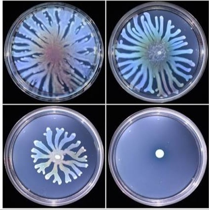
More specifically, I believe the swarming labratory strain of E-coli K-12 to be an appropriate choice. When E. coli K-12 is placed on a moderately soft agar plate, in a rich medium, cells elongate, produce more flagella, become multinucleate, and spread rapidly outward in a thin, highly motile layer.
The E. coli K-12 is nonpathogenic and catogrized under BSL1, removing the health and safety concerns that would arrise if the artwork was made using other pathogenic swarming bacteria (such as Salmonella). Moreover, Unlike in most other swarming species, cells of laboratory strains of E. coli K12 do not secrete surfactants (e.g., lipopeptides or glycolipids). Nevertheless, cells move over the surface of agar in a liquid film, under conditions in which they do not adsorb to the agar or stick to one other (Darnton et al, 2010).
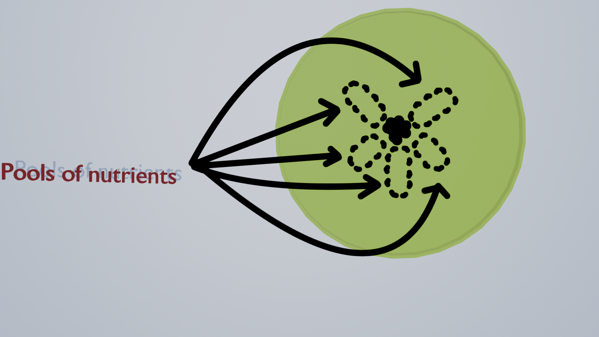
The method that I would use to create the needed artworks from these bacteria colonies is the use of nutriant concentration. I have chosen to paths to take in regards to this method:
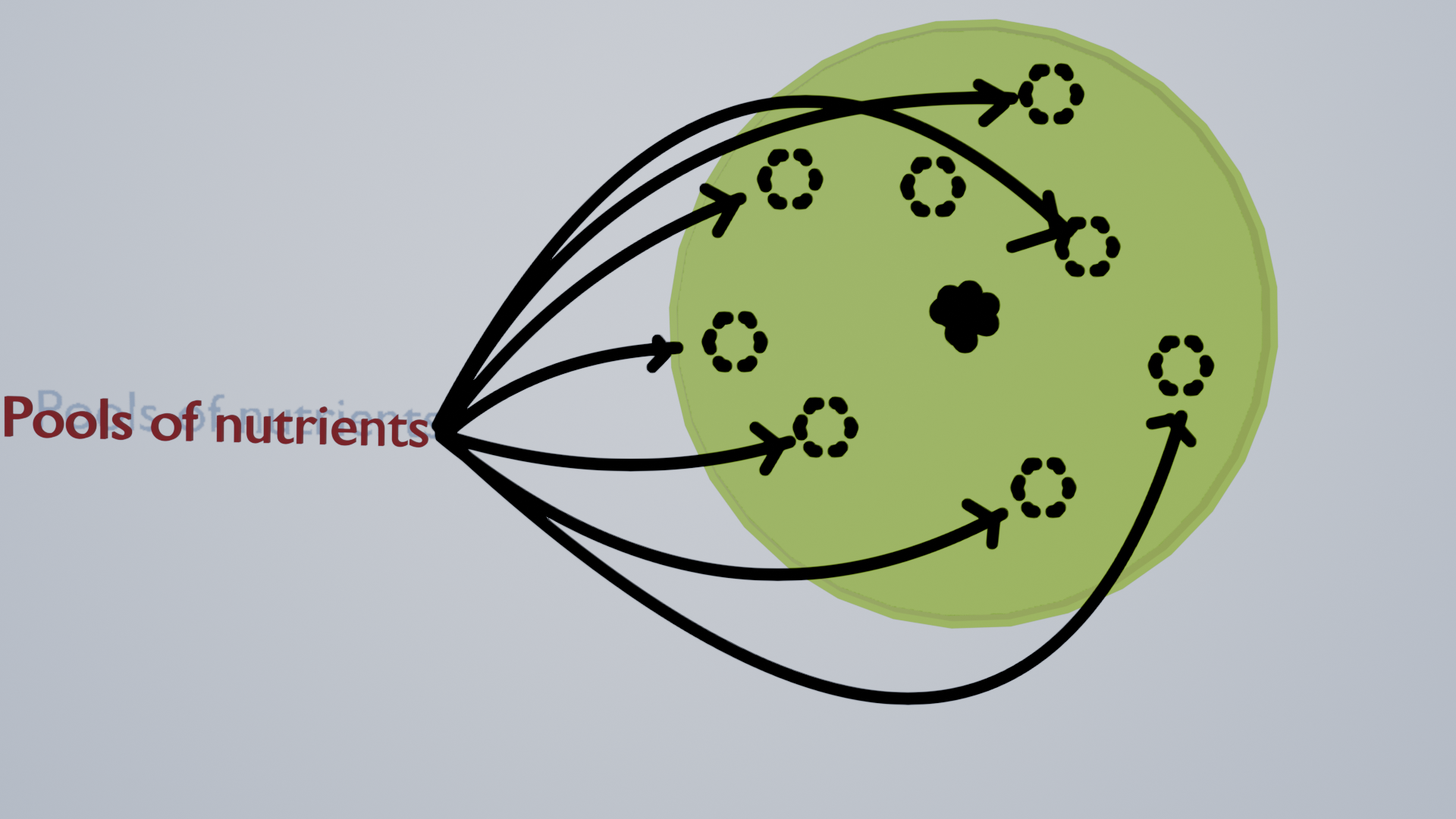
Uncontrolled Chance: In which the nutrients are sprayed onto the plate in a random manner, inducing the bacteria to swarm to different directions and create a specific and unique artwork.
Controlled Chance: In which specific parts of the general design of future artworks are considered, and each plate has a random amount of nutreints deposited on where these parts of art would be.
The form of automation I would use
I would use automation primarily to increase the number of independent manufacturing experiments and systematically record their outputs.
A possible platform is the Opentrons Flex, which supports automated liquid handling with 1-, 8-, and 96-channel pipettes as well as automated movement of laboratory plates and other labware. For this project, automation could be used to standardize the preparation and handling of many parallel experimental inputs, while automated imaging could record the development of the resulting patterns. This is important conceptually as well as practically. I want the manufacturing process to be highly standardized while the biological output remains variable. The automation would therefore control as many non-biological variables as possible without attempting to dictate the final shape.
I would use this system to automate handling and culturing of each plate, as well as the randomness of each plate’s artwork creation.
How much I would make and how long it would take
A For a prototype of this piece, I would create 10 plates of artwork and based on the selection of the human in this loop, I will discontinue two plates and create 8 new plates in the next generation based on the 8 remaining ones of the first round. This process will be repeated until only two artworks remian in the final generation, and one is chosen as the most desirable. Therefore, in this prototype version of the art, I would need to create 30 plates of E-coli K-12 for each peice of this art.
B Given the average time needed for a colony of E. coli to grow, and the number of plates needed for each complete form of this art, I would estimate 1 to 2 weeks per generation, adding up to about 4 to 10 weeks of time needed for a full prototype of this art.
Things don’t know yet
I have not yet fully considered what each artwork would generally look like, and neither if they would be different enough with the methods I had chosen.
I would need to learn more about the process of swarming in bacteria such as E. coli, and how changing the conditions of their growth could be used to manipulate their final form. I also am not yet certain if each new chosen generation will be put in the same conditions of its previous selected design or be an average between two chosen designs.
Week 2 HW: DNA Design
Week 2 HW: DNA Design
0. Preparations:
While waiting for access to Kernel from my node, I thought of what my potential DNA design may be. I initially sought to engineer Tar, a native E. coli chemoreceptor, so that I could directly alter what chemical landscapes the bacteria responded to. I ultimately abandoned this approach because it was solving a problem that the organism already solves naturally: E. coli can detect and chemotax toward a range of nutrient-related attractants. More importantly, changing the sensory system would make the project increasingly about engineering bacterial navigation rather than about observing and recording the variability that emerges from a biological system.
My second idea was far more promising however, being to engineer E. coli to create bioluminescent proteins when exposed to chemicals within the nutrient pools that caused chemotax. This plan, while seemingly more artistic and vanitary than the first, would serve an actual purpose in the designs created by it. Not only would it serve to make each artwork have more of an appealing look, it would also leave evidence of its evolution from each stage to the next through these protiens.
The repository I have created inside of Kernel is named “Daryoosh Afshinjoo HTBAA 2026”, and the folder inside it “Afshinjoo-Factory of Accidents”.
1. Defining my expression target:
My target is a bacterial bioluminescence system encoded by the luxCDABE genes from Photorhabdus luminescens. Rather than producing a single reporter protein, this system produces a small set of proteins that work together to generate light. luxA and luxB encode the two subunits of bacterial luciferase, while luxC, luxD, and luxE encode enzymes involved in producing the aldehyde substrate required for the luciferase reaction. Together, these five genes allow a bacterial cell to generate bioluminescence autonomously.
National Center for Biotechnology Information (NCBI). Photorhabdus luminescens fatty acid reductase (luxC), acyltransferase (luxD), luciferase (luxA), luciferase (luxB), and luxE genes, complete CDS. GenBank accession M90093.1.
My host is Escherichia coli, specifically a K-12-derived laboratory chassis. I chose E. coli because its chemotaxis and motility systems are already well characterized, and because my project relies on allowing the bacteria to interact with a heterogeneous chemical environment rather than engineering movement from scratch. Moreover, E. coli is a non pathogenic BSL-1 bacteria, removing most concerns about the safety of the project.
The luxCDABE proteins need to function inside the bacterial cell, so I am designing for intracellular/cytoplasmic expression rather than secretion.
2. Designing my genetic parts:
Layer
Part
Source
Justification
Transcription start
P_BAD / arabinose-inducible promoter
Existing pBAD/Kernel part
I chose an inducible promoter so that reporter expression is linked to an environmental chemical signal rather than being continuously active.
Translation start
Bacterial RBS
Existing Kernel part / pBAD backbone
I use an E. coli-compatible RBS to provide translation initiation for the reporter genes.
Coding sequence
luxCDABE
Photorhabdus luminescens, GenBank M90093.1
I chose the luxCDABE system because it contains both bacterial luciferase and the enzymes needed to generate its light-producing substrate, allowing the cells to produce light autonomously.
Targeting
None
—
The lux proteins are intended to function within the bacterial cell, so no secretion signal or additional targeting sequence is required.
Transcription stop
Bacterial terminator
Existing Kernel part / pBAD backbone
I use a bacterial terminator to provide a defined endpoint for transcription of the reporter cassette.
Vector context
pBAD-compatible E. coli expression backbone
Existing Kernel backbone
I reuse an established bacterial expression backbone so that replication and selection are already provided rather than redesigning them from scratch.
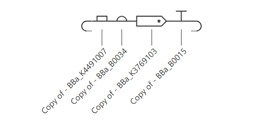
I assembled the construct in the 5′->3′ direction as P_BAD -> RBS -> luxCDABE -> terminator, using an E. coli-compatible expression backbone. The P_BAD promoter provides arabinose-responsive control of the reporter, while the luxCDABE cassette provides the machinery for autonomous bioluminescence. I checked that the components are in the intended orientation and that the coding regions and transcript boundaries are correctly represented in the construct map.
4. Optimizing and quality check:
A) Codon optimization:
Running codon optimization in Vector Builder.
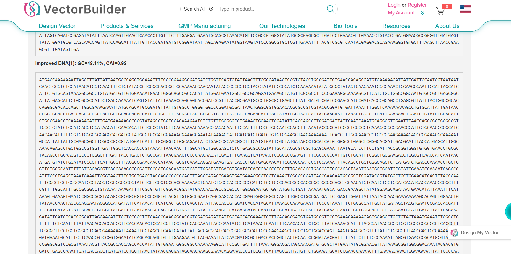
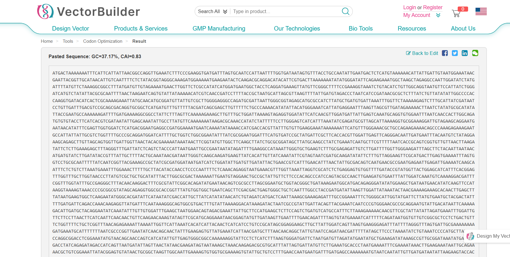
Due to my CDS being a a multigene operon containing five separate coding sequences, I could not run the optimization in Kernel. I ran the luxCDABE coding sequence through VectorBuilder’s codon optimization tool for E. coli. The original sequence had a GC content of 37.17% and a Codon Adaptation Index (CAI) of 0.83. After optimization, the sequence had a GC content of 48.11% and a CAI of 0.92. The nucleotide sequence was therefore substantially altered to better match E. coli’s preferred codon usage while preserving the encoded proteins. The increase in CAI suggests that the optimized sequence is more closely aligned with the host’s codon preferences, which may improve translation efficiency. The increase in GC content also brought the sequence closer to the GC composition more typical of E. coli. I used the tool’s standard optimization for E. coli without imposing additional custom sequence constraints.
B) BLAST:
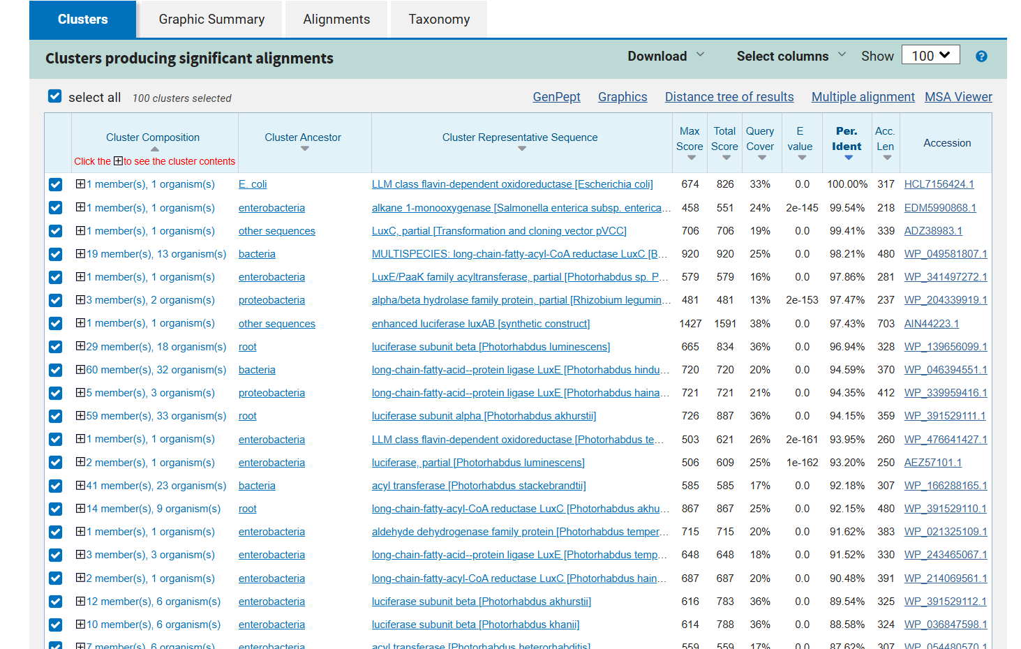
BLAST/sequence search for luxCDABE.
As it can be seen in the image above, the BLAST results for the cassette shows multiple protiens as being close to the search query. The reason behind that is that the cassete contains not only luciferase itself but also the enzymes required to synthesize its substrate, luciferin.
C) Safety screen:
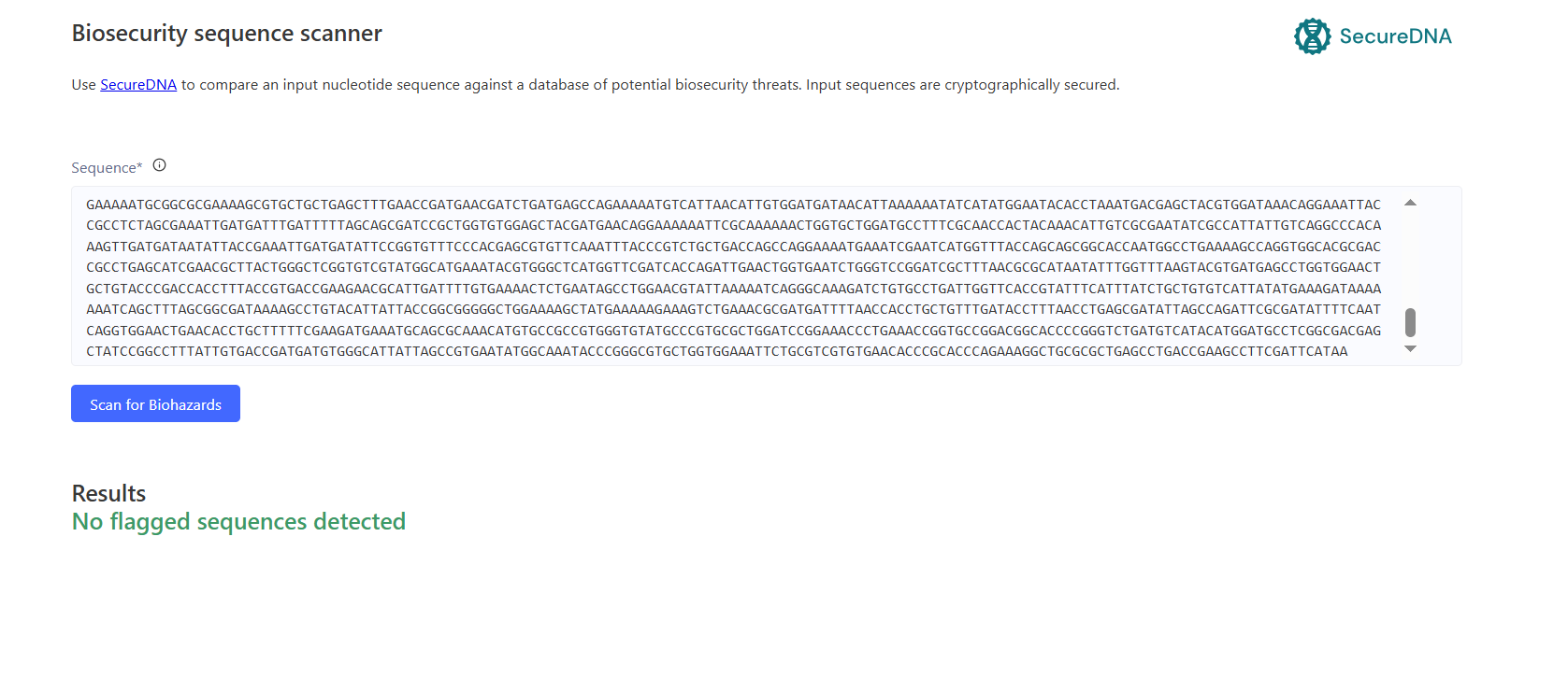
Kernel’s safety screen results.
After having done the biosecurity scan, no flagged sequences were found. Which removes the need for any specific measures for the time being.
5. From design to build:
A) How would I build it?
For the initial prototype, I would order the luxCDABE reporter as synthetic DNA rather than assembling it entirely from short oligonucleotides. My reporter cassette is approximately 5.4 kb long, so it is substantially larger than a typical single coding sequence but still modest compared with the much larger DNA assemblies demonstrated by Gibson et al. Gibson assembly provides a way to join multiple overlapping DNA fragments into a larger construct without requiring the entire sequence to be synthesized as one piece.
For my first construct, I would therefore favor direct synthesis of the reporter sequence and incorporation into an existing E. coli expression backbone. If the complete reporter proved inconvenient to synthesize as a single fragment, I would divide it into several synthetic fragments and use Gibson-style assembly to reconstruct the cassette. This would allow the design to remain modular while avoiding unnecessary redesign of the underlying sequence.
B) What does synthesis constrain?
The main constraint is sequence fidelity. Synthetic DNA is assembled from shorter oligonucleotides, and synthesis errors accumulate as sequences become longer. Carr et al. showed that sequence errors were a major limitation of de novo DNA synthesis and demonstrated protein-mediated error correction that reduced errors by more than 15-fold, illustrating why error correction becomes increasingly important as constructs grow.
My luxCDABE sequence is approximately 5.4 kb and contains five adjacent coding regions, making it more complex than a single reporter gene. However, its optimized GC content is 48.11%, compared with 37.17% for the original sequence, so the optimized version does not appear to have an especially extreme GC composition. I would still expect sequence verification to be important because an error in any of the five coding regions could affect the reporter system as a whole.
C) Prototype vs. scale
For the prototype, I would prioritize simplicity and speed: synthesize the designed reporter sequence, assemble it with the existing expression backbone, and verify the resulting construct.
At a larger scale, the main difference would be the number of constructs rather than the biological product itself. Once a successful construct had been validated, production could shift toward a standardized and repeatable DNA manufacturing workflow. If the project required many different genetic variants, chip-based oligo synthesis and parallel assembly could become more attractive because these methods are designed for high-throughput sequence generation. Kosuri et al. demonstrated this type of parallelization across dozens of genes from large oligonucleotide pools.
6. What I don’t know yet:
1- Will the P_BAD → luxCDABE system produce a useful spatial light signal?
I do not yet know whether the reporter will produce enough light, or an appropriate range of light intensities, to clearly distinguish regions with different levels of environmental exposure. P_BAD regulation and luminescence output may also be nonlinear, so I cannot assume that more arabinose exposure will produce proportionally more light.
How I would find out: I would characterize reporter output across a range of environmental conditions and compare the measured luminescence with the corresponding chemical exposure. This would tell me whether the system provides a useful dynamic range for creating the visual record I want.
2- Will the reporter interfere with the bacteria’s growth or collective movement?
The luxCDABE cassette is relatively large and requires the cell to produce several proteins. This could impose a metabolic burden and potentially change the behavior I am trying to observe. There is therefore a risk that the engineered bacteria will behave differently simply because they are carrying and expressing the reporter.
How I would find out: I would compare the engineered strain with an otherwise equivalent control and measure growth and collective spatial behavior. If expression substantially changes the phenotype, I would investigate alternative expression levels or reporter designs.
3- Will the biological pattern actually provide a meaningful “record”?
The artistic concept depends on the light pattern representing something about the population’s interaction with its environment, rather than simply representing where the bacteria grew most densely. Because arabinose can also affect bacterial physiology, it may be difficult to distinguish signal detection from changes in growth or metabolism.
How I would find out: I would compare spatial luminescence with independent measurements of bacterial distribution and environmental conditions. I would also compare different environmental configurations to determine whether the resulting light patterns consistently reflect the intended chemical landscape.
Final Project
Week-03-HW-Cell-Frees
Subsections of Week-03-HW-Cell-Frees
Week 3 HW: Cell-Free Protocols
Week 3 HW: Cell-Free Protocols
Assignment A:
1.Why does the concentration of potassium glutamate and magnesium glutamate differ between compositions? Which is more sensitive?
Potassium glutamate and magnesium glutamate serve different purposes in a cell-free reaction. Potassium glutamate primarily helps reproduce the ionic environment of the E. coli cytoplasm, where potassium is the predominant intracellular cation. Magnesium glutamate supplies Mg²⁺, which is essential for processes involving ATP, nucleotides, ribosomes, and other components of transcription and translation. The concentrations therefore need to be balanced rather than simply maximized: changing them alters the chemical environment in which the cell-free machinery operates.
The concentrations differ between CFPS formulations because different lysates and reaction chemistries have different optima. For example, PANOx-SP formulations commonly use potassium glutamate at roughly 130–175 mM and magnesium glutamate around 10–20 mM, illustrating that the two salts operate at very different concentration ranges. More recent formulation work likewise treats potassium glutamate and magnesium glutamate as independently tunable variables rather than assuming a fixed ratio.
2.Explain how a PEP+NTP reaction formulation is different from a Ribose+NMP reaction formulation.
A PEP + NTP formulation supplies pre-made nucleotide triphosphates (ATP, GTP, CTP, and UTP) for transcription and uses phosphoenolpyruvate (PEP) as a high-energy phosphate donor to regenerate ATP. In other words, the reaction starts with the expensive, ready-to-use forms of the nucleotides and an energy source that can rapidly regenerate ATP. The course materials identify PEP as an energy-regeneration substrate and NTPs as the building blocks for RNA synthesis. Recent work describes PANOx-SP as a phosphorylated-energy-substrate formulation in which PEP is consumed to generate ATP, producing a rapid initial ATP surge.
A Ribose + NMP formulation takes a more metabolic approach. Instead of supplying all of the nucleotide triphosphates directly, it supplies nucleoside monophosphates (AMP, CMP, GMP, UMP) and relies on enzymes remaining in the E. coli lysate to convert these lower-energy precursors into the NTPs required for transcription. Ribose provides another metabolic precursor that can feed nucleotide-salvage and related pathways. This approach therefore asks the lysate to do more of the work of regenerating the reaction’s nucleotide and energy pools. The 2026 Olsen et al. study found that formulations based on NMPs and inexpensive metabolic substrates could sustain protein production while substantially reducing reagent cost relative to PANOx-SP.
3. Choose two cell-free reaction compositions from the literature, compare and contrast the choice of reagents and the concentration of reagents.
I chose PANOx-SP from Jewett & Swartz (2004) and the optimized reagent formulation (RFopt) from Olsen et al. (2026) because they represent two very different approaches to cell-free protein synthesis.
PANOx-SP is a relatively complex, high-yield formulation based on a phosphorylated energy substrate. It uses PEP at 33 mM to regenerate ATP, along with pre-supplied nucleotide triphosphates (ATP at 1.2 mM and GTP, UTP, and CTP at 0.85 mM each). It also contains potassium glutamate, magnesium glutamate, ammonium glutamate, amino acids, folinic acid, tRNA, NAD, CoA, spermidine, putrescine, and oxalate. The original formulation reported by Jewett and Swartz used 175 mM potassium glutamate and 20 mM magnesium glutamate.
In contrast, RFopt was developed specifically to reduce the cost and complexity of CFPS. Olsen et al. screened 1,231 formulations and arrived at a 12-component optimized formulation that uses non-phosphorylated metabolic substrates rather than PEP. A major change is the replacement of expensive NTPs with nucleoside monophosphates (NMPs), together with potassium glutamate and amino acids. The optimized formulation also incorporates components such as ribose, phosphate, and HEPES rather than relying on the large collection of cofactors and secondary energy substrates used in PANOx-SP.
Assignment B:
Part 1.
My cfps composition table.
K-glutamate — 340 mM
I’m keeping it high because the optimized low-cost formulations found a high-K-glutamate regime useful for protein production.
Mg-glutamate — 8.22 mM
Magnesium is needed by the molecular machinery, but it is more sensitive than potassium. Therefor I decided not to increase it yet.
Amino acids — 3.25 mM
The building blocks of the protien. Needed.
AMP/CMP/GMP/UMP
The lysate has metabolic machinery that can turn them into the triphosphate forms needed for RNA synthesis. That’s one of the main tricks behind the cheap formulation.
I designed my first sfGFP reaction around the low-cost, non-phosphorylated strategy reported by Olsen et al. rather than the PEP-based PANOx-SP formulation. I used potassium glutamate, magnesium glutamate, amino acids, and NMPs while omitting the PEP, NTP, NAD, CoA, and other ancillary components of the conventional formulation. My goal is to reduce reagent cost while maintaining sufficient protein production. I used an RFopt-like composition as my starting point and made a small variation in potassium glutamate concentration to create a distinct candidate for screening. Olsen et al. reported 2.4 ± 0.3 g/L sfGFP at approximately $60/g protein using their optimized formulation.
Part 2.
I upvoted FkpA because it could complement DsbC in the Reteplase reaction by addressing a different part of the protein-folding problem. FkpA is an E. coli chaperone with peptidyl-prolyl isomerase activity. Groff et al. found that adding FkpA to cell-free antibody synthesis reduced aggregation and improved protein solubility and folding, while DsbC primarily improved disulfide-bond formation and assembly. The two therefore addressed different folding bottlenecks and showed complementary effects.
Reteplase is particularly attractive as a target for this idea because it contains nine disulfide bonds and requires an oxidizing CFPS environment with DsbC for correct folding. Although FkpA has not been demonstrated in the cited study specifically for Reteplase, I think it is a worthwhile reagent to test because it could reduce aggregation and improve folding in addition to the disulfide-bond assistance already provided by DsbC. I would therefore treat its benefit for Reteplase as a hypothesis to test, rather than assuming that the antibody results will transfer directly.