I’m Dominic Uriel Orozco Salgado, an undergraduate Biomedical Engineering student at Universidad de Guadalajara (2023–present), with a prior technical degree in Pharmaceutical Chemistry from CETI (2019–2023). I currently work as an Undergraduate Researcher in Biofabrication and Research Assistant in the Department of Translational Bioengineering / Biodigital Innovation Lab.
My work sits at the intersection of biofabrication, embedded systems, and computer vision. A few things I’m currently building or have recently finished:
Undergraduate thesis (in progress): a project on functionalized electrospun fibers, currently exploring fibers that combine electrospinning with cell-free synthetic biology to visually detect hazardous environmental agents (e.g., gaseous formaldehyde) — the direction I’m also developing through this course.
Ocularis: a computer-vision-based augmentative and alternative communication (AAC) system for users with severe motor impairments, using oculomotor tracking (MediaPipe FaceMesh, blink detection, gaze calibration). First-author contribution, finalist at CNIB2026, accepted for publication in the CNIB2026 proceedings (Springer); currently exploring a connectome-constrained neural network (based on the Drosophila optic lobe) as an alternative detection pipeline.
Bioprinting environmental control chamber: designed the electronics and control code (independent PID loops for temperature and humidity) for a low-cost chamber retrofitted onto a Prusa i3, to support consistent bioprinting conditions. Oral presentation accepted at ISBF26.
EEG / neurosemantics: contributing EEG signal processing and spectral analysis to a study on oscillatory traits and contextual states during naturalistic narrative comprehension (manuscript in preparation).
I’m interested in how biofabrication, low-cost embedded hardware, and biology can combine into accessible, real-world tools — whether that’s a communication device, a bioprinting platform, or a sensor you can wear.
Contact info
Homework, Labs, and Projects
See the nav bar on the left to look through my work for How To
Biomanufacture Almost Anything (Fall 2026)!
Subsections of Dominic Orozco — HTBAA Fall 2026
Week 1 HW: Final Project Ideas
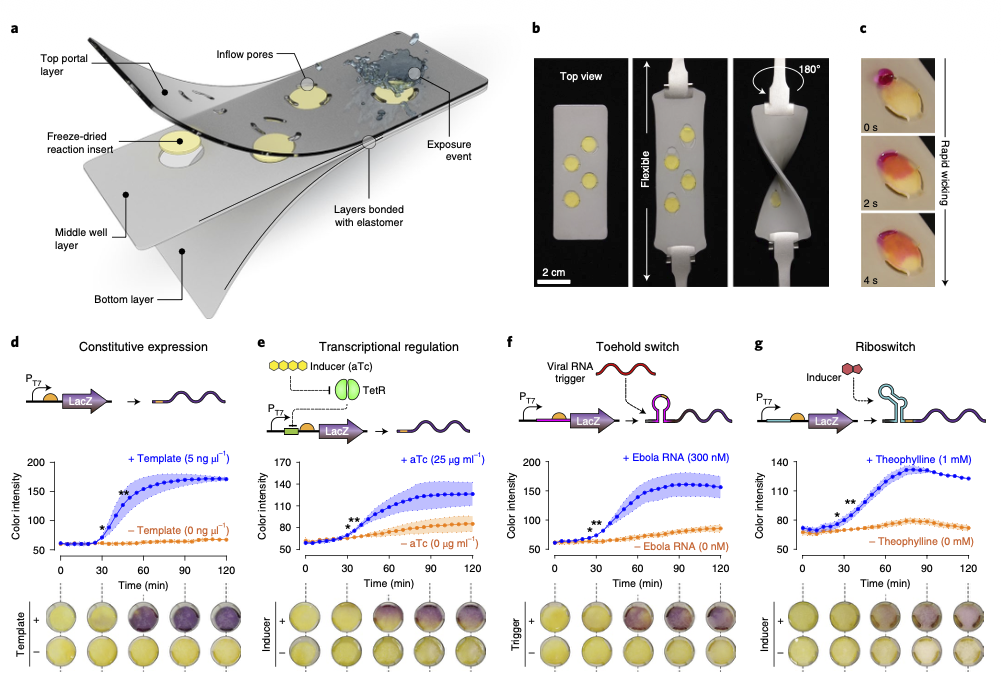
Figure adapted from Nguyen et al. (2021): the wearable freeze-dried cell-free (wFDCF) sensor platform. (a–c) Sensor architecture — a flexible, multi-layer textile insert containing freeze-dried reaction wells that activate on liquid exposure. (d–g) Four genetic-circuit designs (constitutive expression, transcriptional regulation, a toehold switch, and a riboswitch) with their colorimetric readouts over time. Source: Nguyen, P. Q., Soenksen, L. R., Donghia, N. M., Angenent-Mari, N. M., de Puig, H., Huang, A., Lee, R., Slomovic, S., Galbersanini, T., Lansberry, G., Sallum, H. M., Zhao, E. M., Niemi, J. B., & Collins, J. J. (2021). Wearable materials with embedded synthetic biology sensors for biomolecule detection. Nature Biotechnology, 39(11), 1366–1374. https://doi.org/10.1038/s41587-021-00950-3. Reproduced here for educational, non-commercial coursework use — confirm the journal’s reuse terms before publishing this repository publicly.
1. What I would like to biomanufacture and why
I want to biomanufacture an electrospun nanofiber patch that changes color when exposed to gaseous formaldehyde, giving an immediate, electronics-free visual warning of hazardous exposure. The target users are workers with occupational formaldehyde exposure — for example, in leather tanning, footwear and furniture manufacturing, and histology/anatomy labs — industries that are prominent in the region where I live and work, and that rarely have access to continuous, affordable gas monitoring.
Formaldehyde is a recognized human carcinogen with strict occupational exposure limits (e.g., OSHA’s 0.75 ppm time-weighted-average permissible exposure limit). Continuous monitoring today typically relies on electrochemical or photometric instruments that are expensive, need power, and require periodic calibration. A disposable, fiber-based badge — activated only by ambient humidity, needing no battery, and readable by eye — could close a real gap between biomanufacturing and occupational health, especially for small and medium manufacturers that cannot afford commercial gas-detection instrumentation.
This project also lets me combine electrospinning equipment and physical-characterization tools I already have access to with a synthetic-biology sensing layer, connecting directly to my broader undergraduate thesis work on functionalized electrospun fibers.
2. What living system would I use
I would use a cell-free (not cell-based) biomanufacturing chassis: a freeze-dried E. coli transcription–translation (TX-TL) extract carrying a DNA-encoded genetic circuit, embedded in or on the electrospun fiber. This approach has direct precedent: Nguyen et al. (2021) showed that freeze-dried, cell-free synthetic circuits embedded in textiles and other flexible substrates activate by rehydration from an aqueous exposure event and report a target molecule through a colorimetric, fluorescent, or luminescent signal (see figure above) — including circuits for detecting toxic chemical agents. That platform builds on the original paper-based cell-free sensor work of Pardee et al. (2014), which achieved detectable colorimetric outputs in 20–40 minutes at roughly $0.02–0.04 per sensor.
For the specific genetic circuit, I would repurpose E. coli’s native formaldehyde-sensing system, the frmRA(B) operon. The FrmR protein normally represses the Pfrm promoter; in the presence of formaldehyde, FrmR is covalently modified and releases the promoter, allowing transcription of downstream genes (Denby et al., 2016). Concretely:
Input: a TX-TL extract + a DNA template placing Pfrm upstream of a chromoprotein reporter gene (visible by eye, no UV lamp required) + co-expressed FrmR, all freeze-dried onto the fiber.
Process: ambient humidity rehydrates the reaction; gaseous formaldehyde dissolves into that thin aqueous film, modifies FrmR, and derepresses Pfrm.
Output: a visible color change on the fiber once formaldehyde exceeds the design threshold.
As a fallback design (trading the “no living cells” simplicity for stronger signal amplification and longer field stability), whole living bacteria can also be encapsulated directly into electrospun fibers and remain viable for months (Diep & Schiffman, 2023; Liu et al., 2009), including in conductive-biofilm and biomineralized “living ceramic” fiber formats (Massaglia et al., 2021; Schulte-Hermann et al., 2024).
3. How would I use automation
Prototyping and characterization: A desktop liquid-handling robot (e.g., an Opentrons OT-2) would automate DNA assembly (Golden Gate cloning), screening of promoter–reporter combinations, and precise microliter dispensing of the extract + DNA + lyoprotectant mixture onto pre-spun fiber mats — generating reproducible dose–response curves against known formaldehyde concentrations in a controlled gas chamber.
Manufacturing at scale: Single-needle electrospinning (my current setup) suits prototyping but has low throughput (fractions of a gram of fiber per hour). Scaling to production volumes would require transitioning to needle-free or multi-nozzle electrospinning (e.g., Nanospider-type systems), which reach substantially higher throughput — the prototyping and scale-up automation are, deliberately, not the same technology (Petzold & Mukhopadhyay, 2025).
4. How much would I need to make, and how long would it take?
(a) How much?
As a reference scale: supply 1,000 workers with one new patch per week (a reasonable starting scale for one or several small-to-medium tanneries, footwear, or furniture manufacturers). Assumptions:
Each patch is ~4 cm² of functionalized fiber.
Each patch needs ~5 µL of reaction mixture (extract + DNA + lyoprotectant), based on the reaction volumes used in paper-based cell-free sensors (Pardee et al., 2014).
That gives 4,000 cm² of fiber/week and 5 mL of reaction mixture/week. The reaction-mixture volume is trivial to supply — a single cell-free extract batch from 1 L of bacterial culture typically yields hundreds of milliliters of extract, enough for thousands of reactions once diluted.
(b) How long?
A single-needle lab electrospinning setup produces on the order of ~200 cm² of usable mat per 2–4 hour session. Covering 4,000 cm²/week would need roughly 20 sessions/week (~3/day) on one machine — tight but workable at this initial scale. Scaling to, say, 10,000 workers would require the needle-free/industrial electrospinning route described above. Once rehydrated, the sensor’s response time is estimated at 20 minutes to 3 hours, by analogy with the paper- and textile-based cell-free systems reported in the literature (Pardee et al., 2014; Nguyen et al., 2021).
5. What don’t I know yet?
Whether the cell-free components survive electrospinning itself (10–20 kV voltages, organic solvents). Whole living bacteria have been shown to survive electrospinning with specialized protocols and remain viable for months (Diep & Schiffman, 2021, 2023), but a cell-free enzymatic system may be more solvent-sensitive; a safer design might functionalize the fiber after spinning rather than co-spinning the reaction into it.
Real sensitivity and response kinetics on a solid fiber matrix versus in free solution or on flat paper — how gas diffusion into a thin, fiber-supported aqueous film differs, and how much ambient humidity and temperature affect performance.
Shelf life of the freeze-dried reagents once embedded in the fiber at room temperature — this determines whether the weekly production cadence estimated above is realistic, or whether refrigeration/specialized packaging is required.
References
Asin-Garcia, E., Fawcett, J. D., Batianis, C., & Martins dos Santos, V. A. P. (2025). A snapshot of biomanufacturing and the need for enabling research infrastructure. Trends in Biotechnology, 43(5), 1000–1014. https://doi.org/10.1016/j.tibtech.2024.10.014
Denby, K. J., Iwig, J., Bisson, C., Westwood, J., Rolfe, M. D., Sedelnikova, S. E., Higgins, K., Maroney, M. J., Baker, P. J., Chivers, P. T., & Green, J. (2016). The mechanism of a formaldehyde-sensing transcriptional regulator. Scientific Reports, 6, 38879. https://doi.org/10.1038/srep38879
Diep, E., & Schiffman, J. D. (2021). Encapsulating bacteria in alginate-based electrospun nanofibers. Biomaterials Science, 9(12), 4364–4373. https://doi.org/10.1039/D0BM02205E
Diep, E., & Schiffman, J. D. (2023). Electrospinning living bacteria: A review of applications from agriculture to health care. ACS Applied Bio Materials, 6(3), 951–964. https://doi.org/10.1021/acsabm.2c00976
Karim, A. S., & Jewett, M. C. (2021). Toward sustainable, cell-free biomanufacturing. Current Opinion in Biotechnology, 69, 136–144. https://doi.org/10.1016/j.copbio.2020.12.012
Liu, Y., Rafailovich, M. H., Malal, R., Cohn, D., & Chidambaram, D. (2009). Engineering of bio-hybrid materials by electrospinning polymer-microbe fibers. Proceedings of the National Academy of Sciences, 106(34), 14201–14206. https://doi.org/10.1073/pnas.0903238106
Massaglia, G., Sacco, A., Chiodoni, A., Pirri, C. F., & Quaglio, M. (2021). Living bacteria directly embedded into electrospun nanofibers: Design of new anode for bio-electrochemical systems. Nanomaterials, 11(11), 3088. https://doi.org/10.3390/nano11113088
Nguyen, P. Q., Soenksen, L. R., Donghia, N. M., Angenent-Mari, N. M., de Puig, H., Huang, A., Lee, R., Slomovic, S., Galbersanini, T., Lansberry, G., Sallum, H. M., Zhao, E. M., Niemi, J. B., & Collins, J. J. (2021). Wearable materials with embedded synthetic biology sensors for biomolecule detection. Nature Biotechnology, 39(11), 1366–1374. https://doi.org/10.1038/s41587-021-00950-3
Pardee, K., Green, A. A., Ferrante, T., Cameron, D. E., DaleyKeyser, A., Yin, P., & Collins, J. J. (2014). Paper-based synthetic gene networks. Cell, 159(4), 940–954. https://doi.org/10.1016/j.cell.2014.10.004
Petzold, C. J., & Mukhopadhyay, A. (2025). From bench to biofactory: High-throughput technologies and automated workflows to accelerate biomanufacturing. Current Opinion in Biotechnology, 94, 103320. https://doi.org/10.1016/j.copbio.2025.103320
Schulte-Hermann, J., Rießland, H., MacKinnon, N., Korvink, J. G., & Islam, M. (2024). Biomineralization of electrospun bacteria-encapsulated fibers: A relevant step toward living ceramic fibers. ACS Applied Bio Materials, 7(12), 7936–7943. https://doi.org/10.1021/acsabm.4c01046
AI use disclosure
Tool: Claude (Anthropic, Sonnet 5), used through the chat interface.
Prompts (summarized; originally written in Spanish):
Explored possible undergraduate thesis directions related to electrospinning.
Explained that I also needed to complete this MIT course assignment (HTBAA Homework #1) and wanted to combine both, proposing electrospun fibers with added functional properties reactive to specific environmental agents (harmful gases, drink-spiking agents, or damaging UV levels).
Asked to narrow the direction to harmful-gas detection using a cell-free (non-living) biological chassis.
Asked for a report on this idea with credible, foundational supporting literature.
Asked to reformat the material into Markdown for this course repository, following the required front-matter template, and to include the wFDCF figure from Nguyen et al. (2021).
I used the AI’s suggestions to select formaldehyde detection via E. coli’s native FrmR regulator as the specific target, and to identify and verify the literature cited above. The scale assumptions, final structure, and prose were reviewed and adapted by me.
Week 2: Wiring a Formaldehyde Alarm — Circuit Design in Kernel
What molecule am I expressing? Two proteins, working together as a sense-and-report pair, rather than an enzyme that builds a material directly. FrmR is the sensing element — a transcriptional repressor. mCherry is the reporter — a red fluorescent protein that provides the visible/measurable output. Neither protein synthesizes the physical fiber itself (that’s a materials-science step, not a biological one); the biology’s job here is purely informational: turn “formaldehyde present” into “visible red signal.”
What is the sequence, and where does it come from?
FrmR — the native E. coli K-12 gene, sourced from the UniProt entry FRMR_ECOLI (Swiss-Prot). Because this is the same organism as my chassis, no cross-species codon adaptation is conceptually needed (see Part 4).
mCherry — a monomeric red fluorescent protein engineered by directed evolution from DsRed, a naturally pigmented protein from Discosoma sea anemones (Shaner et al., 2004). I did not import mCherry as a standalone sequence; I sourced it, already correctly positioned downstream of its regulated promoter, from the pre-assembled Registry part BBa_K2257007 (see Part 2–3).
What host or chassis? A cell-free system: a freeze-dried E. coli transcription–translation (TX-TL) extract, with no living cells present at the point of use. In practice, though, the plasmid DNA itself still has to be propagated and purified from living E. coli first — there’s no way around that step — so E. coli is both the indirect DNA-production host and the source organism for the cell-free extract itself.
Where does the product need to go? Cytoplasm — both FrmR and mCherry act intracellularly (or, more precisely, in-tube, inside the cell-free reaction volume). No secretion or membrane targeting sequence is needed, since the readout has to be read directly from the reaction/fiber, not exported anywhere.
Part 2: Design Your Genetic Parts
Layer
Part (type, name)
Sequence source
Justification
Transcription start (constitutive)
Promoter + RBS — PfrmR
From BBa_K2257007 (Registry of Standard Biological Parts)
Drives constant baseline expression of FrmR, keeping the circuit always “primed,” mirroring the native operon’s own architecture.
Coding sequence (sensor)
CDS — frmR
E. coli native gene (UniProt: FRMR_ECOLI), delivered within BBa_K2257007
Reuses E. coli’s own evolved formaldehyde-sensing repressor instead of designing a new sensor from scratch, inheriting a mechanism already resolved at the structural level (Denby et al., 2016).
Targeting / verification tag
Peptide tag — FLAG
Included in BBa_K2257007
Lets FrmR expression be confirmed independently (e.g., anti-FLAG Western blot) if the color readout ever fails — not required for sensing itself.
Transcription start (regulated)
Promoter + RBS — PfrmAB
From BBa_K2257007
The actual switch: repressed by FrmR at rest, de-repressed specifically by formaldehyde — this is what makes the whole device formaldehyde-specific rather than constitutively on.
Coding sequence (reporter)
CDS — mCherry
Directed-evolution RFP derived from DsRed (Shaner et al., 2004), delivered within BBa_K2257007
Gives a red color/fluorescence output that’s readable without complex instrumentation, from a fast-maturing, well-characterized reporter.
Transcription stop
Terminator
Included in BBa_K2257007
Cleanly ends the transcript after mCherry so RNA polymerase doesn’t read through into the backbone’s own genes (e.g., AmpR).
Vector context (backbone)
Origin of replication + selection marker — pUC-AmpR v4
Asimov Public part collection (Kernel)
Supplies a pUC-type, high-copy origin (derived from the pMB1/ColE1 lineage; Vieira & Messing, 1982) and AmpR selection — needed to propagate and select for the plasmid in E. coli before extracting it into a cell-free reaction.
No standalone signal peptide was added — the product stays cytoplasmic, so this layer is intentionally absent from the design.
This design continues a long-running strategy in synthetic biology of repurposing natural bacterial repressor proteins as engineered logic elements — the same basic move used in one of the field’s earliest synthetic circuits, a repressor-based oscillator built from TetR, LacI, and λ cI (Elowitz & Leibler, 2000), and later systematized into a general method for mining whole families of natural repressors as circuit parts (Stanton et al., 2014). Here, FrmR plays that role, but its “logic” was borrowed pre-built from nature rather than mined computationally.
Part 3: Assemble Your Construct
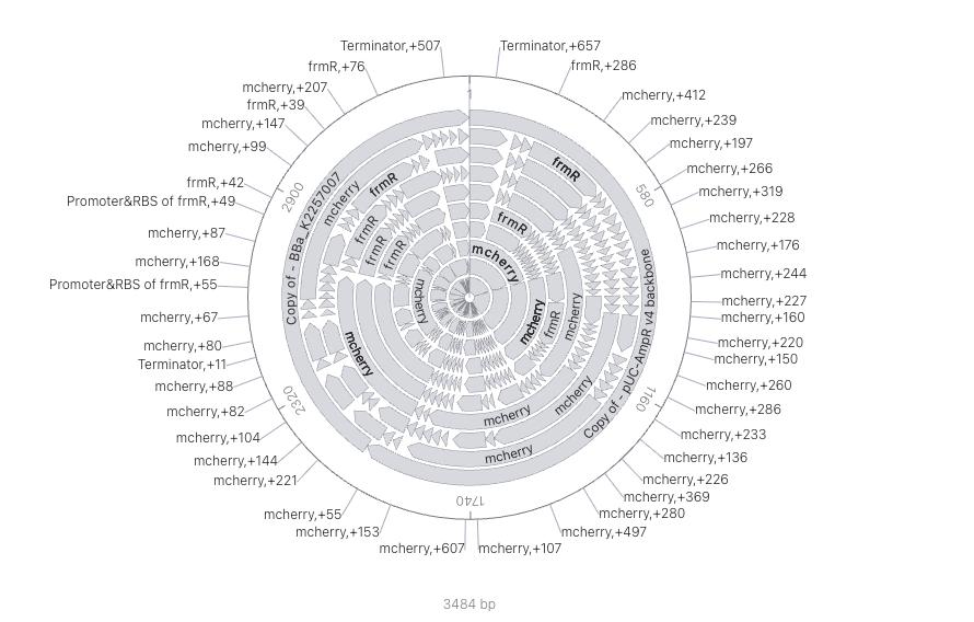
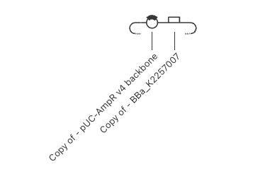
The construct is a single circular plasmid, assembled in Kernel from two blocks: BBa_K2257007 (1,407 bp) and the pUC-AmpR v4 backbone (2,077 bp), for a total of 3,484 bp — matching the sum exactly, a first sanity check that no bases were accidentally duplicated or dropped when the two blocks were joined.
Read 5’→3’ within the sensor module (all elements in the same orientation):
PfrmR (promoter + RBS) → frmR (CDS) → FLAG tag → PfrmAB (promoter + RBS) → mCherry (CDS) → Terminator
…sitting on top of the backbone’s own ori + AmpR cassette, which retains its own internal, independent orientation. I confirmed in Kernel’s sequence viewer that both frmR and mCherry begin with a valid start codon (ATG) and end with a valid stop codon, in-frame, and that the two module junctions (backbone → sensor module, and sensor module back to backbone) have no gaps or overlapping bases.
Part 4: Optimize and Quality-Check
Codon optimization. I ran Kernel’s codon optimizer separately on each CDS for E. coli. In both cases, the resulting protein sequence was translated and compared back against the pre-optimization protein: identical, 0 amino-acid changes in both frmR+FLAG (101 aa) and mCherry (236 aa) — exactly as expected, since codon optimization is only supposed to swap synonymous codons, never change the encoded protein. The only structural change in either sequence was the addition of one extra codon at the very end — a second, in-frame stop codon placed immediately after the first (e.g., mCherry’s stop went from a single TAA to a tandem TGA-TAA). This is a deliberate synthetic-biology safety practice: a tandem stop still terminates translation correctly even if a future point mutation disables the first stop codon, preventing unwanted ribosomal read-through into downstream sequence. The optimizer otherwise consistently shifted codons toward E. coli’s most-used synonyms for a given amino acid (e.g., CGT over CGC/CGA/AGA for Arginine, AAA for Lysine, CAG for Glutamine) — this matters because translation speed depends on how much matching tRNA the host has available for a given codon; rare codons can stall or drop ribosomes mid-transcript, reducing yield or producing truncated/misfolded protein.
Sequence search (pairwise alignment). I ran a pairwise alignment for each CDS’s translated protein against its natural reference, rather than aligning DNA-to-DNA (which would falsely penalize synonymous codon differences):
frmR+FLAG vs. UniProt FRMR_ECOLI (P0AAP3): 90.10% identity, but critically, 0 mutations and 91/91 matches — my 101-aa sequence covers the entire 91-aa native FrmR reference with zero amino acid substitutions; the only difference is 10 extra residues at the C-terminus, which is exactly the FLAG tag (DYKDDDDK) plus a short linker that was deliberately fused on. In other words, the sensing domain itself is unmodified, wild-type FrmR.
mCherry vs. reference (“Designer Cells” construct): 81.00% identity (226/279 matches, 6 mutations, 4 deletions, 43 insertions). The low headline number is misleading on its own — inspecting the alignment shows the reference is 275 aa long, 39 residues longer than my 236-aa mCherry, and that extra C-terminal tail reads EGRGSLLTCGDVEENPGP..., which contains the diagnostic motif of a T2A/P2A viral self-cleaving peptide (...GDVEENPGP) followed by the start of a second, different protein. That means the “reference” I compared against isn’t a standalone mCherry — it’s a multi-protein expression cassette (mCherry-2A-X) from a different construct entirely, and the 43 “insertions” are simply that fused second half, not something missing from my design. Setting that fused tail aside, only a handful of scattered point differences remain, consistent with ordinary variation between different depositors’ mCherry sequences (there are several published mCherry variants with minor substitutions) — not evidence of an error in my construct, especially since my own mCherry independently matches the exact canonical 236-aa published sequence (Shaner et al., 2004) residue-for-residue.
Safety screen. Kernel’s biosecurity screen (SecureDNA) checks a sequence against a database of sequences associated with regulated or select-agent pathogens and toxins before it can be sent out for synthesis. Result: no flagged sequences detected. This is the expected outcome — none of frmR, mCherry, the pUC origin, or AmpR correspond to controlled bio-agent sequences, so a clean screen here confirms there’s nothing in this design that would block ordering synthesis on biosecurity grounds.
Part 5: From Design to Build
How would I build it? At ~3.5 kb and composed entirely of standard, non-repetitive parts, this construct is well within range for direct, clonal full-plasmid gene synthesis rather than requiring in-house fragment assembly. That said, Gibson assembly — isothermal, single-step joining of overlapping DNA fragments via a combined exonuclease/polymerase/ligase reaction (Gibson et al., 2009) — would be my fallback if I wanted to keep reusing this exact pUC-AmpR backbone across future sensor variants (e.g., swapping in a different reporter, or a different chemically-responsive promoter later): ordering backbone and sensor-module insert separately and Gibson-assembling them would let me mix and match modules without re-synthesizing the whole plasmid every time.
What does synthesis constrain? The terminator is deliberately a strong hairpin (that’s mechanically how rho-independent terminators work), and hairpin structures are a known source of synthesis and sequencing errors — exactly the class of error that protein-mediated correction methods, such as MutS-based mismatch filtration, were built to catch before assembly (Carr et al., 2004). A synthesis vendor would likely apply an equivalent error-correction step automatically for a region like this. Outside of the terminator, the design has no unusually repetitive stretches, extreme GC content, or long homopolymer runs that I’d expect to cause major synthesis difficulty.
Prototype vs. scale. For validating this one design, ordering the ~3.5 kb plasmid synthesized and clonally verified by sequencing a handful of colonies is the right prototyping approach — slow per construct, but low overhead for a single test. If I later want to systematically test many variants at once — different promoter strengths, alternative reporters, or other natural regulators mined the way Stanton et al. mined a whole family of prokaryotic repressors for orthogonal logic gates (Stanton et al., 2014) — the more efficient path would be chip-based, multiplexed oligo synthesis: the same parallel, microarray-derived DNA pool strategy described by Tian et al. (2004) for accurate multiplex gene synthesis, and later scaled up with selective amplification from high-fidelity microchip pools (Kosuri et al., 2010) or parallel microfluidic synthesis (Kong et al., 2007). That’s a genuinely different automation technology from what a single prototype needs — mirroring the prototype-vs-scale distinction from Homework #1.
Part 6: What Don’t I Know Yet?
Is the plasmid-encoded FrmR module even necessary, or does native FrmR carried over in the crude cell-free extract already regulate PfrmAB on its own?How I’d find out: run the cell-free reaction with a reporter-only variant (frmR deleted from the construct) side by side with the full construct, and compare formaldehyde-induced signal between the two. If the reporter-only version still responds, native extract FrmR is sufficient and the synthetic frmR cassette is redundant.
Does the freeze-dried, cell-free reaction survive being embedded in an electrospun fiber (high voltage, organic solvents during spinning)? How I’d find out: compare a fiber functionalized after spinning (reaction dispensed onto the finished mat) against one where the reaction is co-spun directly into the fiber, and measure whether formaldehyde-induced signal differs between the two — isolating whether electrospinning itself degrades the reaction.
Does the reporter’s sensitivity and response time on a solid fiber matrix match what’s been characterized in solution/plate format for BBa_K2257007?How I’d find out: run a formaldehyde dose-response curve in a sealed gas chamber, side by side, in a solution tube and on a spotted fiber sample, and compare the detection threshold and kinetics between the two formats.
References
Brophy, J. A., & Voigt, C. A. (2014). Principles of genetic circuit design. Nature Methods, 11(5), 508–520. https://doi.org/10.1038/nmeth.2926
Carr, P. A., Park, J. S., Lee, Y.-J., Yu, T., Zhang, S., & Jacobson, J. M. (2004). Protein-mediated error correction for de novo DNA synthesis. Nucleic Acids Research, 32(20), e162. https://doi.org/10.1093/nar/gnh160
Denby, K. J., Iwig, J., Bisson, C., Westwood, J., Rolfe, M. D., Sedelnikova, S. E., Higgins, K., Maroney, M. J., Baker, P. J., Chivers, P. T., & Green, J. (2016). The mechanism of a formaldehyde-sensing transcriptional regulator. Scientific Reports, 6, 38879. https://doi.org/10.1038/srep38879
Elowitz, M. B., & Leibler, S. (2000). A synthetic oscillatory network of transcriptional regulators. Nature, 403, 335–338. https://doi.org/10.1038/35002125
Gibson, D. G., Young, L., Chuang, R.-Y., Venter, J. C., Hutchison, C. A., & Smith, H. O. (2009). Enzymatic assembly of DNA molecules up to several hundred kilobases. Nature Methods, 6, 343–345. https://doi.org/10.1038/nmeth.1318
Kong, D. S., Carr, P. A., Chen, L., Zhang, S., & Jacobson, J. M. (2007). Parallel gene synthesis in a microfluidic device. Nucleic Acids Research, 35(8), e61. https://doi.org/10.1093/nar/gkm121
Kosuri, S., Eroshenko, N., LeProust, E. M., Super, M., Way, J., Li, J. B., & Church, G. M. (2010). Scalable gene synthesis by selective amplification of DNA pools from high-fidelity microchips. Nature Biotechnology, 28, 1295–1299. https://doi.org/10.1038/nbt.1716
Nguyen, P. Q., Soenksen, L. R., Donghia, N. M., Angenent-Mari, N. M., de Puig, H., Huang, A., Lee, R., Slomovic, S., Galbersanini, T., Lansberry, G., Sallum, H. M., Zhao, E. M., Niemi, J. B., & Collins, J. J. (2021). Wearable materials with embedded synthetic biology sensors for biomolecule detection. Nature Biotechnology, 39(11), 1366–1374. https://doi.org/10.1038/s41587-021-00950-3
Pardee, K., Green, A. A., Ferrante, T., Cameron, D. E., DaleyKeyser, A., Yin, P., & Collins, J. J. (2014). Paper-based synthetic gene networks. Cell, 159(4), 940–954. https://doi.org/10.1016/j.cell.2014.10.004
Shaner, N. C., Campbell, R. E., Steinbach, P. A., Giepmans, B. N. G., Palmer, A. E., & Tsien, R. Y. (2004). Improved monomeric red, orange and yellow fluorescent proteins derived from Discosoma sp. red fluorescent protein. Nature Biotechnology, 22(12), 1567–1572. https://doi.org/10.1038/nbt1037
Stanton, B. C., Nielsen, A. A. K., Tamsir, A., Clancy, K., Peterson, T., & Voigt, C. A. (2014). Genomic mining of prokaryotic repressors for orthogonal logic gates. Nature Chemical Biology, 10, 99–105. https://doi.org/10.1038/nchembio.1411
Tian, J., Gong, H., Sheng, N., Zhou, X., Gulari, E., Gao, X., & Church, G. (2004). Accurate multiplex gene synthesis from programmable DNA microchips. Nature, 432, 1050–1054. https://doi.org/10.1038/nature03151
Vieira, J., & Messing, J. (1982). The pUC plasmids, an M13mp7-derived system for insertion mutagenesis and sequencing with synthetic universal primers. Gene, 19(3), 259–268. https://doi.org/10.1016/0378-1119(82)90015-4
Part:BBa_K2257007 — composite formaldehyde-sensing device (PfrmR–frmR–PfrmAB–mCherry). Registry of Standard Biological Parts (iGEM). https://parts.igem.org/Part:BBa_K2257007
Tool: Claude (Anthropic, Sonnet 5), used through the chat interface.
Prompts (summarized; originally written in Spanish): discussed which E. coli formaldehyde-sensor parts were available in Kernel’s part libraries, asked for help interpreting search results and choosing between candidate parts (settling on BBa_K2257007 and the pUC-AmpR v4 backbone), asked what output the sensor part produces and what the backbone provides, asked for help verifying the assembled construct’s linear logic and orientation, asked for a prompt to verify the plasmid sequence with Kernel’s own AI assistant, and asked for this write-up to be restructured to follow Homework #2’s required sections (Parts 0–6), with reliable, cited supporting literature including the assigned course readings.
I selected the specific parts, built and inspected the construct directly in Kernel, ran (or will run) the codon optimization, BLAST, and safety-screen tools myself, and reviewed/edited the final write-up, table, and citations.