Week 1 HW: Final Project Ideas
WEEK 1
Biomanufacturing a 3D Cervical Transformation-Zone Model for Longitudinal Monitoring and Data-Driven Visualization
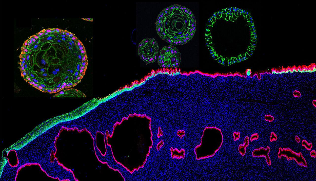 Image credit: Universität Würzburg.
Project at a glance
Goal: Create a standardized 3D cervical transformation-zone model for studying HPV-associated progression
Living system: Cervical transformation-zone/SCJ epithelial cells in 3D culture
Automation: Programmable liquid handling for culture maintenance and longitudinal sampling
Prototype scale: ~80 models
Output: Standardized 3D tissue models + longitudinal biological measurements, with a supplementary data-driven generative visualization
01 - What I would like to biomanufacture and why
I would like to biomanufacture a standardized 3D model of the human cervical transformation zone that could be used to study HPV-associated progression from early cellular abnormalities toward precancer and early invasive cervical cancer. The target product would therefore be a reproducible living research platform rather than a treatment administered directly to patients.
I chose the cervical transformation zone because it is the region where most cervical cancers arise. I am particularly interested in what happens between persistent high-risk HPV-associated changes and the development of invasive cancer. Instead of studying cervical cells only in conventional two-dimensional culture or examining cancer after it has already developed, I want to create a 3D system in which biological changes can be followed longitudinally.
Multiple standardized models could be produced and maintained under control and HPV-associated conditions. Researchers could observe changes in tissue morphology and molecular behavior over time while repeatedly collecting conditioned culture medium. These samples could be analyzed for changes in secreted molecules, extracellular vesicles, or other potential biomarkers that appear as the tissue progresses toward a precancerous or invasive state. My longer-term scientific goal is to investigate whether molecular changes that occur before obvious invasion could eventually contribute to better strategies for understanding or detecting early cervical cancer progression.
I would also like the results generated by this living research platform to have a downstream computational design output. Longitudinal imaging and molecular measurements from each 3D model could be translated into data-driven generative visualizations. Instead of producing an arbitrary image, measured biological variables could control visual properties such as form, structure, density, or movement. As the biological model changes over time, its visual representation would change with it. This would create a creative output directly derived from experimental data and could provide another way to explore and communicate biological progression that is normally difficult to see.
My interest in this project is also personal and social. In my home country, Kyrgyzstan, reproductive health topics such as HPV, cervical cancer, and screening can be difficult to discuss openly. During high school, I gave educational sessions on HPV and cervical cancer prevention, which led me to become more interested in the biology of early disease progression. In particular, I became interested in understanding biological changes that occur before invasive cancer develops, rather than focusing only on established disease.
This project would allow me to connect that interest with mammalian cell biology, tissue engineering, automation, programming, and visual design. Ultimately, I want to explore whether a reproducible living model can help researchers investigate early HPV-associated progression while generating biological data that can be measured, followed over time, and translated into visual form.
02 - What living system would I use?
I would use a mammalian cell culture system based on human cervical epithelial cells grown in 3D. Ideally, I would use primary epithelial cells derived from the cervical transformation zone or squamocolumnar junction (SCJ), since this is the region that I want the model to represent.
The main inputs would be human cervical epithelial cells, a three-dimensional extracellular matrix such as a Matrigel-based basement-membrane matrix, appropriate culture medium and growth factors, and a controlled HPV-associated perturbation such as expression of the high-risk HPV16 oncogenes E6 and E7. Within this environment, the cells would proliferate, differentiate, and organize into three-dimensional tissue structures.
Once the 3D models are established, I would maintain control and HPV-associated models in parallel so that their changes could be compared over time. One possible starting approach would be a controlled expression system for HPV16 E6 and E7. This would allow me to model selected aspects of HPV-associated cellular transformation under defined experimental conditions. However, E6/E7 expression alone would not reproduce the complete process of natural persistent HPV infection, so I would treat it as a simplified experimental model rather than a complete representation of HPV infection.
The primary biological output would be standardized living 3D cervical tissue models representing control and HPV-associated conditions. The system would also generate measurable biological outputs over time. Imaging could provide information about changes in tissue morphology, while conditioned culture medium collected repeatedly from the models could be analyzed for changes in secreted molecules, extracellular vesicles, or other molecular signals.
These longitudinal imaging and molecular measurements would form the biological dataset used for the computational design output described in Section 1. The living system would therefore produce the 3D tissue models and measurable biological data, while a downstream computational process would later translate selected measurements into generative visualizations.
For this initial proposal, I assume that validated primary human cervical transformation-zone or SCJ epithelial cells could be obtained through an established research laboratory or an ethically approved tissue source. Access to an appropriate and reproducible cell source remains an important technical uncertainty. I also assume that an E6/E7-based system would reproduce enough relevant features of HPV-associated transformation to allow meaningful comparisons over time, although this would need to be experimentally validated.
03 - How would I use automation?
I would combine the 3D cervical tissue system with a programmable liquid-handling robot, such as an Opentrons platform. The main purpose of automation would be to make the production, maintenance and longitudinal monitoring of multiple 3D tissue models more standardized and reproducible.
The robot could automate repetitive liquid-handling steps such as adding culture medium and reagents, performing scheduled media changes, and collecting conditioned-medium samples from multiple models at predefined time points. This would be particularly useful because I want to follow HPV-associated changes longitudinally rather than analyze the tissue only at a single endpoint. Control and HPV-associated models would need to be treated and sampled consistently so that observed differences are less likely to result from variation in manual handling or sampling time.
For example, conditioned medium could be automatically collected from each model at regular intervals and transferred into labeled wells for later analysis of selected secreted molecular signals, such as proteins or extracellular-vesicle-associated signals. In parallel, imaging would be performed using a separate imaging system at corresponding time points to record changes in tissue morphology. The liquid-handling robot would therefore automate culture maintenance and sample collection, while imaging would represent a separate measurement stream. Measurements from both streams would remain associated with the specific model, experimental condition, and time point from which they were collected, creating a structured longitudinal dataset.
I would then use this dataset to generate the computational design output introduced in Section 1. Selected biological measurements could first be normalized relative to appropriate control values and then translated through a simple rule-based mapping into visual parameters. For example, a change in a molecular signal could influence the density or intensity of visual elements, while a quantitative morphological measurement could influence their form or structure. The same mapping rules would be applied across all models and time points, so changes in the resulting visualization would be driven by measured biological differences rather than arbitrary artistic choices.
At the prototype stage, I would use the liquid-handling robot with multiwell 3D cultures to automate culture maintenance and longitudinal sampling while developing the computational visualization pipeline in parallel. If the system were later expanded to much larger numbers of models, more integrated automation could combine plate handling, incubation, imaging, sampling, measurement, and data management. In this way, automation would support both the reproducibility of the biological experiment and the reproducible conversion of its measurements into a creative visual output.
04 - How much would I need to make, and how long?
(a) How much?
Because my target product is a research platform rather than a medicine administered to patients, I would define a meaningful initial scale as the number of standardized 3D cervical models required for one experimental campaign.
For an initial prototype, I would compare two experimental conditions: a control condition and an HPV-associated condition. I would examine the models at five experimental endpoints and use eight biological replicates for each condition at each endpoint.
2 conditions × 5 endpoints × 8 biological replicates = 80 models
Therefore, I would initially aim to manufacture approximately 80 standardized 3D cervical models.
This design would fit within the capacity of one 96-well plate, leaving 16 wells available for additional controls, optimization, or models that fail quality-control criteria. I assume approximately one standardized experimental model per well. This assumption would need to be validated because 3D cervical cultures may vary in size, number, and growth between wells.
Conditioned culture medium could be collected repeatedly from each model during its lifetime, providing longitudinal molecular measurements without requiring a new model for every sampling event. The five endpoints in my calculation would instead allow models to be removed at different stages for endpoint tissue imaging or other analyses that may not be compatible with continued culture.
Each experimental model would therefore generate two connected forms of data: morphological measurements from imaging and molecular measurements from conditioned medium. These measurements would also provide the input for the computational design pipeline described above, meaning that the same 80-model experiment could generate a collection of data-driven visual outputs representing different biological conditions and stages.
If the prototype were reproducible, I would consider a larger research-scale campaign of approximately 1,000 models. Assuming one model per well:
1,000 models ÷ 96 wells per plate = 10.4 plates
I would therefore need approximately 11 × 96-well plates, providing capacity for up to:
11 × 96 = 1,056 models
At this scale, the additional capacity could support more biological replicates, experimental conditions or perturbations rather than simply reproducing the same experiment.
(b) How long?
The main time constraint would be the biology of establishing the cervical 3D models rather than the speed of the liquid-handling robot. A published protocol for patient-derived human cervical organoids reports approximately 4–6 weeks for organoid generation. I would therefore use 4–6 weeks as my initial estimate for establishing and expanding the 3D cervical culture system (Gurumurthy et al. 2022).
I would then assume approximately 2–3 additional weeks for the experimental monitoring phase, during which control and HPV-associated models would be maintained, conditioned medium would be collected at predefined intervals, and imaging measurements would be acquired.
My approximate manufacturing timeline would therefore be:
4–6 weeks for model establishment and expansion
+ 2–3 weeks for longitudinal experimental monitoring
= approximately 6–9 weeks per experimental campaign
The 2–3 week monitoring period is an engineering assumption for this initial proposal rather than an established biological requirement and would need to be adjusted based on how quickly reproducible HPV-associated changes appear in the model.
Importantly, the models would be cultured in parallel rather than sequentially. Therefore, producing 80 models would not require 80 times the growth period. Increasing the scale would instead increase the required plate capacity, incubator space, culture reagents, imaging capacity, and automated liquid-handling workload.
For the prototype, I would use a programmable liquid-handling robot such as an Opentrons Flex with multiwell cultures to automate media changes and conditioned-medium sampling. A 96-channel Flex pipette is designed to work across 96-well plates, making this format compatible with parallel liquid-handling workflows. The imaging measurements would be collected using a separate imaging system, while the resulting imaging and molecular measurements would be organized computationally for the generative visualization pipeline.
At a scale of approximately 1,000 models, eleven 96-well plates could theoretically provide sufficient well capacity, but plate capacity alone would not guarantee practical manufacturing at that scale. Incubation, imaging, sterile handling, sample storage, and data management could become bottlenecks. More integrated automation for plate movement, incubation, imaging, sampling, and measurement would therefore likely be needed.
The computational visualization step should scale differently from the biological manufacturing process. Once a rule-based mapping between biological measurements and visual parameters has been established, the same computational pipeline could be applied automatically to datasets from additional models. Therefore, I expect the main scale-up limitations to come from producing and measuring reproducible living models rather than from generating their visual representations.
05 - What don’t I know yet?
The first major unknown is whether I can obtain and maintain an appropriate and reproducible source of human cervical transformation-zone or squamocolumnar-junction epithelial cells. My proposed system assumes that these cells can be obtained through an established research laboratory or an ethically approved tissue source and expanded while retaining the biological characteristics needed to represent the transformation zone. I would need to determine how much the cells change during expansion and whether models produced from different batches or donors remain sufficiently comparable. This could be evaluated by comparing relevant cellular markers, morphology, and growth characteristics across passages, batches, and donor-derived cultures.
The second major unknown is whether the HPV-associated model accurately reproduces the biological changes that I want to study. A controlled HPV16 E6/E7 expression system could model important aspects of HPV-driven cellular transformation, but it would not reproduce the complete process of natural persistent HPV infection. I would therefore need to determine whether control and HPV-associated models develop reproducible differences in morphology and molecular outputs over time, and whether these changes are relevant to progression toward precancer and early invasion. This could be investigated by comparing morphological and molecular trajectories between control and HPV-associated models at multiple time points and determining when consistent differences become measurable.
The third major challenge is whether the system can remain standardized and biologically reproducible as it is scaled up and measured longitudinally. My manufacturing estimate assumes that comparable 3D models can be produced across many wells and plates, but variation in model size, viability, morphology, extracellular matrix behavior, or molecular output could make comparisons difficult. I would measure well-to-well, plate-to-plate, and batch-to-batch variability using replicate cultures and repeated measurements to determine whether automation actually improves reproducibility at larger scale.
This reproducibility is also important for the computational design output. Before using biological measurements to generate visualizations, I would need to determine which measurements are sufficiently reliable and biologically meaningful to use as visual inputs. The mapping itself would be rule-based and reproducible, but the visualization would only be informative if the underlying measurements consistently reflect biological differences rather than experimental noise. Therefore, I would first validate the biological measurements and only then define the final mapping between those measurements and visual parameters.
AI Use
I used OpenAI ChatGPT as a learning and feedback tool while developing this proposal. I used it primarily to evaluate technical assumptions, compare possible experimental approaches, identify limitations in my proposed workflow and refine the scaling and automation strategy. I also used it to help develop the connection between longitudinal biological measurements and the computational design output. The core project concept and research direction were developed from my own interests, and I reviewed and revised the AI-assisted material to ensure that I understood the concepts included in the proposal.
Prompts used during proposal development:
“Could a standardized 3D cervical transformation-zone model be used to follow HPV-associated progression over time, and what are the main limitations of this idea?”
“Would HPV16 E6/E7 expression be a reasonable way to create an HPV-associated condition in this model, and what would it fail to reproduce?”
“How could I use automation, such as an Opentrons liquid-handling robot, for repeated culture maintenance and longitudinal sampling?”
“How should I estimate a meaningful experimental scale for this research platform, including the number of models, replicates, plates, and manufacturing time?”
“What are the biggest technical assumptions or bottlenecks in this proposed 3D cervical biomanufacturing system?”
“I want to add a creative design output without changing the biomedical focus of the project. How could real imaging and molecular measurements from the models be translated into data-driven generative visualizations?”
References
Maru, Y., Kawata, A., Taguchi, A., et al. (2020). Establishment and molecular phenotyping of organoids from the squamocolumnar junction region of the uterine cervix. Cancers, 12(3), 694. https://doi.org/10.3390/cancers12030694
Koster, S., Gurumurthy, R. K., Kumar, N., et al. (2022). Modelling Chlamydia and HPV co-infection in patient-derived ectocervix organoids reveals distinct cellular reprogramming. Nature Communications, 13, 1030. https://doi.org/10.1038/s41467-022-28569-1
Gurumurthy, R. K., Koster, S., Kumar, N., Meyer, T. F., & Chumduri, C. (2022). Patient-derived and mouse endo-ectocervical organoid generation, genetic manipulation and applications to model infection. Nature Protocols, 17, 1658–1690. https://doi.org/10.1038/s41596-022-00695-6
Petzold, C. J., & Mukhopadhyay, A. (2025). From bench to biofactory: High-throughput technologies and automated workflows to accelerate biomanufacturing. Current Opinion in Biotechnology, 94, 103320. https://doi.org/10.1016/j.copbio.2025.103320
Opentrons. Flex Pipettes: 96-Channel Pipette. Opentrons Flex System Description. https://docs.opentrons.com/flex/system-description/pipettes/