Week 2 HW: DNA Design
Synthetic Vascularization Encoded by PDGF-BB for Bioprinted Liver Tissue
Emmanuel Cárdenas Barceló
Barranquilla, Colombia
emmanuel2003cardenas@gmail.com
Part 1: Define your expression target
What molecule are you expressing?
Given the needs of the product, the molecule best aligned for expression is PDGF-BB (Platelet-Derived Growth Factor BB), a secreted growth factor that in my system will be produced by the endothelial cells lining the vascular channel. Its role is to recruit pericytes to the abluminal surface of the newly formed endothelium, a step needed to prevent vessel regression and local hemorrhage inside the printed channel (Zhang et al., 2021). Another option for this work is VEGF-A (Vascular Endothelial Growth Factor A), the factor that triggers the initial stage of the vascular cascade by activating endothelial cells to migrate, proliferate and form the lumen inside the printed channel (Son et al., 2025; Cai et al., 2025). Although it was a viable option, I prioritized PDGF-BB over VEGF-A because sustained overexpression of VEGF-A has been reported to cause leaky and unstable vessels (Son et al., 2025), whereas PDGF-BB acts specifically in the pericyte recruitment and adhesion stage that my design needs to accelerate.
What is the sequence?
The sequence corresponds to the human PDGFB gene, UniProt accession P01127, a 241-amino-acid precursor that the cell processes before releasing the active form. The full precursor has four segments. A signal peptide at positions 1 to 20 directs the nascent protein to the endoplasmic reticulum and the secretory pathway. An N-terminal propeptide at positions 21 to 81 and a second, C-terminal propeptide at positions 191 to 241 are both removed by furin-mediated proteolysis during transit through the Golgi. Finally, the mature chain at positions 82 to 190 is the biologically active form of PDGF-B, the one that homodimerizes to form PDGF-BB and the one that will actually recruit pericytes in the system. Within this mature chain, the UniProt annotation reports two receptor-binding residues (positions 108 and 111) and a network of disulfide bonds (97 to 141, 130 to 178 and 134 to 180, plus two interchain disulfides at positions 124 and 133) that stabilize the characteristic fold of this cytokine family. I will keep this in mind when evaluating whether codon optimization or the chosen host could compromise correct folding.
For the construct design, the ideal choice is to encode the CDS of the full precursor (all 241 positions) and not only the mature chain fragment. The reason is that the signal peptide and the propeptides are needed for the cell to process and secrete the protein correctly. Expressing only region 82 to 190 without its native processing machinery would produce an intracellular, non-secreted and potentially misfolded protein. The reference amino acid sequence comes from the official UniProtKB/Swiss-Prot record (entry version 263, accessed in September 2026). The DNA sequence of the CDS corresponds to the RefSeq transcript NM_002608 (variant 1, protein NP_002599.1), 726 nucleotides long including the stop codon, and I entered it manually in Kernel as a custom part. I verified that its translation matches the 241 amino acids of UniProt P01127 letter by letter, with a single stop codon at the end and no internal stops.
What host or chassis would you use?
The chassis that best fits the needs is HUVEC (Human Umbilical Vein Endothelial Cells), primary human endothelial cells isolated from the umbilical vein and widely used as a standard system to model the formation and maturation of vascular networks in vitro (Shiwarski et al., 2025; Son et al., 2025). It is the reference option in most published bioprinted vasculature systems, with well-established culture and characterization protocols, which makes it appropriate for validating a first expression design like this one.
Another alternative I considered was iPSC-derived endothelial cells, which have the advantage of being patient-specific and therefore relevant for an eventual autologous application (Son et al., 2025). However, they require a prior differentiation protocol that introduces additional batch-to-batch variability, which would complicate this exercise, so I discarded them for this design and chose HUVEC as the formal chassis.
Where does the product need to go?
PDGF-BB must be secreted into the extracellular medium toward the lumen of the channel, where it acts in a paracrine way on the pericytes located on the abluminal surface of the forming vessel (Son et al., 2025). There is no need to design an artificial signal peptide or a membrane anchoring sequence, because the native signal peptide of the gene already directs the protein along that pathway.
Part 2: Design your genetic parts
Since the chosen chassis is HUVEC (a primary human endothelial cell, a eukaryotic host), the expression construct is defined only on the eukaryotic host column of the assignment table. The prokaryotic column does not apply.
Decision table
| Layer | Chosen part | Justification |
|---|---|---|
| Transcription start | EF-1α promoter | It sustains stable expression in non-immortalized primary cells such as HUVEC. CMV is discarded because of its progressive silencing by methylation in this type of culture. |
| Translation start | 5’ UTR with Kozak (GCCACCATGG) | Standard mammalian Kozak sequence that ensures efficient translation initiation at the ATG codon of the CDS. |
| Coding sequence | Full PDGFB CDS (241 aa), UniProt P01127 / RefSeq NM_002608 | It encodes the full precursor, which is necessary because the native signal peptide and propeptides are what allow correct processing and secretion of the mature protein. |
| Targeting (signal peptide) | Native PDGFB signal peptide (aa 1 to 20) | Already included within the CDS itself, it directs the protein to the secretory pathway without needing an exogenous signal peptide. |
| Transcription stop | Terminator with SV40 or bGH polyadenylation signal | Standard mammalian terminator that ensures a clean end of transcription. |
| Vector context | Standard mammalian expression backbone (origin of replication + selection marker) | An existing one from the library is reused instead of designing it from scratch, and it provides replication and selection for cloning. |
Part 3: Assemble your construct
I built the construct PDGFB-HUVEC-EF1a-v1 in Kernel, inside the folder Emmanuel-PDGFB-HUVEC, with four parts in forward orientation and in this 5’ to 3’ order: Short HsEef1a1 promoter, 5’ UTR Kozak (GCCACC), PDGFB-CDS-native and bGH poly(A) signal.
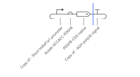
I verified the assembly in Kernel’s sequence viewer.
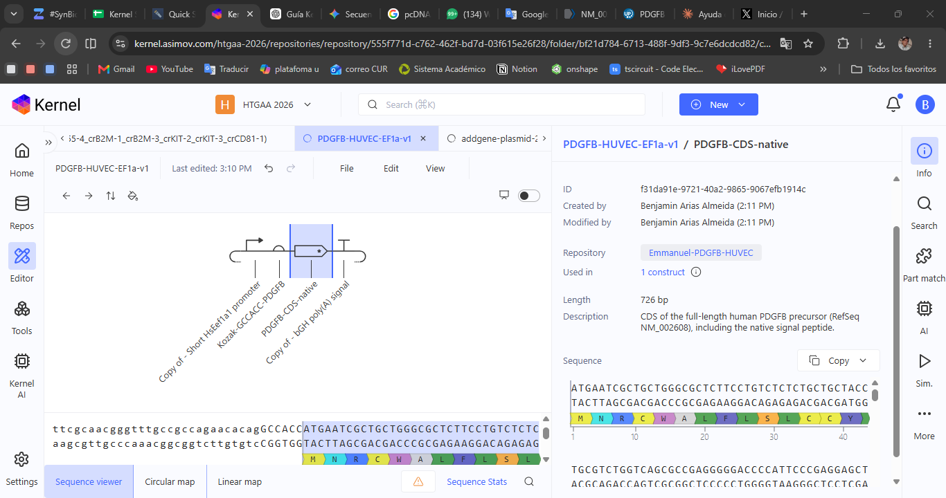
Kozak to CDS junction. The search for GCCACCATG gave a single match (1 of 1), followed by AATCGCTGC, that is, ATGAATCGCTGC, which is the expected start of PDGFB (M, N, R, C). There is no duplicated ATG and the reading frame is preserved.
End of the CDS and poly(A). The search for GAGCCTAGCTGTGCC gave a single match (1 of 1). The CDS ends in the TAG stop codon and the bGH poly(A) starts immediately after it. The translation Kernel shows at the end of the gene reads E, T, L, G, A and an asterisk, that is, it ends in ETLGA with its stop.
The four elements appear in the order and orientation planned in the construct schematic, and the Sequence Stats indicator showed green.
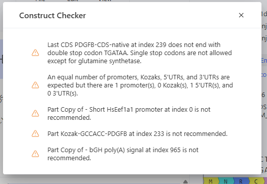
Kernel’s Construct Checker. When I checked PDGFB-HUVEC-EF1a-v1 it showed five warnings. First, the last CDS (PDGFB-CDS-native, index 239) does not end in the double stop codon TGATAA, and Kernel states that single stop codons are not allowed except for glutamine synthetase. Second, Kernel expects an equal number of promoters, Kozaks, 5’ UTRs and 3’ UTRs, and it found 1 promoter, 0 Kozaks, 1 5’ UTR and 0 3’ UTRs. This is because my Kozak part is typed as a 5’ UTR and not as a Kozak, and because I did not include a separate 3’ UTR. Third, it marked the parts Copy of - Short HsEef1a1 promoter (index 0), Kozak-GCCACC-PDGFB (index 233) and Copy of - bGH poly(A) signal (index 965) as “not recommended”, and I have not yet found the reason for that flag. Because of time constraints I did not modify the design, since none of the warnings prevents the analyses in this assignment. In a later iteration I would add a second stop codon (TGATAA), review the type assigned to the Kozak part and ask Kernel AI why those parts are not recommended.
Part 4: Optimize and quality-check
Codon optimization
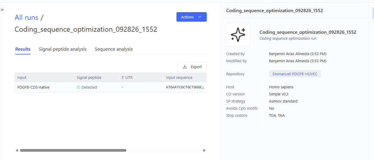
I used Kernel’s Optimize coding sequences tool (Actions menu of the PDGFB-CDS-native part, 726 bp) in the run Coding_sequence_optimization_092826_1552. Before running it I saved a view of the native sequence, with the CDS highlighted in the schematic and its translation MNRCWALFLSL, which matches the expected start of PDGFB.
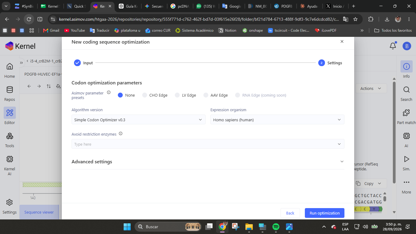
Kernel’s default expression organism is Cricetulus griseus (Chinese hamster, CHO), and I changed it to Homo sapiens because HUVEC is a primary human cell. The result is therefore an approximation and not an optimization specific to HUVEC, and I do not describe it that way in this report. The remaining parameters were left as they were. There was no Asimov preset (None), the algorithm was Simple Codon Optimizer v0.3, no restriction enzymes were avoided, CpG motifs were not avoided, TGA and TAA were allowed as stop codons, and the signal peptide strategy was Asimov standard, which Kernel ignored because it had already detected the signal peptide in my sequence. I did not apply additional constraints on restriction sites or GC content.
What it returned. The run registered Homo sapiens as the host and Simple v0.3 as the version, and Kernel detected the native signal peptide with 90% confidence, with the cleavage point around residues 19 and 20, in line with positions 1 to 20 reported by UniProt. This indicates that the native signal peptide is preserved and that the protein will still enter the secretory pathway.
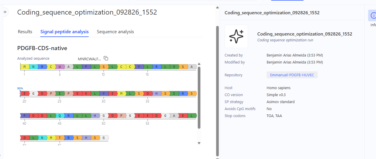
The optimized sequence is 729 bp long compared with 726 bp for the native one. It starts with ATGAACAGATGCTGGGCC (the native one started with ATGAATCGCTGC) and translates to MNRCWALFLSLC, the same protein start, which means the codons changed and the amino acids did not. The 3 additional bp could correspond to a second stop codon, since the run allows TGA and TAA, but I have not verified this yet.

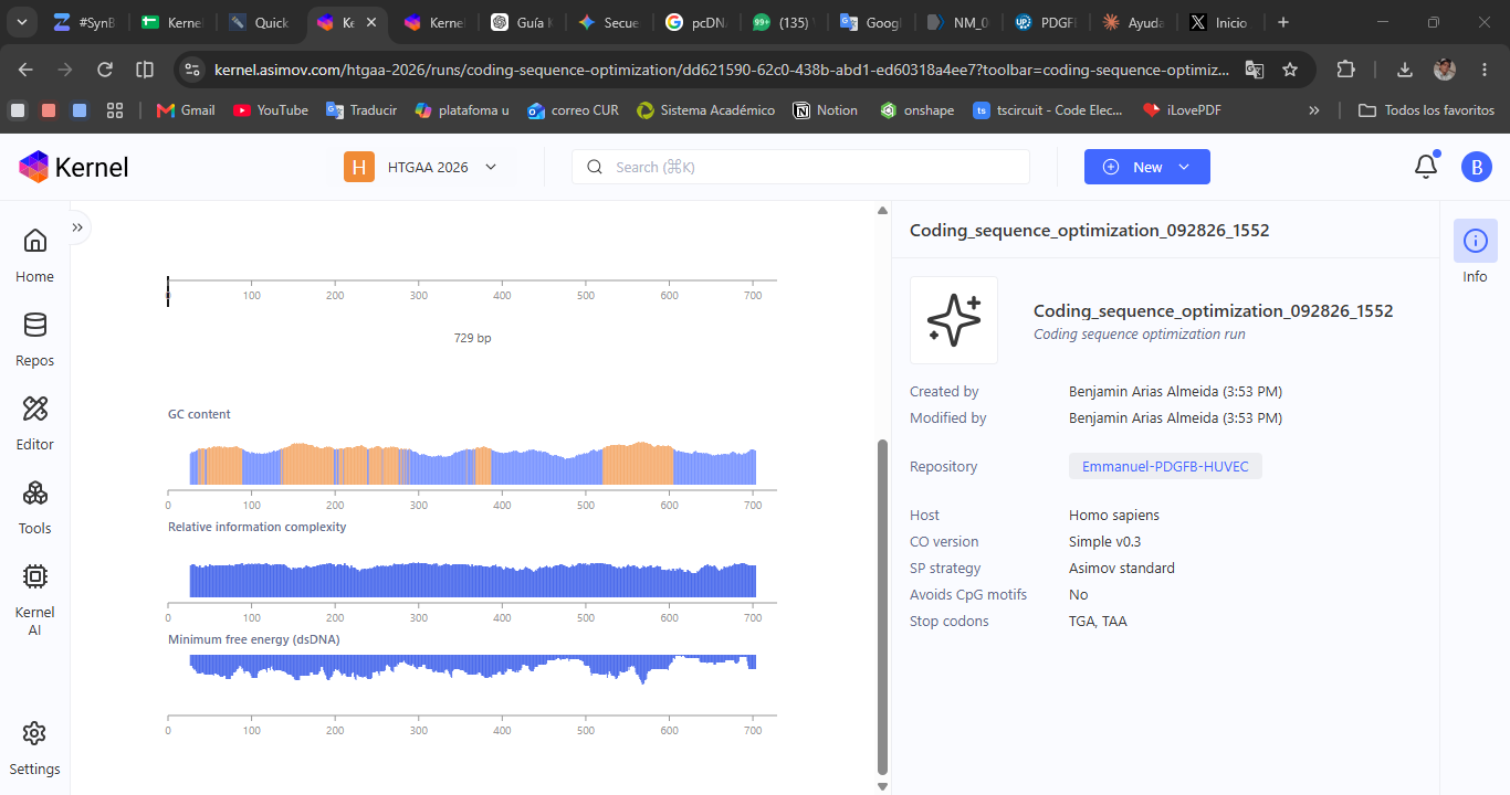
The Sequence analysis tab showed graphs of GC content, relative information complexity and minimum free energy of the dsDNA along the 729 bp, without numeric CAI values.
Why codon usage matters. The genetic code is redundant, and each organism uses some synonymous codons more than others, in relation to the abundance of its tRNAs. A gene with rarely used codons can be translated more slowly or with lower yield. In a secreted protein with several disulfide bonds such as PDGF-B, the pace of translation could also influence folding. Since PDGFB is already a human gene and HUVEC is a human cell, I expect the gain from optimizing to be moderate, and I leave that as a hypothesis to measure by ELISA.
BLAST
BLAST will be run on the native 726 bp version, not the optimized one, to check that the CDS is human PDGFB, because optimization changes many codons without changing the protein. As a starting reference, its translation matches UniProt P01127 and its calculated GC content is 62.3%. I will record the best match, score, E-value, identity and coverage.
Part 5: From design to build
How would you build it?
The full PDGFB CDS, including its native signal peptide, is 726 base pairs long (confirmed in Kernel), within the range of a single gene synthesis and without the need for fragment assembly. For that reason I propose ordering it directly as double-stranded synthetic DNA (for example a gBlock or a gene synthesized by a commercial provider), instead of using multi-piece Gibson assembly, which is better justified for constructs of tens of thousands of base pairs (Gibson et al., 2009).
What does synthesis constrain?
The PDGFB gene does not contain unusually repetitive regions or extreme GC content that I have identified so far, so I do not anticipate major synthesis difficulty. Even so, since the accuracy of standard chemical synthesis is around 1 error per 100 to 1000 bases, it would be worth applying MutS-type error correction before proceeding to the final cloning, to reduce the error rate to a range of 1 in 10,000 to 1 in 100,000 (Carr et al., 2004).
Prototype vs. scale
For the prototype in this assignment, ordering a single construct is enough to validate expression and secretion in HUVEC. If in the future it were necessary to scale to multiple variants (for example, different promoters or expression levels to find the optimum), multiplexed on-chip synthesis would allow many variants of the same gene to be manufactured in parallel before selecting the best one (Kosuri et al., 2010).
Partial verification. I checked the native 726 bp CDS and it contains no sites for EcoRI, BamHI, HindIII, XbaI or NotI. Its GC content is 62.3%, moderate and without extremes.
Part 6: What don’t you know yet?
First, I do not know whether the EF-1α promoter will give the right expression level specifically in HUVEC, since Kernel’s simulation models seem to be calibrated on standard production cell lines such as CHO or HEK293 and not on primary endothelial cells. To find out, I would measure the concentration of secreted PDGF-BB by ELISA after transient transfection in HUVEC, and compare it against the level reported as necessary for effective pericyte recruitment.
Second, I do not know whether the native signal peptide of the gene is efficient enough in the specific context of secretion into a printed channel under flow, compared with a stronger exogenous signal peptide such as the IgK one. To find out, I would compare the titer of secreted protein between both signal peptide variants in the same system.
AI
For this assignment I used Consensus to compare the scientific evidence behind the protein candidates considered (VEGF-A, Angiopoietin-1, PDGF-BB, Claudin-5 and VE-cadherin) and to support the choice of PDGF-BB as the molecule to express. I then used its findings as a reference to focus this assignment around current trends in the field, which allowed me to track academic references. I also used Claude (Anthropic, Sonnet 5.5) as an assistant to structure the reasoning of each part of the assignment, to clarify the difference between HUVEC and iPSC-derived cells as a host, and to verify the real capabilities and limitations of the Kernel software before planning the parts design.
The exact prompt used in Consensus was:
I want to understand which proteins are typically used to induce or accelerate the maturation of synthetic vascular networks in bioprinted tissue engineering—specifically in systems where endothelial cells (HUVECs or iPSC-derived) form a monolayer within a perfusable channel and recruit pericytes to stabilize the vessel wall.
Please compare the following candidates and their specific biological roles in this process:
VEGF-A: Angiogenesis and induction of endothelial formation. Angiopoietin-1 (ANG1): Vascular stabilization and pericyte recruitment. PDGF-BB: Pericyte recruitment and adhesion to the vessel wall. Claudin-5 and VE-cadherin: Formation of tight junctions and adherens junctions that render the endothelial barrier impermeable.
For each one, please specify:
The exact role it plays in the sequence of events leading from an empty channel to a functional vessel with an impermeable barrier and stabilized pericytes. Whether there is reported evidence—citing specific studies—that its exogenous overexpression in the endothelial cells or pericytes themselves, within a bioprinted system, accelerates or improves vascular maturation. Any reported limitations or adverse effects associated with its overexpression (e.g., permeable or unstable vessels due to excess VEGF-A).
Finally, provide your recommendation regarding the best single candidate—or combination of two—to accelerate the formation of a stable endothelial barrier with pericyte recruitment in a bioprinted, vascularized liver tissue, including full references.
On the other hand, to make the work in the design software easier, I used ChatGPT (OpenAI) to create a step-by-step guide of what I had to do inside Kernel to complete the project requirements. The exact prompt, written originally in Spanish, was:
Necesito que me crees una guía paso a paso, lo más explícita posible, para trabajar en Kernel, el software de diseño genético de Asimov. Es la primera vez que uso un programa de este tipo, así que quiero instrucciones concretas de qué botón o menú usar y en qué orden, sin asumir que conozco la interfaz.
MATERIALES QUE TE ENTREGO
- La documentación oficial completa de Kernel en https://docs.kernel.asimov.com/ . El índice completo de páginas está en https://docs.kernel.asimov.com/llms.txt y cada página tiene versión Markdown si se le agrega .md al final. Debes leer las páginas relevantes una por una y no basarte solo en la página de bienvenida.
- Un archivo con la transcripción limpia de una clase del curso HTBAA/HTGAA (MIT) donde Tracy Haddock, de Asimov, demuestra Kernel. Solo es relevante la parte desde que ella toma la palabra (secciones sobre Asimov, Kernel y la guía práctica). Ignora todo lo relacionado con Cello, DAMP Lab y microfluídica.
- El enunciado de la Tarea 2 (HTBAA Homework 2, DNA Design).
- Opcionalmente, mi archivo con las partes que ya elegí para la Parte 2.
MI PROYECTO Voy a expresar PDGF-BB humano (gen PDGFB, UniProt P01127, RefSeq NM_002608) en células endoteliales primarias humanas (HUVEC), como parte de un módulo de tejido hepático vascularizado y bioimpreso. El CDS es el precursor completo con su péptido señal nativo, para que la proteína se secrete. Mi cassette de expresión usa promotor EF-1α, 5’ UTR con Kozak (GCCACCATGG), CDS de PDGFB, terminador con señal poliA (SV40 o BGH) y un backbone de expresión en mamífero ya existente.
QUÉ QUIERO QUE HAGA LA GUÍA
- Que cubra únicamente lo que es estrictamente necesario hacer dentro de Kernel para completar la Tarea 2. Esto corresponde a la Parte 0 (crear repositorio y carpeta, y anotar sus nombres), la Parte 2 (crear o importar cada parte con su tipo correcto), la Parte 3 (armar el constructo y verificarlo) y la Parte 4 (optimizar codones, buscar con BLAST y correr el escáner de bioseguridad SecureDNA). Las Partes 1, 5 y 6 se responden en el documento escrito y no en Kernel, así que no las incluyas salvo para indicar qué dato debo tener listo antes de abrir el programa.
- Que esté orientada a mi proyecto. Cada paso debe usar mis partes reales (EF-1α, Kozak, PDGFB, terminador, backbone) y no ejemplos genéricos. Para cada parte indícame qué tipo de parte debo asignar, de dónde obtener su secuencia (Asimov Public, UniProt Swiss-Prot, Addgene o importación manual) y qué alternativa usar si no aparece en la biblioteca.
- Que sea secuencial y numerada, con el nombre exacto de cada botón, menú o pestaña tal como aparece en la documentación oficial, y con el resultado esperado después de cada paso para que yo pueda confirmar que lo hice bien.
- Que en cada herramienta de la Parte 4 me diga qué debo revisar en el resultado y qué debo anotar o capturar en pantalla para mi informe (antes y después del optimizador de codones, mejor coincidencia de BLAST, resultado del escáner de seguridad).
- Que incluya una lista de verificación final con los entregables que debo tener al terminar en Kernel (nombre del repositorio y carpeta, partes registradas, constructo armado, esquema exportado, capturas de cada herramienta).
- Que incluya una sección corta de errores comunes y qué hacer si algo falla, por ejemplo si una parte no está en la biblioteca, si el verificador marca una advertencia o si el marco de lectura se rompe.
REGLAS IMPORTANTES
- No inventes botones, menús, formatos ni funciones. Si un paso no está confirmado en la documentación oficial ni en la transcripción, dilo explícitamente en lugar de suponerlo. Ya hubo un intento previo con otra IA que inventó pasos que no existen, así que necesito que verifiques todo contra la documentación.
- Si la transcripción y la documentación se contradicen, prevalece la documentación oficial, pero avísame de la diferencia. Como referencia, en la clase se dijo que el verificador de constructos está calibrado para mamífero y que el simulador demostrado era solo de E. coli. Confírmalo con la documentación.
- Si algo de mi diseño puede no funcionar tal como está en Kernel (por ejemplo, que el backbone o el promotor no estén en las bibliotecas públicas), avísame antes de dar el paso a paso y propón la alternativa.
- Escribe en español, con lenguaje directo y conciso. Evita guiones largos y el uso de dos puntos para presentar argumentos, porque me parecen rasgos de escritura de IA.
- Entrégame la guía como un documento descargable (.md o .txt) para poder seguirla mientras trabajo en Kernel.
References
Cai, B., Kilian, D., Ghorbani, S., Roth, J. G., Seymour, A. J., Brunel, L., Mejía, D., Rios, R. J., Szabo, I. M., Iranzo, S. C., Perez, A., Rao, R. R., Shin, S., & Heilshorn, S. (2025). One-step bioprinting of endothelialized and self-supporting arterial and venous networks. Biofabrication, 17. https://doi.org/10.1088/1758-5090/adab26
Carr, P. A., Park, J. S., Lee, Y.-J., Yu, T., Zhang, S., & Jacobson, J. M. (2004). Protein-mediated error correction for de novo DNA synthesis. Nucleic Acids Research, 32, e162. https://doi.org/10.1093/nar/gnh160
Gibson, D. G., Young, L., Chuang, R.-Y., Venter, J. C., Hutchison, C. A., & Smith, H. O. (2009). Enzymatic assembly of DNA molecules up to several hundred kilobases. Nature Methods, 6, 343-345. https://doi.org/10.1038/nmeth.1318
Kosuri, S., Eroshenko, N., Leproust, E. M., Super, M., Way, J., Li, J. B., & Church, G. M. (2010). Scalable gene synthesis by selective amplification of DNA pools from high-fidelity microchips. Nature Biotechnology, 28, 1295-1299. https://doi.org/10.1038/nbt.1716
NCBI Reference Sequence Database. Homo sapiens platelet derived growth factor subunit B (PDGFB), transcript variant 1, mRNA, NM_002608. https://www.ncbi.nlm.nih.gov/nuccore/NM_002608
Ritola, K. (depositor). Addgene plasmid #230820, EF1a-MCS-WPRE-SV40 - GCH13. University of North Carolina at Chapel Hill. https://www.addgene.org/230820/
Shiwarski, D. J., Hudson, A. R., Tashman, J., Bakirci, E., Moss, S., Coffin, B., & Feinberg, A. W. (2025). 3D bioprinting of high-resolution, collagen-based, internally perfusable scaffolds for engineering fully biological tissue systems. Science Advances, 11. https://doi.org/10.1126/sciadv.adu5905
Son, J., Li, S., & Jeong, W. (2025). Bioprinting clinically relevant vascularized constructs, hydrogel system design for biological maturation. Gels, 11. https://doi.org/10.3390/gels11080636
The UniProt Consortium. (2026). UniProt: the Universal Protein Knowledgebase. Nucleic Acids Research. PDGFB_HUMAN (P01127), Platelet-derived growth factor subunit B, entry version 263 (accessed September 2026). https://www.uniprot.org/uniprotkb/P01127
Zhang, Y., Liu, J., Zou, T., Qi, Y., Yi, B., Dissanayaka, W. L., & Zhang, C. (2021). DPSCs treated by TGF-β1 regulate angiogenic sprouting of three-dimensionally co-cultured HUVECs and DPSCs through VEGF-Ang-Tie2 signaling. Stem Cell Research & Therapy, 12. https://doi.org/10.1186/s13287-021-02349-y