I’m a Bioinformatician from Zambia with a background in biology, bioinformatics and data science. I enjoy building things and figuring out how technology can be used to solve practical problems, especially in biology and healthcare.
I’ve worked mostly with biological data, but I’m also interested in synthetic biology and biomanufacturing. I’m interested in learning more about how we can actually engineer biological systems to make useful things, particularly in places like Zambia where resources can be limited.
What is a Biological −20°C Low-Cost Fluorescent Time–Temperature Indicator Sticker?
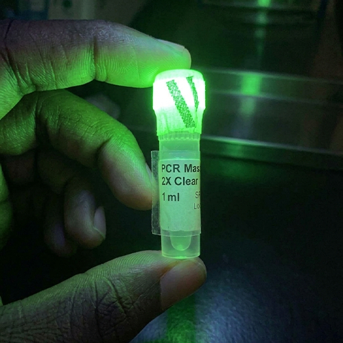
This a sticker that can be attached to temperature-sensitive laboratory reagents and provide an early visual warning when they experience inappropriate warming.
The indicator would ideally respond to both the temperature reached and the duration of exposure. Under appropriate storage conditions, it would remain in a low-fluorescence state. When the temperature rises above the −20°C storage threshold, it would begin producing a detectable fluorescent signal. A longer or more severe temperature excursion would produce a stronger signal, providing an indication of the severity of the exposure.
Why make this?
My motivation comes from a recent laboratory incident in which a number of reagents were lost after they were left outside their required refrigerated storage conditions. The problem was not only that the reagents were exposed to an inappropriate temperature, but that there was no simple way to know when the exposure had occurred or how long it had lasted.
The proposed sticker is intended to provide an early warning so that laboratory personnel can identify reagents that have experienced an inappropriate temperature excursion and investigate or return them to proper storage before assuming that they are lost. The indicator would not determine whether a particular reagent has been damaged; rather, it would provide a record that its storage conditions were exceeded.
The primary users would be laboratory scientists and technicians working with temperature-sensitive reagents such as enzymes and other biological materials. The same concept could potentially be adapted for other cold-chain applications.
What living system would you use?
Escherichia coli as microbial manufacturing chassis to produce an engineered AsLOV
I would initially use Escherichia coli as the microbial manufacturing chassis to produce an engineered AsLOV2-based temperature-sensing protein.
AsLOV2 is a light-responsive protein domain from Avena sativa that undergoes a conformational change involving the Jα helix. Recent research has demonstrated that engineered AsLOV2-containing proteins can also exhibit tunable temperature-dependent switching in E. coli, providing a basis for investigating AsLOV2 as the sensing component of this project. However, the demonstrated temperature range is much warmer than −20°C, so shifting the response toward the desired storage temperature would remain a major engineering challenge.
The engineered AsLOV2 would function as the temperature-sensing component, rather than the entire indicator. A separate fluorescent reporting system would convert the temperature-dependent molecular change into a visible signal.
The final sticker would therefore contain a biological sensing material rather than living bacteria. This would allow the manufacturing organism and the final product to be physically separated.
My initial assumption is that E. coli would be suitable because recombinant AsLOV2 production in E. coli has already been demonstrated.
At a high level, the proposed manufacturing process would be:
Engineered AsLOV2 design → E. coli produces the protein → sensing protein is recovered → protein is incorporated into an indicator material → indicator is assembled into a fluorescent sticker
How would I use automation?
The most important application of automation would be screening different AsLOV2 variants and temperature conditions. I would use an automated liquid-handling platform such as the Opentrons Flex to automate the design–build–test cycle for the sensor.
Variant Testing
The workflow would conceptually be:
AsLOV2 variants → automated preparation → parallel testing at different temperatures → fluorescence measurement → automated data analysis → identify the best-performing variant
The Opentrons Flex supports 1-, 8-, and 96-channel pipetting and can handle liquid volumes from 1–1,000 μL depending on the pipette configuration. It also supports modular temperature-control equipment and automated movement of labware.
For this project, automation would allow many candidate sensor variants and exposure conditions to be tested in parallel rather than manually testing each one.
For example, the system could compare candidate variants across a temperature range surrounding the desired −20°C threshold and measure their fluorescence. The resulting data could be used to identify variants with the clearest distinction between the normal-storage and excursion states.
At a later manufacturing stage, the automation required would be different. Large-scale production would use larger fermentation and formulation/assembly systems, while the Opentrons-type system would mainly be valuable during research, development, optimization, and quality-control screening.
How much would I need to make, and how long would it take?
Production and Manufacturing
(a) How much?
For an initial meaningful prototype scale, I would aim to produce 10,000 indicator stickers, representing approximately the number that could be used across a laboratory or small laboratory network. I will assume that each sticker requires approximately 10 μg of the biological sensing material.
Therefore:
10,000 stickers × 10 μg/sticker = 100,000 μg = 100 mg of sensing material
I would therefore target approximately 100 mg of recoverable sensing material for the initial 10,000-sticker production run.
This is a planning assumption rather than an experimentally established requirement. The actual amount needed per sticker would need to be determined experimentally based on the sensitivity of the fluorescent system.
For the production calculation, I can make a further engineering assumption of 10 mg of recoverable sensing protein per litre of production culture.
Therefore:
100 mg ÷ 10 mg/L = 10 L of production culture
Under these assumptions, approximately 10 L of culture would be required to obtain enough sensing protein for 10,000 stickers.
The actual yield could be substantially different, so this calculation would need to be revised after experimentally measuring protein production and recovery.
(b) How long?
I would divide the production timeline into:
Sensor production
Recovery/formulation of the sensing material
Sticker assembly
Quality-control testing
For the prototype, I would estimate approximately one week per production cycle, allowing time for biological production, recovery/formulation, assembly, and quality-control testing.
The exact production time would depend heavily on the final protein yield and the stability of the sensing material.
At larger scale, production could be parallelized using multiple production vessels and automated downstream processing. The prototype automation system would therefore be used primarily to optimize the sensor, while larger-scale manufacturing would use dedicated bioprocessing and manufacturing equipment.
The 10,000-sticker estimate is therefore not intended as a commercial manufacturing specification; it is a back-of-the-envelope scale that gives the project a measurable manufacturing target.
What don’t you know yet?
1. Can AsLOV2 be engineered to respond around −20°C?
This is the largest technical uncertainty. Existing work demonstrates temperature-responsive AsLOV2 systems, but the demonstrated switching range is approximately 37–41°C rather than −20°C.
I would therefore need to determine whether modifications to the AsLOV2 system can shift its useful thermal response sufficiently toward the −20°C range.
2. Can the temperature response be converted into a useful fluorescent signal?
The sensing system needs to distinguish between normal −20°C storage and an excursion above that temperature. I would need to determine whether the molecular change can produce a sufficiently large and reproducible fluorescence difference.
There is also an important design issue because AsLOV2 naturally responds to blue light. The final system would therefore need to distinguish the desired temperature response from unintended light activation. Existing AsLOV2 research has demonstrated that modifying the native photocycle can help reduce unwanted light activation, providing a possible direction for addressing this issue.
3. Can the indicator record both temperature and duration?
A useful sticker should not simply respond to the temperature at one instant. It should integrate temperature × exposure duration so that a brief minor excursion can be distinguished from a prolonged or severe excursion.
I would therefore need to experimentally determine the relationship between temperature, exposure time, and fluorescence intensity, and determine whether the resulting signal remains detectable after the reagent returns to −20°C.
Ultimately, the project would need to demonstrate that the biological sensor can be converted into a stable, inexpensive sticker that provides a sufficiently reliable warning to be useful in real laboratory cold-chain storage.
Provides continuous expression of the engineered thermal transcriptional regulator so it is available to respond when temperature changes.
Sensor
BBa_B0034
RBS
Kernel
Enables efficient translation of the engineered AsLOV2-EL222 HTH fusion in E. coli.
Sensor
Engineered AsLOV2-I532A + EL222 HTH Domain
CDS
Designed
Combines the engineered temperature-responsive AsLOV2-I532A module with the EL222 DNA-binding domain to couple temperature-dependent conformational changes to transcriptional regulation.
Sensor
Terminator
Terminator
Kernel
Real-Time Output
C120
Promoter
Kernel
Provides the promoter activated by the EL222 HTH domain to drive the temperature-responsive reporter.
Real-Time Output
BBa_B0034
RBS
Kernel
Enables translation of the fluorescent reporter in E. coli.
Real-Time Output
GFP-ssrA
CDS
Kernel
Provides a fluorescent signal while the thermal response is active, with the ssrA tag promoting degradation after the signal is switched off.
Real-Time Output
Terminator
Terminator
Kernel
Terminates transcription of the GFP reporter cassette.
Memory
C120
Promoter
Same as output
Memory
BBa_B0034
RBS
Kernel
Provides a well-characterized ribosome-binding site for efficient translation initiation in E. coli.
Memory
amilCP
CDS
Kernel
Uses the same thermal-responsive regulatory input so that cumulative colour production begins under the same temperature conditions as the fluorescent response.
Memory
Terminator
Terminator
Kernel
Provides reliable transcription termination, helping define the end of each expression cassette and reduce unwanted read-through transcription.
Vector
E.Coli Backbone
Backbone
Existing vector
Provides the replication origin and selectable marker required to maintain the construct in E. coli.
Sequences before Fusing both to create Chimeric Protein
I would initially build the construct using commercially synthesized DNA fragments followed by Gibson assembly. The construct contains several functional modules: the engineered AsLOV2-I532A–EL222 HTH regulatory fusion, the C120/GFP-ssrA reporter cassette, and the C120/amilCP memory cassette. Rather than synthesizing the entire construct as one long sequence, these modules could be ordered as separate DNA fragments and assembled into an appropriate E. coli backbone. This approach makes it easier to modify or replace individual modules during prototyping.
What does synthesis constrain?
DNA synthesis can introduce sequence errors and may be more difficult for long, repetitive, or structurally challenging sequences. My design is not expected to contain unusually repetitive DNA, but the complete multi-cassette construct is more complex than a single gene. I would therefore use sequence-verified synthetic fragments and verify the assembled construct before testing. If a fragment proved difficult to synthesize accurately, it could be divided into smaller fragments and assembled using Gibson assembly, reducing the length of each individual synthesis target.
Prototype vs. scale
For the prototype, I would build and test a small number of sequence-verified constructs, particularly the I532A AsLOV2 variant, to determine whether the proposed temperature-responsive system produces the expected transcriptional and reporter response.
For larger-scale production, the emphasis would shift toward a standardized, sequence-verified construct and a reproducible DNA manufacturing process. Once the design and regulatory architecture were validated, the same construct could be produced using a scalable synthetic-DNA supplier rather than individually assembling each copy. This separates the exploratory prototype stage from the eventual manufacturing process.
References
Guerois R, Nielsen JE, Serrano L (2002). Predicting changes in the stability of proteins and protein complexes: a study of more complex cases. Journal of Molecular Biology, 320(2), 369–387.
Schymkowitz J, Borg J, Stricher F, Nys R, Rousseau F, Serrano L (2005). The FoldX web server: an online force field. Nucleic Acids Research, 33(Web Server issue), W382–W388.
Harper SM, Neil LC, Gardner KH (2003). Structural basis of a phototropin light switch. Science, 301(5639), 1541–1544.
Halavaty AS, Moffat K (2007). N- and C-terminal flanking regions modulate light-induced signal transduction in the LOV2 domain of the blue light sensor phototropin 1 from Avena sativa. Biochemistry, 46(49), 14001–14009.
Zayner JP, Antoniou C, Sosnick TR (2012). The amino-terminal helix modulates light-activated conformational changes in AsLOV2. Journal of Molecular Biology, 419(1–2), 61–74.
Motta-Mena LB et al. (2014). An optogenetic gene expression system with rapid activation and deactivation kinetics. Nature Chemical Biology, 10, 196–202. (EL222 characterisation)
Strickland D et al. (2008). Rationally improving LOV domain–based photoswitches. Nature Methods, 5, 267–273. (AsLOV2 fusion design principles)
Week 3 HW: Cell Free
A. COMPARE CELL-FREE REACTION COMPOSITIONS (20 minutes)
1. Why does the concentration of potassium glutamate and magnesium glutamate differ between
compositions? Which is more sensitive?
Magnesium Glutamate is more sensitive when compared to Potassium Glutamate, small deviations from the optimal concentration can lead to significant drops in protein production [20] where as research has shown that protein production is relatively independent of potassium glutamate concentration over a wide range
[20]. Magnesium (Mg²⁺) binds to ATP and other nucleotides, and phosphate can pull it out of solution. So a recipe with more nucleotides or phosphate needs more Mg²⁺ to keep enough free for the ribosomes and T7 RNA polymerase. Potassium (K⁺) mostly sets the overall ionic strength and helps ribosomes and translation factors work, so it can vary over a wide range, often roughly 80-200 mM.
2. Explain how a PEP+NTP reaction formulation is different from a Ribose+NMP reaction formulation.
PEP acts as a high-energy phosphate donor, allowing for the regeneration of ATP from ADP and GTP from GDP via endogenous pyruvate kinase
[7] while Ribose+NMP system utilizes NMPs instead of expensive NTPs which are significantly more cost-effective. The cell extract’s endogenous metabolic pathways (such as the pentose phosphate pathway or glycolysis) are harnessed to phosphorylate NMPs into NTPs using energy derived from the catabolism of ribose, glucose, or maltodextrin
[9][15][16]
3. Choose two cell-free reaction compositions from the literature, compare and contrast the choice of
reagents and the concentration of reagents.
Composition A: Li et al. (2022)
The reaction mixture used by Li et al. is optimized for supramolecular protein assembly[2].
Glutamate Salts: 175 mM potassium glutamate, 10 mM magnesium glutamate, and 10 mM ammonium glutamate.
Energy Source: 3 mM PEP, 1.2 mM ATP, and 0.86 mM each of CTP, GTP, and UTP.
Other Reagents: Includes 2.7 mM potassium oxalate monohydrate (to inhibit PEP consumption by competing pathways), 1.5 mM spermidine, 1 mM putrescine, 0.33 mM NAD, 0.27 mM coenzyme A, and folinic acid.
Special Components: T7 RNA polymerase and a high concentration of amino acids (specified as 50 mM each of 19 amino acids without glutamic acid)[2].
Composition B: Vezeau & Salis (2021)
This baseline composition was designed to control the dynamics and time-delay of gene expression[5].
Glutamate Salts: 100 mM total potassium glutamate and 8.67 mM total magnesium glutamate.
Energy Source: 30 mM PEP, 1.5 mM each of ATP and GTP, and 0.9 mM each of CTP and UTP.
Other Reagents: 50 mM HEPES, 1 mM putrescine, 0.26 mM CoA, 0.33 mM NAD, 0.75 mM cAMP, and 0.068 mM folinic acid.
Amino Acids: 1.5 mM each (with leucine at 1.25 mM)[5].
Contrast and Discussion
The two compositions represent different optimization goals. Li et al. use a significantly higher potassium glutamate concentration (175 mM vs 100 mM) and a much lower PEP concentration (3 mM vs 30 mM). The inclusion of potassium oxalate in the Li et al. formulation is a notable difference, intended to stabilize the PEP energy source [2]. Conversely, the Vezeau & Salis composition includes cyclic AMP (cAMP), which is often used to stimulate transcription in systems using endogenous RNA polymerase, whereas Li et al. utilize exogenous T7 RNA polymerase [2], [5]. The extreme difference in reported amino acid concentrations (50 mM vs 1.5 mM) suggests that the Li et al. system is geared toward high-intensity synthesis, though such high concentrations are atypical for standard TX-TL reactions [2],[5],[11].
B. DESIGN ONE REACTION COMPOSITION FOR EACH OF THE THREE PROTEINS (40 minutes)
Bonus question: design a T7 helper plasmid that you could add to the cell-free reaction to improve the
yield of functional reteplase (general chaperones, disulfide-bond machinery, etc).
References:
*[1]
F. Guzmán-Chávez et al., “Constructing Cell-Free Expression Systems for Low-Cost Access,” ACS Synthetic Biology, vol. 11, no. 3, pp. 1114–1128, Mar. 2022, doi: 10.1021/acssynbio.1c00342.
[2]
Z. Li et al., “Supramolecular protein assembly in cell-free protein synthesis system,” Bioresources and Bioprocessing, vol. 9, no. 1, pp. 1–14, Mar. 2022, doi: 10.1186/s40643-022-00520-8.
[4]
A. C. Hunt et al., “A rapid cell-free expression and screening platform for antibody discovery,” Nature Communications, vol. 14, no. 1, July 2023, doi: 10.1038/s41467-023-38965-w.
[5]
G. E. Vezeau and H. M. Salis, “Tuning Cell-free Composition Controls the Time-delay, Dynamics, and Productivity of TX-TL Expression,” bioRxiv, Apr. 2021, doi: 10.1101/2021.04.02.438196.
[6]
F. Ba, Y. Zhang, X. Ji, W.-Q. Liu, S. B. Ling, and J. Li, “Expanding the toolbox of probiotic Escherichia coli Nissle 1917 for synthetic biology,” bioRxiv, June 2023, doi: 10.1101/2023.06.05.543671.
[7]
Y. Liu, B. R. Fritz, M. J. Anderson, J. A. Schoborg, and M. C. Jewett, “Characterizing and alleviating substrate limitations for improved in vitro ribosome construction.,” ACS Synthetic Biology, vol. 4, no. 4, pp. 454–462, Apr. 2015, doi: 10.1021/SB5002467.
[8]
Y. Zhang et al., “Protocell Arrays for Simultaneous Detection of Diverse Analytes,” bioRxiv, Feb. 2021, doi: 10.1101/2021.02.13.431022.
[9]
K. Calhoun and J. R. Swartz, “An economical method for cell-free protein synthesis using glucose and nucleoside monophosphates.,” Biotechnology Progress, vol. 21, no. 4, pp. 1146–1153, Sept. 2008, doi: 10.1021/BP050052Y.
[10]
J. Failmezger, R. Nitschel, A. Sánchez-Kopper, M. Kraml, and M. Siemann-Herzberg, “Site-Specific Cleavage of Ribosomal RNA in Escherichia coli-Based Cell-Free Protein Synthesis Systems.,” PLOS ONE, vol. 11, no. 12, Dec. 2016, doi: 10.1371/JOURNAL.PONE.0168764.
[11]
R. B. Ferguson, “Probing metabolism in anE. coli-based cell-free system reveals a trade-off between transcription and translation,” Mar. 2023, doi: 10.1101/2023.03.22.533877.
[12]
J. Failmezger, M. Rauter, R. Nitschel, M. Kraml, and M. Siemann-Herzberg, “Cell-free protein synthesis from non-growing, stressed Escherichia coli,” Scientific Reports, vol. 7, no. 1, pp. 16524–16524, Nov. 2017, doi: 10.1038/S41598-017-16767-7.
[13]
C. J. Whitfield et al., “Cell-free genetic devices confer autonomic and adaptive properties to hydrogels,” bioRxiv, Dec. 2019, doi: 10.1101/2019.12.12.872622.
[14]
J. Shin and V. Noireaux, “Study of messenger RNA inactivation and protein degradation in an Escherichia coli cell-free expression system,” Journal of Biological Engineering, vol. 4, no. 1, pp. 9–9, July 2010, doi: 10.1186/1754-1611-4-9.
[15]
C. Albayrak and J. R. Swartz, “Using E. coli-based cell-free protein synthesis to evaluate the kinetic performance of an orthogonal tRNA and aminoacyl-tRNA synthetase pair,” Biochemical and Biophysical Research Communications, Feb. 2013, doi: 10.1016/J.BBRC.2012.12.108.
[16]
“Development of a cell-free strategy for the directed evolution of enzymes for high-value natural products,” Dec. 2022, doi: 10.22024/unikent/01.02.99209.
[18]
C. A. Elena-Real et al., “The structure of pathogenic huntingtin exon 1 defines the bases of its aggregation propensity,” bioRxiv, vol. 30, no. 3, pp. 309–320, Oct. 2022, doi: 10.1038/s41594-023-00920-0.
[19]
M. Neerathilingam, S. Bairy, and S. Mysore, “Deciphering Mode of Action of Functionally Important Regions in the Intrinsically Disordered Paxillin (Residues 1-313) Using Its Interaction with FAT (Focal Adhesion Targeting Domain of Focal Adhesion Kinase),” PLOS ONE, vol. 11, no. 2, Feb. 2016, doi: 10.1371/JOURNAL.PONE.0150153.
[20]
J. Shin and V. Noireaux, “Efficient cell-free expression with the endogenous E. Coli RNA polymerase and sigma factor 70.,” Journal of Biological Engineering, vol. 4, no. 1, pp. 8–8, June 2010, doi: 10.1186/1754-1611-4-8.*