Week 1 HW: Final Project Ideas
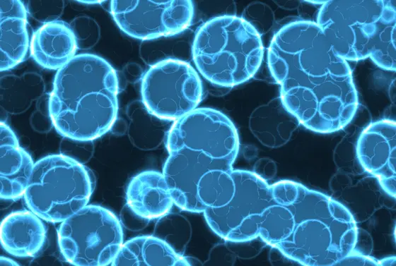
HTBAA Homework #1 – 3D Diffuse Large B-Cell Lymphoma (DLBCL) Microtumors for Drug Screening
(1) What would you like to biomanufacture and why?
I would like to biomanufacture standardized 3D microtumors of diffuse large B-cell lymphoma (DLBCL). Each microtumor consists of lymphoma cells encapsulated in a hydrogel that recreates key features of the lymph node environment. They are produced in multi-well plates so that many drugs and doses can be tested in parallel.
I believe this could be achieved by encapsulating commercially available DLBCL cell lines in a synthetic hydrogel functionalized with adhesion peptides, using 3D-printed molds and inserts to standardize each microtumor, and an automated liquid handler to dispense them reproducibly.
Part of the problem is how drugs are tested. Lymphoma cells are usually screened in 2D suspension culture, floating freely in medium. This ignores the lymph node microenvironment, where the extracellular matrix and adhesion signals activate survival pathways in lymphoma cells through integrin receptors. This phenomenon, known as cell adhesion-mediated drug resistance, means that 2D assays can overestimate drug efficacy. Tian et al. (2015) showed that integrin-binding hydrogels alter the proliferation and drug response of B-cell lymphoma cells compared with 2D culture, which suggests that a more realistic model could better identify drugs that will actually work.
My motivation is also personal: a close family member went through DLBCL. I am also based in Peru, where resources for biomedical research are limited and access to specialized equipment is difficult. Coming from biomedical engineering with experience in medical imaging, 3D printing, and design, this project is a way to enter cancer research using the skills I already have, while exploring how low-cost fabrication could make this type of research more feasible in resource-limited settings.
(2) What living system would you use?
The chassis would be mammalian cells, specifically commercially available human DLBCL cell lines such as SU-DHL-4 or OCI-Ly1. A human stromal cell line such as HS-5 could later be added to mimic the supportive cells of the lymph node.
The central process is cell encapsulation: lymphoma cells are mixed into a liquid hydrogel precursor, which is then solidified so that the gel forms around them and holds them in three dimensions. This approach suits DLBCL cells, which grow in suspension and do not naturally attach to scaffolds. Several hydrogels could be used, ranging from synthetic materials that can be precisely tuned and enriched with adhesion signals, to simpler and cheaper natural materials useful for early testing.
In broad terms, cells, culture medium, and hydrogel components go into the system. Inside the gel, the cells grow over a few days into small tumor-like aggregates, which are then exposed to different drugs. What comes out are 3D microtumors together with viability and imaging data that show how each drug affects the cancer cells.
This design rests on several assumptions. I use cell lines rather than patient cells because they are easier to obtain, more reproducible, and ethically simpler. I keep serum in the medium, although a serum-free formulation, like those discussed by Yap et al., would be a future improvement. Finally, I assume access to a laboratory with a biosafety cabinet and a CO₂ incubator through a collaboration.
(3) How could automation help you make it?
During prototyping, automation would combine three tools. The first is my Formlabs Form 4B resin printer. It would fabricate the custom hardware around them. This includes molds for casting agarose microwell arrays, in which suspension cells settle into uniform aggregates without ever touching the resin; rings that give each hydrogel droplet the same diameter and height; pillar lids with standard plate dimensions that allow gels to be transferred between plates; and adapters for the liquid handler and microscope. Parts in contact with cells would be printed in biocompatible BioMed resin, then washed, post-cured, leached, sterilized, and tested for cytotoxicity before use.
The second tool is an Opentrons liquid handler (OT-2 or Flex), which would automate the steps most prone to human error: dispensing the viscous cell-hydrogel mixture with identical volumes, preparing serial drug dilutions, and adding the viability reagent (CellTiter-Glo 3D, which measures ATP released by living cells). The third is automated microscopy combined with image analysis in Python, where my imaging background contributes most. Segmentation would measure microtumor size and shape as well as live/dead staining in every well.
The reason to automate these steps is reproducibility. Small differences in gel volume, cell number, or gel geometry could distort dose-response curves, and manual pipetting of hundreds of viscous droplets cannot be consistent.
flowchart TD
    subgraph A["A. Devices (Form 4B)"]
        A1["CAD design<br/>molds / inserts / adapters"] --> A2["Print in BioMed resin"]
        A2 --> A3["IPA wash + post-cure"]
        A3 --> A4["Leaching in PBS + sterilization"]
        A4 --> A5["Cytotoxicity test"]
    end
    subgraph B["B. Cells"]
        B1["Thaw DLBCL cell line"] --> B2["Suspension expansion<br/>~6 days"]
        B2 --> B3["Count and viability check"]
    end
    A5 --> C1
    B3 --> C1
    subgraph C["C. Encapsulation"]
        C1["Prepare hydrogel precursor<br/>+ adhesion peptide (RGD)"] --> C2["Mix cells with precursor"]
        C2 --> C3["Dispense with Opentrons<br/>10 µL per well"]
        C3 --> C4["Gelation<br/>chemical or light-triggered"]
        C4 --> C5["Add culture medium"]
    end
    C5 --> D["D. 3D maturation<br/>3 days, 37 °C, 5% CO₂"]
    D --> E["E. Drug treatment<br/>dilutions with Opentrons, 72 h"]
    E --> F["F. Readout<br/>automated imaging + CellTiter-Glo 3D"]
    F --> G["G. Analysis<br/>segmentation, dose-response curves, IC50<br/>2D vs 3D comparison"]
    G -.->|"Learn and redesign"| A1
    G -.->|"Adjust gel / cell density"| C1
The main output of the analysis is the IC50, the drug concentration that reduces viability by 50%. Comparing IC50 values between 2D and 3D cultures shows whether the hydrogel recovers the resistance that the lymph node environment confers.
(4) How much would you need to make, and how long would it take?
(a) How much?
I define a meaningful scale as a drug-repurposing screen of approved oncology drugs across a panel of DLBCL cell lines, large enough to produce a full dataset that could identify candidates for further study.
| Parameter | Value | Source / assumption |
|---|---|---|
| Drugs | 150 | Similar to the NCI Approved Oncology Drugs set (~150–180 compounds) |
| Doses per drug | 8 | Standard dose-response design |
| Replicates | 3 | Statistical minimum |
| Cell lines | 5 | Covering the GCB and ABC molecular subtypes |
| Controls | +10% | Assumption (untreated and vehicle controls) |
| Cells per microtumor | 5,000 | Typical assumption for 3D models |
| Gel volume per well | 10 µL | Assumption for 384-well plates |
Each cell line requires 150 × 8 × 3 = 3,600 wells. Across five lines and including controls, the screen needs 3,600 × 5 × 1.1 ≈ 19,800 wells, equivalent to about 52 plates of 384 wells. At 5,000 cells per microtumor, this amounts to roughly 1 × 10⁸ cells (≈ 2 × 10⁷ per line) and about 200 mL of hydrogel. By comparison, a prototype on a single 96-well plate is about 200 times smaller, which explains why manual work is acceptable for prototyping while automation becomes essential at scale.
(b) How long?
Starting from a vial of about 2 × 10⁶ cells, each line must grow roughly 11-fold to reach 2.2 × 10⁷ cells (including a 10% margin). This requires log₂(11) ≈ 3.5 doublings, which at an assumed doubling time of ~40 hours takes about 6 days. At a typical density of 1 × 10⁶ cells/mL, each line needs about 22 mL of culture, or one to two T75 flasks, and all five lines can be expanded in parallel.
On a single Opentrons Flex, I estimate about 15 minutes per plate for seeding, 10 minutes for drug addition, and 10 minutes for the readout reagent. Across 52 plates this totals about 30 hours of robot time, or roughly four working days. Automated imaging at about one second per well adds around 6.5 minutes per plate, or about 6 hours overall. The biological steps, 3 days of 3D maturation followed by 3 days of drug exposure, overlap with seeding because plates are processed in staggered batches.
Altogether, the full screen would take about three weeks: 6 days of expansion, about 4 days of staggered seeding, 6 days of culture and treatment, and 1–2 days of readout and analysis. The bottleneck is biology rather than automation, so adding robots would barely shorten the timeline; faster-growing cells or shorter assays would have a much larger effect.
Prototyping and scale-up would look quite different. The prototype would use one cell line and one 96-well plate, Form 4B parts, and manual pipetting or an OT-2, taking about two weeks. It would test R-CHOP drugs in their active in vitro forms: doxorubicin, vincristine, 4-hydroperoxycyclophosphamide, and prednisolone, since cyclophosphamide and prednisone are prodrugs activated in the liver. Rituximab would be left for later, because it acts mainly through complement and immune cells that are absent from the gel. The full screen, in contrast, would run on a cloud lab or high-throughput platform with 384-well plates and commercial consumables.
(5) What don’t you know yet?
The first and most important unknown is whether the 3D model actually outperforms 2D culture. I do not yet know whether these microtumors reproduce clinically relevant drug resistance, or whether the difference from 2D is large enough to matter. To find out, I would compare 2D and 3D dose-response curves for the R-CHOP drugs and check them against published data for the same cell lines.
The second unknown concerns the Form 4B parts. Printed resin could release uncured residues that harm cells, and I do not know the smallest pillar or channel size I can print reliably. An extract cytotoxicity test based on ISO 10993-5 would address the first issue: printed parts are incubated in medium for 24 hours, cells are cultured in that medium, and their viability is compared with fresh medium. For resolution, I would print calibration parts with features of different sizes and measure how far they deviate from the CAD design.
The third unknown is whether robot-dispensed hydrogel can be made reproducible. Viscous gels may clog or gel inside pipette tips, and if gelation is too slow, cells sink before being trapped and lose their 3D distribution. I would measure the coefficient of variation in gel volume and cell number between wells, aiming for less than 15%, and compare fast chemical gelation (PEG-4MAL) with on-demand light-triggered gelation (GelMA), which allows more time for dispensing.
Beyond these three points, cell lines do not capture the diversity of real patients, and the model contains no immune cells. Later versions would need patient-derived cells and immune components to evaluate antibody therapies such as rituximab.
References
- Tian, Y. F. et al. (2015). Integrin-specific hydrogels as adaptable tumor organoids for malignant B and T cell lymphomas. Biomaterials, 73, 110–119.
- Yap, W. S., Choudhury, D. & Suntornnond, R. (2023). Towards biomanufacturing of cultured meat. Trends in Biotechnology, 41(3), 292–294.
- Zhang, Y.-H. P., Sun, J. & Ma, Y. (2017). Biomanufacturing: history and perspective. J Ind Microbiol Biotechnol, 44, 773–784.
- Figure 1: Ayda P (2022). Cell culture-fig.png. Wikimedia Commons.
AI
I used Claude (Anthropic, Claude Opus 5 model) through claude.ai. The main prompt, originally in Spanish, were:
- “I need help to improve redaction to complete my homework. I am a biomedical engineer with experience in medical imaging and 3D printing… I want to research DLBCL… evaluate CAR-T vs. hydrogel encapsulation with microfluidics and, if they are too difficult, propose an alternative. Answer in markdown.”