Week 2 HW: DNA Design
Exploratory optogenetic DNA design for cortical organoids
Ilknur Icke
29 September 2026
My HW1 organoid platform uses electrical stimulation and electrode recording as its baseline. For HW2, I designed a ChR2(H134R)–EYFP expression construct to explore an optional optical-input interface for the same cortical tissue. Adopting this interface would require a demonstrated experimental benefit. The saved V1 is a 6,225 bp circular reconstruction of Addgene plasmid #26973. Its sequence, assembly, codon-optimization comparison, sequence search, and safety screen have been checked in software. Expression and functional performance in organoids remain experimental questions.
Optogenetic design options considered
I considered four related genetic design options. Their labels identify questions about input delivery, expression timing, and activity-dependent gene expression. V1 is the only construct assembled and analyzed in Kernel. V2–V4 are ideas considered; they are not a required development sequence or a commitment for the final project.
V1 Reference optical input candidate. The hSyn promoter drives ChR2(H134R)–EYFP, providing a candidate blue-light input channel with a fluorescent expression readout. V1 asks whether a defined genetic module can make neurons optically addressable. This is the reference-based construct assembled and analyzed in Kernel. [2]
V2 Red-shifted input for three dimensional tissue. This proposal would replace the V1 actuator/reporter cassette with a red-shifted excitatory opsin such as ChrimsonR and a suitable reporter. It asks whether changing the optical input improves access and response uniformity across a 3D organoid. Any advantage over V1 would require measurement.
V3 Developmentally timed input. This proposal adds inducible control of opsin expression so that availability of the input channel can be studied at different maturation stages. The working architecture uses a TRE3G-controlled opsin cassette with the rtTA transactivator supplied by the host or a second cassette. Expression timing and background expression would need validation.
V4 Activity-responsive gene expression. This concept would connect neural activity to an activity-responsive regulatory element driving a reporter and, potentially later, a selected plasticity or developmental effector. It asks whether structured input can influence a lasting biological state. The regulatory sequence, output, and feedback behavior remain to be defined.
I selected V1 for this exploratory study because it has a traceable reference sequence and a manageable architecture. The final platform would adopt it only if testing justified the additional genetic and optical requirements. In the sequence comparisons, “baseline” means the unchanged Addgene reference; the platform baseline remains electrical stimulation. The codon-recoded sequence in Part 4 is a comparison within V1, not V2. V2–V4 have not been built or experimentally validated here.
Part 0 Kernel workspace
My work is in the HTGAA 2026 workspace, repository HTBAA26_Genspace, folder Ilknur Icke / HW2 / v1. I used the existing personal folder and retained the existing ChR2(H134R)-EYFP CDS part. Individual parts hold sequence and function records; the construct links them in their intended arrangement.
Open the saved V1 construct in Kernel
Part 1 Expression target
Connection to HW1. My HW1 project proposes manufacturing human cortical organoids toward defined functional phenotypes through controlled stimulation, recording, and culture conditions. Its baseline uses camera-derived electrical stimulation and electrode recording. HW2 explores a genetic optical-input alternative within that system. The design question is whether neurons could express a defined actuator to support controlled optical excitation. The larger manufactured product remains the organoid system, and electrical input remains the baseline unless a comparison establishes a reason to change it. [1]
Molecule and source. I selected the engineered humanized ChR2(H134R) protein fused at its C terminus to EYFP. The exact 1,662 bp CDS comes from Addgene #26973, full-sequence record 199313, bases 456–2117. It encodes 553 amino acids followed by a stop, including the source PAAAT linker. “Humanized” describes the engineered expression sequence, not a naturally occurring human gene. [2]
Host and destination. The intended expression host is neuronal cells within human iPSC-derived cortical organoids. Kernel’s selected host is Homo sapiens. The desired destination is the neuronal membrane, where ChR2 should mediate light-dependent electrical responses. EYFP supports expression and localization measurements. I retained the reference fusion without adding a secretion signal; membrane localization and activity will be tested rather than inferred from fluorescence alone.
Part 2 Genetic parts
All eight parts use exact segments of Addgene full-sequence record 199313. Each Kernel record contains its DNA, part type, source coordinates, and rationale. Coordinates below are 1-based and inclusive in the original source. The complete sequences are also preserved in the companion GenBank file and parts FASTA. [2]
| Part | Kernel type | bp | Source bases | Reason for selection |
|---|---|---|---|---|
| hSyn promoter | Promoter | 448 | 6190–6225; 1–412 | Selects a neuronal expression context; its performance in the intended organoids remains to be measured. |
| 5UTR-Kozak | 5′ UTR with Kozak | 43 | 413–455 | Retains the reference upstream leader and the GCCACC sequence immediately before the CDS. |
| ChR2(H134R)–EYFP CDS | CDS with start and stop | 1662 | 456–2117 | Provides the intended optical actuator and fluorescent fusion, including ATG, PAAAT linker, and TAA. |
| CDS-WPRE junction | Miscellaneous | 24 | 2118–2141 | Preserves reference spacing after the stop codon; no separate regulatory role is assumed. |
| WPRE | 3′ UTR | 589 | 2142–2730 | Retains the source posttranscriptional regulatory element as part of the baseline expression context. |
| WPRE-polyA junction | Miscellaneous | 31 | 2731–2761 | Preserves the reference junction before the polyadenylation region. |
| hGH polyA | Poly(A) signal | 477 | 2762–3238 | Provides the reference transcript 3′ processing context. |
| pAAV vector context | Backbone | 2951 | 3239–6189 | Retains both annotated AAV terminal repeats, bacterial replication and AmpR selection features, and intervening context. |
Boundary and vector decisions
The promoter crosses the original plasmid origin, so its two annotated intervals are joined in the part record. The upstream part ends before ATG. The full Kozak annotation overlaps the first coding bases, which are included only in the CDS, avoiding duplication at the boundary. The upstream-part boundary is an operational design boundary, not a measured transcription start site.
The vector-context part is a composite region containing both terminal repeats and the bacterial backbone between them. Detailed annotations retain the individual features. The full plasmid length includes bacterial propagation sequences and must not be interpreted as the packaged AAV genome length. No new secretion tag or independent targeting sequence is added.
Part 3 Assembled construct
V1_hSyn_ChR2_H134R_EYFP_baseline contains the eight linked parts in the order shown below. The circular display origin is moved to source base 6190 to place hSyn first. Every source base is retained exactly once; the circular DNA sequence is unchanged.
The CDS occupies assembled bases 492–2153, starts with ATG, ends with TAA, has no internal stop codons, and retains R134 and the PAAAT linker. DNA copied back from Kernel matched the prepared 6,225 bp sequence exactly. The saved part links persisted after reload, and Kernel’s construct checker reported 0 errors and 0 warnings. The checker is separate from the SecureDNA screen in Part 4.
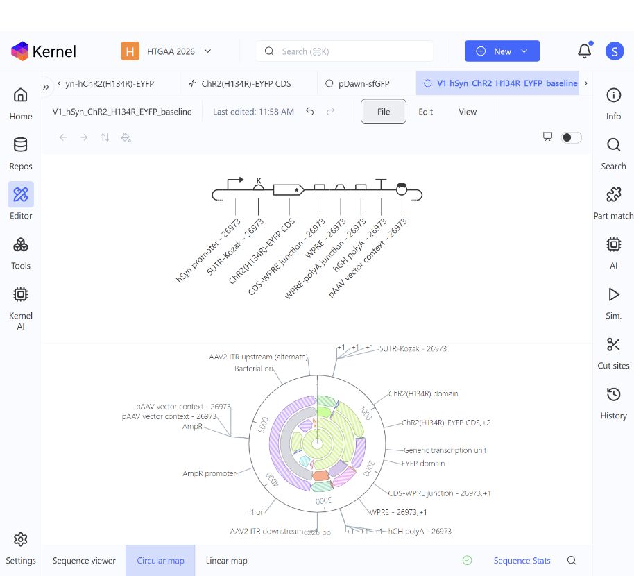
Figure 1. Saved Kernel schematic and circular map for V1. The schematic shows all eight linked parts; the bottom status area shows the construct-checker result. A screen capture is used because the raw schematic PNG export clipped its angled labels.
Part 4 Optimization and quality checks
Codon optimization
I ran Kernel SIMPLE_0_3 on the complete 1,662 bp CDS for Homo sapiens. Settings were no Asimov preset, no restriction-enzyme exclusions or additional avoided motifs, no 5′ UTR input, and no explicit GC target. Signal-peptide handling and CpG avoidance were off, and a single TAA stop was retained. The resulting DNA changed at 218 positions across 186 of 553 sense codons. Independent translation confirmed the same 553-amino-acid protein, with no internal stops. The metrics below were calculated from the actual input and output sequences. [3]
| Measure | Source CDS | Optimized comparison |
|---|---|---|
| DNA length | 1,662 bp | 1,662 bp |
| Protein length excluding stop | 553 aa | 553 aa |
| GC content | 57.04% | 62.64% |
| Start and stop codons | ATG / TAA | ATG / TAA |
| Internal stops | 0 | 0 |
| Protein identity to source | Reference | 100% |
Codon preferences can influence expression in a host, but synonymous recoding alone does not establish a biological improvement. Kernel displayed N/A for both its Asimov Codon Usage Index and RNA structure score. I retained the source CDS in V1 and kept the recoded sequence as a separate candidate for an empirical comparison. Figure 2 records the completed run and settings. [3]
BLAST and sequence identity
I searched the full baseline CDS against Kernel’s indexed constructs and parts, including Addgene Plasmids. The public pAAV-hSyn-hChR2(H134R)-EYFP reference was among several tied full-length matches: 100.0% identity, query bases 1–1662, subject bases 456–2117, bit score 3070.3, and displayed E-value 0.00e+0. This supports the identity and boundaries of the imported engineered fusion. It is an existing engineered sequence, not an unmodified natural gene or a newly invented CDS. The displayed E-value is limited by application precision. This was a search of Kernel’s indexed collections, not an exhaustive natural-genome or off-target analysis. Figure 3 shows representative results. [2]
Safety screen
I screened the complete 6,225 bp baseline construct with Kernel’s SecureDNA tool. The submitted input matched the saved V1 exactly. The tool describes comparison against a database of potential biosecurity threats using cryptographically secured input. It returned “No flagged sequences detected.” That is the result for this sequence under the available screen; it does not establish cell health, delivery suitability, or experimental safety. The recoded comparison was not incorporated into this construct and was not part of this screen. Figure 4 records the result.
Part 5 From design to build
Build strategy
For the unchanged V1 prototype, I would obtain the existing Addgene #26973 reference plasmid through an appropriate laboratory and verify it against the saved sequence. Dividing the reference into reusable Kernel parts does not require resynthesizing every part. This is a direct route to the baseline whose sequence I analyzed. [2]
To test the recoded comparison, I would order its 1,662 bp CDS as sequence-verified synthetic DNA and replace the corresponding CDS in the existing vector context. The order would also need appropriate flanking assembly sequences, so its total length would exceed 1,662 bp. I would preserve the promoter, leader, WPRE, poly(A) region, and backbone, making synonymous CDS sequence the intended variable. Exact fragment boundaries and overlaps remain to be designed before ordering.
Gibson et al. describe assembly of overlapping DNA fragments through combined exonuclease, polymerase, and ligase activities. I would apply this principle to a small number of physical fragments for the CDS replacement. The eight Kernel parts are functional design modules and do not dictate eight separate synthesis or assembly fragments. [4]
Synthesis constraints and verification
The 448 bp hSyn promoter is 71.21% GC, calculated from the saved sequence. The CDS changes from 57.04% GC to 62.64% GC after recoding, and the vector contains annotated inverted terminal repeats. I would flag these GC-rich and repetitive regions for supplier review. These are sequence features requiring attention, not observed synthesis failures or vendor rejection results. I would preserve functional promoter and terminal-repeat sequences and reuse the existing context where possible.
If the recoded CDS cannot be supplied as one verified fragment, smaller fragments are a fallback after sequence review. I would verify the finished plasmid across the CDS, junctions, regulatory regions, and terminal repeats using methods that resolve those regions adequately.
Carr et al. used mismatch-binding MutS to reduce errors in synthetic DNA. Their result motivates treating synthesis fidelity separately from successful assembly. Error reduction can improve the input material, but final sequence verification is still needed. I would review the supplier’s documented quality checks rather than assume every provider uses this specific method. [5]
Prototype and scale
An optical-input pilot would use one verified reference and, if justified, one verified recoded comparison. Only if that pilot supported adopting optical input would I plan DNA or delivery-vector production at the scale of the HW1 experiment. That production would require documented identity, quality, and lot consistency. More organoids using the same design would require more material and consistent delivery, not a different synthetic gene for each organoid.
An oligo pool would add complexity to this small comparison; it could become useful for a future large library of distinct designs. Required DNA or delivery-vector quantity remains uncertain until delivery method, organoid stage, and pilot requirements are established. The 6,225 bp plasmid includes bacterial backbone sequence. Delivery selection and any later vector production require separate specifications; no serotype or vector-production process has been finalized here.
Part 6 Unknowns and proposed tests
Expression in the intended neurons
If V1 advances to an experimental pilot, delivery, maturation, and cell composition could produce variable hSyn-driven expression within the organoids. I would quantify the fraction of neuronal-marker-positive cells with EYFP signal, per-cell signal distribution, its location across depth, and its stability over time, alongside cell health. If signal is weak, measurements of delivery and transcript abundance would help distinguish failure to reach the cells from failure to express the cassette.
I would compare source and recoded CDS versions under matched conditions before adopting the optimized output. The unchanged protein sequence and higher GC content do not demonstrate an expression benefit. If an optical-input pilot revealed a need to control when the opsin is expressed, V3 would be an optional follow-up, requiring measurements of background expression and induction timing.
Membrane localization and useful light responses
EYFP fluorescence does not by itself prove functional channel activity. I would examine membrane-associated localization, measure light-evoked currents or firing in individual cells, and then use multielectrode-array recordings to assess organoid responses. The readouts would include response probability, latency, repeatability, and changes during repeated stimulation.
Controls would include V1-expressing tissue without stimulation and expression-negative tissue receiving the same light exposure. I would compare functional responses and health alongside fluorescence, rather than select the brightest construct automatically.
Reproducible input across a three dimensional organoid
Responses may vary with tissue depth and geometry even if individual cells express a functional opsin. Repeated illumination or sustained expression could also change baseline activity or tissue health. I would map expression and evoked responses by location, track their stability over time, and measure viability, spontaneous firing, and network-burst patterns before and after repeated stimulation.
Expression-negative/light-exposed and expression-positive/unstimulated controls would help separate optical exposure from construct-related effects. If the optical approach warranted further work but tissue access limited responses, V2 and illumination geometry would be possible comparisons. V3 would be considered separately if sustained expression proved limiting. Neither extension is assumed necessary for the organoid platform.
The HW1 comparison of structured, matched-random, and no stimulation can proceed with electrical input. V1 would merit adoption only if a comparison showed a useful benefit relative to the additional delivery and optical requirements. Demonstrating light-evoked responses alone would not establish improved organoid function. Acceptance criteria and replication would be set with an experimental collaborator. V4 remains an exploratory question about activity-responsive gene expression, outside the current implementation.
References and AI disclosure
[1] Icke, I. (2026). Week 1 HW Final Project Ideas. Biomanufacturing Neural Organoids Toward Defined Functional Phenotypes. HW1 project page
[2] Addgene. pAAV-hSyn-hChR2(H134R)-EYFP, plasmid #26973, deposited by Karl Deisseroth. Sequence source: Addgene full-sequence record 199313. Plasmid record
[3] Asimov Kernel. Codon Optimization documentation. Actual run settings and results are recorded in Part 4 and Figure 2. Kernel documentation
[4] Gibson, D. G., Young, L., Chuang, R.-Y., Venter, J. C., Hutchison, C. A. III, and Smith, H. O. (2009). Enzymatic assembly of DNA molecules up to several hundred kilobases. Nature Methods 6, 343–345. Assigned reading. doi:10.1038/nmeth.1318
[5] Carr, P. A., Park, J. S., Lee, Y.-J., Yu, T., Zhang, S., and Jacobson, J. M. (2004). Protein-mediated error correction for de novo DNA synthesis. Nucleic Acids Research 32, e162. Assigned reading. doi:10.1093/nar/gnh160
AI use
I used OpenAI ChatGPT and Codex to develop the V1–V4 design discussion, organize the homework, assist with Kernel and Addgene browser operations, calculate sequence comparisons, and draft this write-up.
Sequence and analysis records
The exact DNA is available in the linked Kernel construct and the companion V1_hSyn_ChR2_H134R_EYFP_baseline.gb and parts.fasta files. Part 4 metrics were independently calculated from the source and exported optimized CDS; the optimized sequence was not adopted into V1.
Saved V1 construct | Completed human codon-optimization comparison
Appendix Codon optimization evidence
Run: Ilknur_HW2_V1_ChR2_H134R_EYFP_Human_Comparison. The completed tool output shows Homo sapiens, SIMPLE_0_3, a single TAA stop, and N/A for the displayed codon-usage and RNA-structure scores. GC percentages and change counts in Part 4 were computed independently.
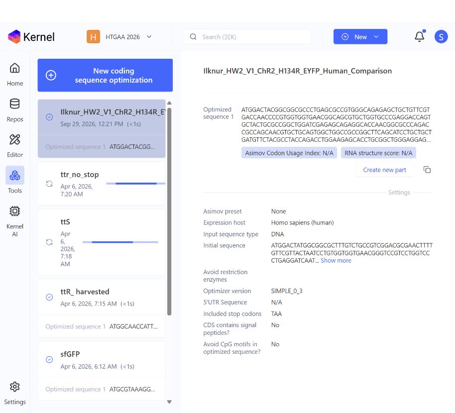
Figure 2. Completed optimization run with the selected host and settings. The recoded CDS remains a separate comparison output.
Appendix Sequence search evidence
The query was the complete 1,662 bp baseline CDS. The screenshot shows representative top matches at 100.0% identity. The original pAAV-hSyn-hChR2(H134R)-EYFP reference was also present in the rendered results, with query bases 1–1662 and subject bases 456–2117; it is below the visible rows in this screenshot.
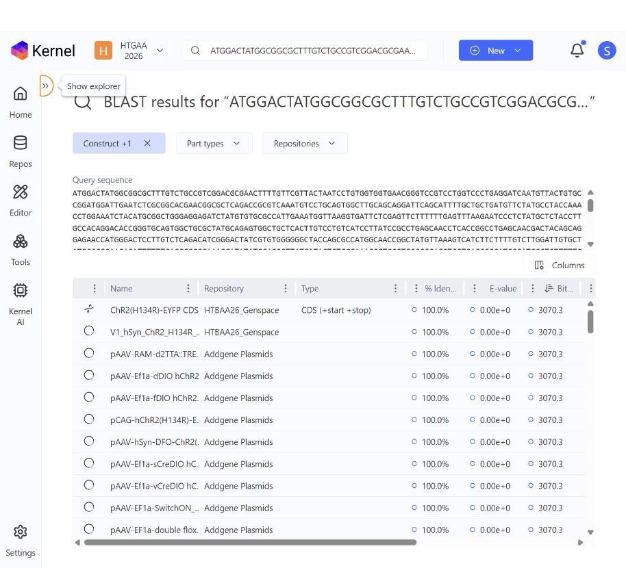
Figure 3. Kernel BLAST results for the baseline CDS. Several records share the highest score; this screenshot does not establish a total hit count.
Appendix Safety screening evidence
The screened sequence was the complete 6,225 bp saved V1 baseline. A local sequence comparison verified that the submitted input matched that construct exactly.

Figure 4. SecureDNA returned “No flagged sequences detected” for V1. This is a sequence-screening result, not a functional or experimental-safety assessment.