I am a fourth year medical student from Taiwan with a focus on orthopedics. I speak three languages: English, Mandarin, and German. My favorite artist is Salvador Dalí! I am a curious person who asks a lot of questions, and I can’t wait to ask even more in this course!!
See the nav bar on the left to look through my work for How To
Biomanufacture Almost Anything (Fall 2026)!
Subsections of Ivy Hung — HTBAA Fall 2026
Week 1 HW: Final Project Ideas
Project: Bone Implant Surfaces, Grown Microbially 🦴
01
🧬 1. What do you want to make?
Titanium implants are mechanically strong and osseointegrate well in many patients, but they are biologically inert at the interface. In a meaningful subset of cases, aseptic loosening and fibrous encapsulation still cause failure. Plasma-sprayed hydroxyapatite is already used clinically, but the high-temperature process and rapid cooling cause instability in vivo. At the other extreme, hydroxyapatite sintered at high temperature is highly crystalline but brittle, and osteoclasts can barely resorb it, so it can persist in the defect for years and resist remodeling 1.
I propose bypassing that tradeoff: bacterial biomineralization deposits calcium phosphate on a scaffold at physiological temperature, with no sintering step. This may allow crystallinity and resorption rate to be tuned through bacterial and culture parameters rather than heat.
bacterial biomineralization deposits calcium phosphate on a scaffold at physiological temperature, with no sintering step.
Bacterial alkaline phosphatase (ALP) activity has been shown to precipitate low crystallinity, ion substituted hydroxyapatite 2. Whether bacterially produced mineral actually behaves more like native bone than sintered synthetic grafts, in terms of resorption, mechanical integration, or in vivo performance, is an open question this project is built to test. That research paper used Serratia marcescens, but as a Gram negative opportunistic pathogen it is not a viable candidate for an implantable material. The mechanism it demonstrates, bacterial ALP driving bone-like mineralization, is what this project builds on. The organism itself is not.
A central question here is what living cells contribute beyond the enzyme itself. Purified ALP can also drive mineralization in solution 3, so this project includes a cell-free ALP control. If bacteria produce thicker, better-adhered, or more bone-like mineral than the enzyme alone, that justifies the microbial approach. If the enzyme alone matches them, that is also a useful result, because it gives the same mineral with no bacterial residue to clean up.
This approach is a biological variant of an established chemical technique called biomimetic deposition, developed by Kokubo in 1990. In that method, a pre-functionalized substrate is soaked in simulated body fluid so apatite precipitates spontaneously, mimicking physiological bone mineralization. My proposal replaces the chemical bath with living bacteria that create local pH and ion shifts through ALP activity (or ureolysis), executing the same kind of mineral biologically instead of purely chemically.
The target is a biomineralized surface coating for osseointegrated implants, the percutaneous titanium stems used in limb loss prosthetics, which are surgically anchored directly into residual bone. The clinical failure point in these systems is not the titanium itself but the bone-implant interface, which is exactly where this coating would be applied.
02
🧫 2. What living system could make it?
A paper from 2023 found that Escherichia coli produces hydroxyapatite precipitation through alkaline phosphatase (ALP) activity, the same enzyme class that osteoblasts themselves use 4.
That result was on a solid plate. A follow up study used a 10 mM concentration of Ca²⁺, which led to biofilm mineralization in both solid and liquid mediums and mineralized faster than the plate condition 5.
Standard E. coli is Gram negative, meaning it carries lipopolysaccharide (LPS) in its outer membrane, and LPS residue can trigger a strong inflammatory response if left on an implant surface after sterilization. Rather than switching to a different organism that requires a change in the mineralization mechanism, I propose using strains that reduce the endotoxin concern at the organism level, like the engineered endotoxin-free E. coli strains described by Mamat et al.7. The cell-free ALP control described below is the fallback if that cleanup proves impractical.
03
🤖 3. How could automation help you make it?
Programmable microfluidics could screen a lot of variables at once.
Ca and phosphate ratio
inducer concentration for ALP expression
pH
incubation time
strain variant
submerged vs. semi-solid air-liquid interface format for mineralizing onto a scaffold
cell-based vs. cell-free ALP (purified enzyme with a phosphate source) as the mineralizing system
(generated by ai)
Once I know the best chemical and genetic conditions, Cloud Lab could automate producing centimeter-scale 3D scaffold replicates under those conditions, plus downstream characterization like imaging, microCT, and compression testing to measure mineral density and mechanical strength. (generated by ai)
A lot of these parameters aren't actually new. They're already used to study chemical biomimetic deposition, things like solution ionic composition, immersion time, resulting Ca/P ratio, crystallinity, and morphology. So this screen is really asking one question. Can microbially driven nucleation match or beat what's already known about those structure-property relationships?
04
🏭 4. What would it take to make it at a meaningful scale?
The scale I'm targeting is a 2 to 3 cm segment of a titanium implant stem, the part that sits inside the bone in an osseointegration implant. That's the spot where bone to metal integration currently underperforms, so that's where the coating needs to work.
For calcium, I'm also considering a 10 mM concentration, since it's already been shown to cause biofilm mineralization in both liquid and solid medium. That study got a visible layered structure after 5 days, with a mineralized layer around 20 to 40 μm thick. The two layer separation got clearer after 7 to 10 growing days, each layer around 40 to 60 μm. This gives me a rough timeline to work from for the scale I'm aiming for. 5
Day
Observation
5
Visible layered structure, ~20–40 μm mineral layer
7–10
Two-layer separation clearer, each layer ~40–60 μm
For the manufacturing path, microfluidic screening finds the best Ca and phosphate ratio, pH, and incubation time for mineral density. Cloud Lab then produces titanium coupons at scale under those conditions.
For evaluation, I'd use microCT to check mineral density and thickness, and pull-out and shear testing to check strength at the coating-substrate interface, since that's the same failure point that limits current osseointegration implants. Every measurement above is run on both arms: bacterial and cell-free. Comparing mineral density, thickness, crystallinity, and interface strength between them shows what the living cells actually add. I want to tie these results to real standards instead of just reporting numbers on their own. ISO 13779-2 recommends a crystallinity degree greater than 45% for calcium phosphate coatings used in bone implants 6. I'm treating 45% as a stability floor, not the goal. The real question is whether microbial mineral can clear that floor while remaining less crystalline than sintered HA, and so resorbing more readily. If it can't reach 45%, that is still a useful result, because it tells me where the biological approach falls short of the standard.
I'm treating 45% as a stability floor, not the goal.
Fig. — projected crystallinity vs. the ISO stability floor (illustrative)
05
❓ 5. What don't you know yet?
a.
Can the bacteria and their residue be removed completely without damaging the new mineral? Implants that pass through the skin already carry infection risk, so a bacteria-derived coating has to be shown to be completely clean, including live cells, dead cell debris, DNA, and proteins. If cleanup isn't achievable, the cell-free ALP arm becomes the main route instead of the control. Either way, the comparison between the two arms is informative, as it shows what living cells add beyond the enzyme.
b.
How would the bacteria behave on surfaces other than petri dishes? How would they grow in porous or trabecular titanium?
c.
Which scaffold surface treatment works best for getting the biofilm to attach before mineralization starts? Roughened titanium, gelatin precoated titanium, I'm not sure which one wins yet.
d.
How long production actually takes at this scale? The 5 to 10 day benchmark I have is from biofilms on a different substrate, so it might not carry over directly.
e.
Real bone isn't just mineral, it's mineral plus a collagen matrix, and hydroxyapatite on its own only covers the mineral part. My next step is to precoat the titanium with a thin collagen or gelatin layer before mineralization, so the bacteria are mineralizing onto something organic instead of bare metal, closer to how it actually happens in real bone. Or maybe genetically engineer the bacteria to secrete it itself or just secrete a mimetic, since E. coli can't natively fold triple-helical collagen. And this is actually really important too, one of the key properties that make real bone less brittle than ceramics...
💻 6. AI usage
I used Claude to summarize papers, identify gaps in my thinking and possible ways to address them, and convert my draft to markdown for this website. The items marked "(generated by ai)" in section 3 were also generated with Claude. I skimmed most of the cited papers and read the key ones in full (4, 5, and 6). I checked my key claims against the original sources. The cell-free ALP control was suggested by Claude, and I found published research for it in 3.
📚 7. References
[1] Trzaskowska, M., Vivcharenko, V., & Przekora, A. (2023). The Impact of Hydroxyapatite Sintering Temperature on Its Microstructural, Mechanical, and Biological Properties. International journal of molecular sciences, 24(6), 5083. https://doi.org/10.3390/ijms24065083
[2] Paramasivan, M., Sampath Kumar, T. S., Kanniyappan, H., Muthuvijayan, V., & Chandra, T. S. (2023). Biomimetic ion substituted and Co-substituted hydroxyapatite nanoparticle synthesis using Serratia Marcescens. Scientific reports, 13(1), 4513. https://doi.org/10.1038/s41598-023-30996-z
[3] Foley, B., Guibert, C., Selmane, M., Mezzetti, A., Lefebvre, C., El Kirat, K., & Landoulsi, J. (2022). Tunable Enzyme-Assisted Mineralization of Apatitic Calcium Phosphate by Homogeneous Catalysis. International journal of molecular sciences, 24(1), 43. https://doi.org/10.3390/ijms24010043
[4] Zorzetto, L., Scoppola, E., Raguin, E., Blank, K. G., Fratzl, P., & Bidan, C. M. (2023). Induced Mineralization of Hydroxyapatite in Escherichia coli Biofilms and the Potential Role of Bacterial Alkaline Phosphatase. Chem. Mater., 35(7), 2762–2772. https://doi.org/10.1021/acs.chemmater.2c02969
[5] Bauer, L. J., Zorzetto, L., Scoppola, E., Wagener, Y., Mantouvalou, I., & Bidan, C. M. (2026). Zinc and hydroxyapatite co-localize during in vitro E. coli biofilms mineralization. Scientific reports, 16(1), 2059. https://doi.org/10.1038/s41598-025-33942-3
[6] Montesissa, M., Tommasini, V., Rubini, K., Boi, M., Baldini, N., & Boanini, E. (2025). State of Art and Perspective of Calcium Phosphate-Based Coatings Coupled with Bioactive Compounds for Orthopedic Applications. Nanomaterials (Basel, Switzerland), 15(15), 1199. https://doi.org/10.3390/nano15151199
[7] Mamat, U., Wilke, K., Bramhill, D., Schromm, A. B., Lindner, B., Kohl, T. A., Corchero, J. L., Villaverde, A., Schaffer, L., Head, S. R., Souvignier, C., Meredith, T. C., & Woodard, R. W. (2015). Detoxifying Escherichia coli for endotoxin-free production of recombinant proteins. Microbial cell factories, 14, 57. https://doi.org/10.1186/s12934-015-0241-5
Week 1 HW: Final Project Ideas
Project: Bone Implant Surfaces, Grown Microbially 🦴
🧬 1. What do you want to make?
Titanium implants are mechanically strong and osseointegrate well in many patients, but they are biologically inert at the interface. In a meaningful subset of cases, aseptic loosening and fibrous encapsulation still cause failure. Plasma-sprayed hydroxyapatite is already used clinically, but the high-temperature process and rapid cooling cause instability in vivo. At the other extreme, hydroxyapatite sintered at high temperature is highly crystalline but brittle, and osteoclasts can barely resorb it, so it can persist in the defect for years and resist remodeling 1.
I propose bypassing that tradeoff: bacterial biomineralization deposits calcium phosphate on a scaffold at physiological temperature, with no sintering step. This may allow crystallinity and resorption rate to be tuned through bacterial and culture parameters rather than heat.
Bacterial alkaline phosphatase (ALP) activity has been shown to precipitate low crystallinity, ion substituted hydroxyapatite 2. Whether bacterially produced mineral actually behaves more like native bone than sintered synthetic grafts, in terms of resorption, mechanical integration, or in vivo performance, is an open question this project is built to test. That research paper used Serratia marcescens, but as a Gram negative opportunistic pathogen it is not a viable candidate for an implantable material. The mechanism it demonstrates, bacterial ALP driving bone-like mineralization, is what this project builds on. The organism itself is not.
A central question here is what living cells contribute beyond the enzyme itself. Purified ALP can also drive mineralization in solution 3, so this project includes a cell-free ALP control. If bacteria produce thicker, better-adhered, or more bone-like mineral than the enzyme alone, that justifies the microbial approach. If the enzyme alone matches them, that is also a useful result, because it gives the same mineral with no bacterial residue to clean up.
This approach is a biological variant of an established chemical technique called biomimetic deposition, developed by Kokubo in 1990. In that method, a pre-functionalized substrate is soaked in simulated body fluid so apatite precipitates spontaneously, mimicking physiological bone mineralization. My proposal replaces the chemical bath with living bacteria that create local pH and ion shifts through ALP activity (or ureolysis), executing the same kind of mineral biologically instead of purely chemically.
The target is a biomineralized surface coating for osseointegrated implants, the percutaneous titanium stems used in limb loss prosthetics, which are surgically anchored directly into residual bone. The clinical failure point in these systems is not the titanium itself but the bone-implant interface, which is exactly where this coating would be applied.
🧫 2. What living system could make it?
A paper from 2023 found that Escherichia coli produces hydroxyapatite precipitation through alkaline phosphatase (ALP) activity, the same enzyme class that osteoblasts themselves use 4.
That result was on a solid plate. A follow up study used a 10 mM concentration of Ca²⁺, which led to biofilm mineralization in both solid and liquid mediums and mineralized faster than the plate condition 5.
Standard E. coli is Gram negative, meaning it carries lipopolysaccharide (LPS) in its outer membrane, and LPS residue can trigger a strong inflammatory response if left on an implant surface after sterilization. Rather than switching to a different organism that requires a change in the mineralization mechanism, I propose using strains that reduce the endotoxin concern at the organism level, like the engineered endotoxin-free E. coli strains described by Mamat et al.7. The cell-free ALP control described below is the fallback if that cleanup proves impractical.
🤖 3. How could automation help you make it?
Programmable microfluidics could screen a lot of variables at once.
Ca and phosphate ratio
inducer concentration for ALP expression
pH
incubation time
strain variant
submerged vs. semi-solid air-liquid interface format for mineralizing onto a scaffold
cell-based vs. cell-free ALP (purified enzyme with a phosphate source) as the mineralizing system
(generated by ai)
Once I know the best chemical and genetic conditions, Cloud Lab could automate producing centimeter-scale 3D scaffold replicates under those conditions, plus downstream characterization like imaging, microCT, and compression testing to measure mineral density and mechanical strength. (generated by ai)
A lot of these parameters aren’t actually new. They’re already used to study chemical biomimetic deposition, things like solution ionic composition, immersion time, resulting Ca/P ratio, crystallinity, and morphology. So this screen is really asking one question. Can microbially driven nucleation match or beat what’s already known about those structure-property relationships?
🏭 4. What would it take to make it at a meaningful scale?
The scale I’m targeting is a 2 to 3 cm segment of a titanium implant stem, the part that sits inside the bone in an osseointegration implant. That’s the spot where bone to metal integration currently underperforms, so that’s where the coating needs to work.
For calcium, I’m also considering a 10 mM concentration, since it’s already been shown to cause biofilm mineralization in both liquid and solid medium. That study got a visible layered structure after 5 days, with a mineralized layer around 20 to 40 μm thick. The two layer separation got clearer after 7 to 10 growing days, each layer around 40 to 60 μm. This gives me a rough timeline to work from for the scale I’m aiming for. 5
For the manufacturing path, microfluidic screening finds the best Ca and phosphate ratio, pH, and incubation time for mineral density. Cloud Lab then produces titanium coupons at scale under those conditions.
For evaluation, I’d use microCT to check mineral density and thickness, and pull-out and shear testing to check strength at the coating-substrate interface, since that’s the same failure point that limits current osseointegration implants. Every measurement above is run on both arms: bacterial and cell-free. Comparing mineral density, thickness, crystallinity, and interface strength between them shows what the living cells actually add. I want to tie these results to real standards instead of just reporting numbers on their own. ISO 13779-2 recommends a crystallinity degree greater than 45% for calcium phosphate coatings used in bone implants 6. I’m treating 45% as a stability floor, not the goal. The real question is whether microbial mineral can clear that floor while remaining less crystalline than sintered HA, and so resorbing more readily. If it can’t reach 45%, that is still a useful result, because it tells me where the biological approach falls short of the standard.
❓ 5. What don’t you know yet?
a.Can the bacteria and their residue be removed completely without damaging the new mineral? Implants that pass through the skin already carry infection risk, so a bacteria-derived coating has to be shown to be completely clean, including live cells, dead cell debris, DNA, and proteins. If cleanup isn’t achievable, the cell-free ALP arm becomes the main route instead of the control. Either way, the comparison between the two arms is informative, as it shows what living cells add beyond the enzyme.
b.How would the bacteria behave on surfaces other than petri dishes? How would they grow in porous or trabecular titanium?
c.Which scaffold surface treatment works best for getting the biofilm to attach before mineralization starts? Roughened titanium, gelatin precoated titanium, I’m not sure which one wins yet.
d.How long production actually takes at this scale? The 5 to 10 day benchmark I have is from biofilms on a different substrate, so it might not carry over directly.
e.Real bone isn’t just mineral, it’s mineral plus a collagen matrix, and hydroxyapatite on its own only covers the mineral part. My next step is to precoat the titanium with a thin collagen or gelatin layer before mineralization, so the bacteria are mineralizing onto something organic instead of bare metal, closer to how it actually happens in real bone. Or maybe genetically engineer the bacteria to secrete it itself or just secrete a mimetic, since E. coli can’t natively fold triple-helical collagen. And this is actually really important too, one of the key properties that make real bone less brittle than ceramics…
💻 6. AI usage
I used Claude to summarize papers, identify gaps in my thinking and possible ways to address them, and convert my draft to markdown for this website. The items marked “(generated by ai)” in section 3 were also generated with Claude. I skimmed most of the cited papers and read the key ones in full (4, 5, and 6). I checked my key claims against the original sources. The cell-free ALP control was suggested by Claude, and I found published research for it in 3.
📚 7. References
[1] Trzaskowska, M., Vivcharenko, V., & Przekora, A. (2023). The Impact of Hydroxyapatite Sintering Temperature on Its Microstructural, Mechanical, and Biological Properties. International journal of molecular sciences, 24(6), 5083. https://doi.org/10.3390/ijms24065083
[2] Paramasivan, M., Sampath Kumar, T. S., Kanniyappan, H., Muthuvijayan, V., & Chandra, T. S. (2023). Biomimetic ion substituted and Co-substituted hydroxyapatite nanoparticle synthesis using Serratia Marcescens. Scientific reports, 13(1), 4513. https://doi.org/10.1038/s41598-023-30996-z
[3] Foley, B., Guibert, C., Selmane, M., Mezzetti, A., Lefebvre, C., El Kirat, K., & Landoulsi, J. (2022). Tunable Enzyme-Assisted Mineralization of Apatitic Calcium Phosphate by Homogeneous Catalysis. International journal of molecular sciences, 24(1), 43. https://doi.org/10.3390/ijms24010043
[4] Zorzetto, L., Scoppola, E., Raguin, E., Blank, K. G., Fratzl, P., & Bidan, C. M. (2023). Induced Mineralization of Hydroxyapatite in Escherichia coli Biofilms and the Potential Role of Bacterial Alkaline Phosphatase. Chem. Mater., 35(7), 2762–2772. https://doi.org/10.1021/acs.chemmater.2c02969
[5] Bauer, L. J., Zorzetto, L., Scoppola, E., Wagener, Y., Mantouvalou, I., & Bidan, C. M. (2026). Zinc and hydroxyapatite co-localize during in vitro E. coli biofilms mineralization. Scientific reports, 16(1), 2059. https://doi.org/10.1038/s41598-025-33942-3
[6] Montesissa, M., Tommasini, V., Rubini, K., Boi, M., Baldini, N., & Boanini, E. (2025). State of Art and Perspective of Calcium Phosphate-Based Coatings Coupled with Bioactive Compounds for Orthopedic Applications. Nanomaterials (Basel, Switzerland), 15(15), 1199. https://doi.org/10.3390/nano15151199
[7] Mamat, U., Wilke, K., Bramhill, D., Schromm, A. B., Lindner, B., Kohl, T. A., Corchero, J. L., Villaverde, A., Schaffer, L., Head, S. R., Souvignier, C., Meredith, T. C., & Woodard, R. W. (2015). Detoxifying Escherichia coli for endotoxin-free production of recombinant proteins. Microbial cell factories, 14, 57. https://doi.org/10.1186/s12934-015-0241-5
Week 2 HW: DNA Design
Building the CsgA-PhoA Bone-Mimetic Circuit 🦴
Week 2Chassis: E. coliBone-mimetic biofilm project
Bauer et al. used E. coli K-12 W3110 for their alkaline phosphatase biofilm mineralization work, but didn’t explain why. [1]
Mamat et al. originally used KPM318 for endotoxin-free protein production, but it had growth defects above 40°C. They switched to a more robust strain, KPM335. [2]
Justification for using KPM335. KPM335 is a later strain in the same detoxification series as KPM318. It carries the same lipid A modifications that reduce endotoxicity, but was selected because it doesn’t have the temperature-sensitive growth defect of KPM318. Mamat et al. tested LPS from KPM318, KPM335, and KPM404 side by side in human macrophages and confirmed KPM335 keeps the reduced immunostimulatory LPS profile while growing normally at higher temperatures. [2] That’s the paper to cite for justification, since it’s the same source that generated the strain. I haven’t found a separate paper that independently used KPM335 for curli or biofilm work, so right now the only real citation is Mamat et al. itself.
💡 Motivation
Last week TA Ada asked me whether my construct would be too brittle since it’s kind of like a ceramic. Thank you Ada! It made me think more about that.
Bone is made of organic and inorganic substrates. Type I collagen makes up a large part of the organic side.
That made me wonder if bacteria could secrete collagen too, or at least something mimetic. Osteoblasts build bone in two phases: first secrete a layer of collagen, then mineralize it.
Wild type E. coli can’t produce collagen. It lacks the enzymes needed for secondary modification. Prolyl-4-hydroxylase (P4H) is a tetramer, and simple cells like E. coli don’t have the machinery to make it. So scientists have turned to collagen-like proteins that don’t need hydroxylation, like curli. [3]
Bacterial curli reacts with the immune system. In a lot of diseases curli seems to initiate or accelerate pathogenic processes. [4] Still, for this project I'll use curli as a placeholder, and think about how to solve the brittleness later, either by getting rid of it or changing it to a form humans can tolerate.
To mimic actual bone production, the DNA shouldn’t be transcribed within the same operon. It’s better for the genes to have separate sequences so they can replicate independently and be isolated in time. In the future I’d also like to add quorum sensing so the cells automatically detect when to continue.
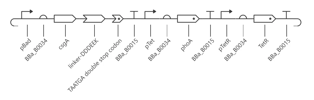
pBad turns on when you add arabinose. I used it to control CsgA-DDDEEK.
pTet turns on when you add aTc. I used it to control phoA.
🦴 Why (DDDEEK)₃ on the CsgA linker?
DDDEEK shows excellent adsorption on the hydroxyapatite (HA) surface. Yang et al. showed DDDEEKC-modified tannic acid coats HA surfaces effectively, using (DDDEEKC)₃. [5] The full text is paywalled, so I’m going off the abstract. I’ll try DDDEEK for now, and leave DDDEEKC for the future.
🛑 Why double stop codon instead of single, and why TAATGA instead of TGATAA or TGATGA?
Ribosomes sometimes skip single stop codons by accident. It’s harder to skip two. Chance decision for now. I still need to ask someone or test it myself. Not sure if TAATAA would work better than TAATGA, but my hypothesis is TAATGA performs better.
🔀 Why pTet instead of something like the lac operon?
I don’t know for sure yet. I asked Claude AI and it says pTet is generally considered “tighter” (better OFF state, cleaner ON switch) for a two-gene circuit like this. I can’t find papers backing that up online, so it looks anecdotal and experience-based rather than something I can cite.
🧪 Plasmid source
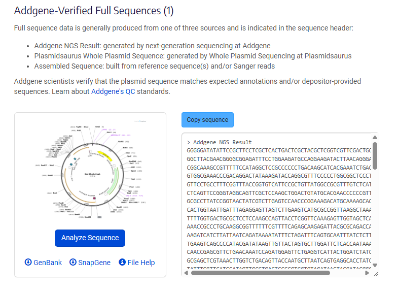
Instead of pulling parts from public repositories separately, I used a plasmid that already expresses both csgA and phoA together. Addgene plasmid #170787, Rec-PhoA/CsgA, a gift from Urartu Seker. [6][7]
🛠️ Build steps
Step 1. Got the plasmid sequence. Rec-PhoA/CsgA, Addgene #170787, downloaded as a GenBank file.
Rec-PhoA/CsgA on Addgene (#170787)
Addgene-verified full sequence
Step 2. Imported it into Kernel.
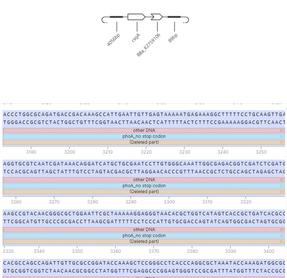
csgA imported and annotated in Kernel
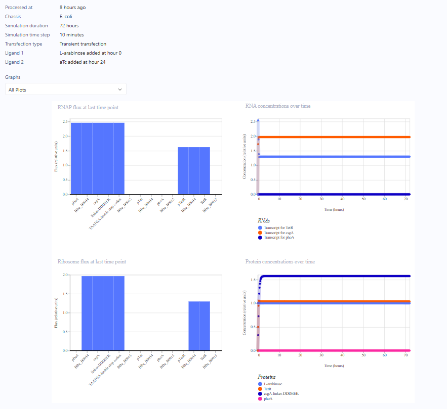
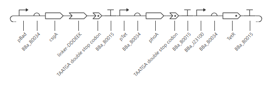
Step 3. Isolated phoA and csgA as parts, then merged them into a full plasmid. Simulation caught a bug here, see Debugging the simulation below for the fix.
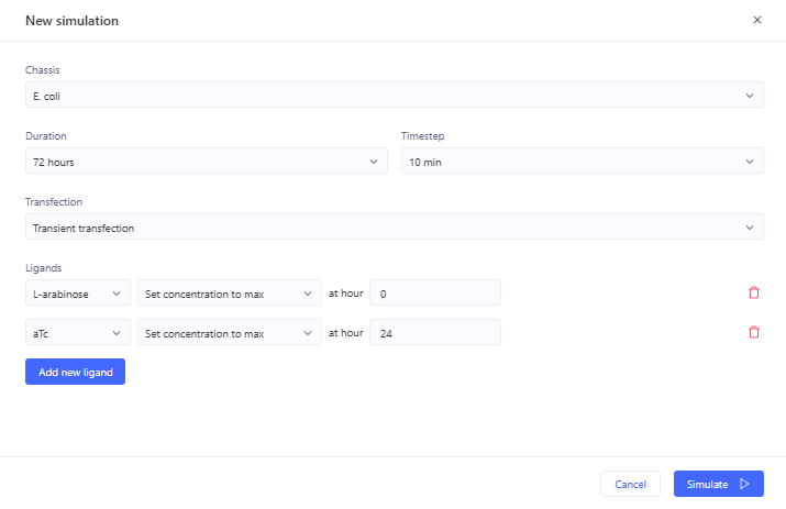
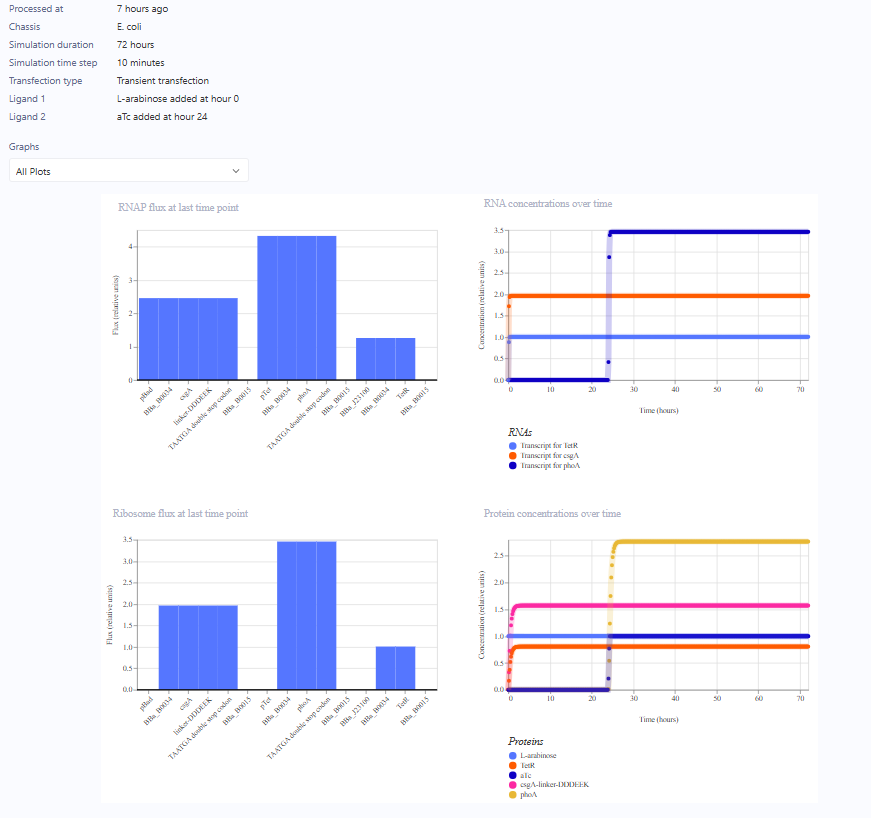
Simulation setup: E. coli, 72 h, arabinose at 0 h + aTc at 24 h
Circuit map, first attempt
Result: phoA never turns on (the bug)
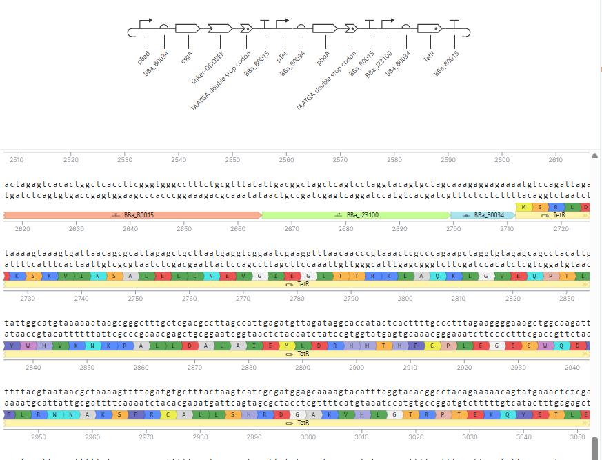
Circuit map, fixed (pTetR swapped for BBa_J23100)
Result: phoA turns on at hour 24 as intended
Fixed construct, annotated sequence
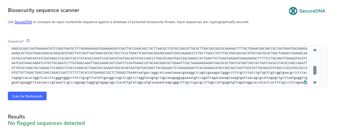
Step 4. Final check. Ran the finished sequence through a biosecurity scan before moving on. No flagged sequences.
SecureDNA biosecurity scan: no flagged sequences
🐛 Debugging the simulation
🐛 Bug found. Ran the simulation, it didn’t work.
TetR part description says it binds the TetO operator sequence within the Tet promoter, also called pTet or pTetR. That means Kernel’s part library treats pTet and pTetR as the same regulatory element, both containing TetO. That’s not what I designed for.
My intended architecture was pTetR as a plain constitutive promoter driving TetR continuously, unaffected by TetR itself, and pTet as the actual TetR-repressible promoter driving phoA, repressed until aTc shows up.
Checked the pTetR part description directly. It also says TetO, repressed by TetR, also called pTet. Confirmed. pTetR and pTet are literally the same repressible promoter, and I used it for both jobs. My TetR-supply cassette was self-repressing. At the start no TetR exists yet, so its own promoter is unrepressed, some TetR gets made, then that TetR starts repressing its own promoter too, not just phoA’s. That caps TetR production instead of giving the clean constitutive supply I wanted.
This likely explains why the combined-ligand run broke specifically. If TetR levels ended up lower or mistimed, there might not have been enough TetR to properly repress pTet/phoA before the arabinose-driven cassette started pulling ribosomes and RNAP toward csgA-DDDEEK, straining shared transcription and translation capacity in a way the single-ligand run never had to deal with.
✅ Fix. Replace pTetR in cassette 3 with a genuine constitutive promoter, something like a J23-series promoter from the iGEM registry, commonly used for supplying constitutive repressor expression.
Also removed the original TAA stop codon from the phoA sequence and put in the double stop codon instead.
❓ Open question
Simulation should run clean now. After it succeeds I need to import it into a chassis. Plasmid or direct genome integration? Still confused about this.
Plasmids are faster to build and test, give higher copy number and stronger expression, and are easy to swap out if a cassette needs revision, which matters while I’m still debugging things like the pTetR issue above. The tradeoff is plasmid loss without selection pressure and burden on the cell from maintaining and replicating extra DNA copies.
Genome integration gives single-copy, stable, uniform expression without needing antibiotic selection, and is more realistic for anything meant to persist over many generations, like an engineered biofilm. The tradeoff is it’s slower to build and harder to modify once integrated.
Given I’m still in the iteration and debugging phase, plasmid first makes sense. Move to genome integration once the circuit logic is confirmed stable, especially before adding quorum sensing later.
🤖 AI usage
AI (Claude) was used to:
teach me how to use Asimov Kernel
do a bit of literature search for me (I did check the references and skim the papers)
answer my intensive questions preliminarily
transform my ugly markdown into a beautiful one with CSS and HTML :)
On that last point, yes, that’s accurate. The badges, callout boxes, image grids, and jump links throughout this page were added by Claude on top of my original text.
📚 References
[1] Bauer, L. J., Zorzetto, L., Scoppola, E., Wagener, Y., Mantouvalou, I., & Bidan, C. M. (2026). Zinc and hydroxyapatite co-localize during in vitro E. coli biofilms mineralization. Scientific Reports, 16(1), 2059. https://doi.org/10.1038/s41598-025-33942-3
[2] Mamat, U., Wilke, K., Bramhill, D., Schromm, A. B., Lindner, B., Kohl, T. A., Corchero, J. L., Villaverde, A., Schaffer, L., Head, S. R., Souvignier, C., Meredith, T. C., & Woodard, R. W. (2015). Detoxifying Escherichia coli for endotoxin-free production of recombinant proteins. Microbial Cell Factories, 14(1). https://doi.org/10.1186/s12934-015-0241-5
[3] Ramshaw, J. A. M., Glattauer, V., & Werkmeister, J. A. (2025). Progress on production of collagen-like proteins by expression in Escherichia coli. Progress in Biomedical Engineering, 7(3), 032007. https://doi.org/10.1088/2516-1091/ade106
[4] Miller, A. L., Bessho, S., Grando, K., & Tükel, Ç. (2021). Microbiome or Infections: Amyloid-Containing Biofilms as a Trigger for Complex Human Diseases. Frontiers in Immunology, 12. https://doi.org/10.3389/fimmu.2021.638867
[5] Yang, X., Huang, P., Wang, H., Cai, S., Liao, Y., Mo, Z., Xu, X., Ding, C., Zhao, C., & Li, J. (2017). Antibacterial and anti-biofouling coating on hydroxyapatite surface based on peptide-modified tannic acid. Colloids and Surfaces B: Biointerfaces, 160, 136-143. https://doi.org/10.1016/j.colsurfb.2017.09.006
[6] Ahan, R. E., Saltepe, B., Apaydin, O., & Seker, U. O. S. (2019). Cellular biocatalysts using synthetic genetic circuits for prolonged and durable enzymatic activity. ChemBioChem, 20(14), 1799-1809. https://doi.org/10.1002/cbic.201800767
[7] Rec-PhoA/CsgA was a gift from Urartu Seker (Addgene plasmid # 170787 ; http://n2t.net/addgene:170787 ; RRID:Addgene_170787)
Week 3 HW: Cell-Free Protocols
AI assistance disclosure: I used an AI assistant (Claude) for brainstorming and feedback on this assignment, including ideas for how to approach the composition design and help with formatting this page. The written answers below are my own.
Note on this submission: Note on this submission: I have an exam on Thursday and ran out of time this week. The three compositions are first pass submissions, not optimized designs, and some of the questions are only partly answered or left for next week. I’d like to keep working on this course, so I’ll replace the placeholders with properly calculated compositions and complete the remaining answers by next week.
Goal
Using the collective intelligence of the HTBAA community and accessible AI systems, design novel CFPS reaction compositions for each of the three target proteins (sfGFP, PETase, and Reteplase) to reduce the cost per gram of functional protein. Each design should be compatible with the experimental framework and should reflect the distinct expression and folding requirements of that protein.
A. Compare cell-free reaction compositions
1. Why does the concentration of potassium glutamate and magnesium glutamate differ between compositions? Which is more sensitive?
When I saw this question, it sounded eerily similar to how Ca2+ is highly regulated in cells while K+ is much more abundant, because it helps maintain cellular homeostasis. My guess was that Mg2+ and K+ in the reaction would work the same way, and after searching the literature, it confirmed my hypothesis. In E. coli, K+ is present at high concentrations and plays a role in osmoregulation, whereas Mg2+ is kept at much lower concentrations and mainly plays a role in ribosome and nucleic acid stability. (I still need to fill in the exact concentration ranges.) (I think the concentration ranges matter a lot here, and it raises a question for me: should a cell-free reaction mimic the concentrations of a living cell, or go beyond what a cell would tolerate to maximize protein output? I also think this connects to the next question. My guess is that PEP+NTP reactions may need tighter tuning of Mg2+ than Ribose+NMP reactions, but this is just a hypothesis and I’d want to check it.)
Not only that, in a paper by Shin and Noireaux (J Biol Eng 4, 8, 2010; https://doi.org/10.1186/1754-1611-4-8), they said that Mg has to be tightly regulated for optimal protein synthesis. They also showed that Mg2+ and K+ need to be optimized for the specific plasmid you use.
So if I had to pick which one is more sensitive, I would guess Mg2+, since it’s the lower-concentration ion that matters so much for the ribosome.
That made me learn that I also have to calibrate my concentrations to the plasmid I use, instead of just copying them from another composition.
2. Explain how a PEP+NTP reaction formulation is different from a Ribose+NMP reaction formulation.
PEP+NTP is faster but more expensive. PEP is a direct energy source and the NTPs are ready-made building blocks for transcription, so the reaction doesn’t have to do much work first. Ribose+NMP is much cheaper, but the lysate has to convert the ribose and NMPs into ATP and NTPs on its own, which introduces more variables (and probably more variability between lysates).
Choose two cell-free reaction compositions from the literature, compare and contrast the choice of reagents and the concentration of reagents.
Note: A recommended starting point would be to review at least one published formulation, such as PANOx-SP (Jewett & Swartz 2004), a cost-optimized formulation from Olsen et al. (2026), and/or an AI-designed formulation from Smith et al. (2026).
B. Design one reaction composition for each of the three proteins
Sign in to coopetition.htgaa.org and publish your first unique 20 µL CFPS reaction composition for each of the following (you can resubmit later): sfGFP, PETase, Reteplase.
sfGFP
PETase
Reteplase
Community Reagents
C. Asimov Kernel (optional)
Build T7 compatible plasmids for cell-free expression using the amino acid coding sequences for sfGFP, PETase, and Reteplase.
Bonus: design a T7 helper plasmid that you could add to the cell-free reaction to improve the yield of functional reteplase (general chaperones, disulfide-bond machinery, etc).