Week 2 HW: DNA Design
- My Repository at Kernel is Campos-PLAHB and the Folder with the Homework has the same name: Campos-PLAHB
Homework #2 — Expression Design
Sucrose utilization module (cscABK) for a PLA-co-3HB producing E. coli platform
Kernel construct: cscABK_annotated_v2, repository Campos-PLAHB · 6,252 bp
Context
My HW1 project is a microbial platform producing poly(lactate-co-3-hydroxybutyrate), P(LA-co-3HB), designed to be manufactured inside sugar mills rather than in dedicated biotech facilities. The rationale is that mills already have cheap fermentable feedstock, low-cost energy and process infrastructure.
That rationale has a biological precondition the current strain does not meet. The production host is an E. coli K-12 derivative, and K-12 lineages cannot metabolize sucrose. Sugar mill feedstock — cane juice, molasses — is predominantly sucrose. The platform cannot eat the feedstock that justifies its deployment model.
This assignment therefore designs the enabling module: a cscABK operon conferring a Suc⁺ phenotype on the production host.
Part 1 — Expression target
What molecule am I expressing?
A set of three enzymes forming a minimal sucrose catabolic pathway. The HW1 product is a polymer, so per the assignment instructions I design for the proteins that produce it — here, the enzymes that deliver carbon into that pathway.
| Gene | Enzyme | EC | Function |
|---|---|---|---|
| cscB | Sucrose permease | — | Proton symporter; imports sucrose across the inner membrane |
| cscA | Sucrose hydrolase (invertase) | 3.2.1.26 | Hydrolyzes sucrose to glucose + fructose |
| cscK | Fructokinase | 2.7.1.4 | Phosphorylates fructose to fructose-6-phosphate |
The three are strictly sequential: transport, cleave, phosphorylate. CscB alone imports a sugar the cell cannot cleave; CscA alone acts on a substrate that never enters; CscK is needed because K-12’s native fructose handling does not efficiently process the free intracellular fructose CscA generates.
The native csc regulon is repressed by CscR. This design omits cscR and replaces the native promoter with a constitutive synthetic one, converting a regulated catabolic operon into an always-on module.
What is the sequence?
| Element | Source | Identifier | Verified length |
|---|---|---|---|
| cscA | E. coli W (ATCC 9637) csc regulon | Genome accession CP002185 | 1,434 bp / 477 aa |
| cscB | E. coli W csc regulon | CP002185 | 1,248 bp / 415 aa |
| cscK | E. coli W csc regulon | CP002185 | 915 bp / 304 aa |
| J23110 | Anderson constitutive promoter family | iGEM Registry BBa_J23110 | 35 bp |
| B0034 | Ribosome binding site | iGEM Registry BBa_B0034 | 12 bp |
| SAY backbone | Internal propagation vector | — | 2,419 bp |
CDS coordinates were determined by ORF detection on the assembled sequence and cross-checked against reference protein lengths. CscA at 477 aa matches the E. coli W reference exactly. For CscB, two in-frame start codons exist (positions 1515 and 1584); position 1584 yields 415 aa, matching the reference, and was adopted.
All coding sequences are natural bacterial genes — not engineered variants, not de novo designs. The function required is well-characterized wild-type activity.
What host or chassis?
E. coli JU15, a K-12 MG1655 derivative: ΔpflB, ΔadhA, ΔfrdA, ΔxylFGH, Δreg 27.3 kb, gatC S184L (Utrilla et al., 2012). The deletions remove competing mixed-acid fermentation routes, making the strain homofermentative for D-lactate — the monomer precursor for the lactate fraction of the copolymer.
The host drives the design in three concrete ways:
- It creates the requirement. K-12 lineages lack the csc regulon entirely; that absence is the gap this module fills.
- It sets the codon context. The genes come from E. coli W — a different strain of the same species — so codon usage is already near-native. This is the basis for the optimization argument in Part 4.
- It constrains selection. The production strain already carries a kanamycin-marked polymer plasmid, so the eventual integration vector must use an orthogonal marker.
Where does the product need to go?
This is the design-critical question here, and the answer differs per protein.
- CscA (hydrolase) — cytoplasm, soluble. No targeting sequence.
- CscK (fructokinase) — cytoplasm, soluble. No targeting sequence.
- CscB (permease) — inner membrane. A polytopic integral membrane protein of the Major Facilitator Superfamily, with 12 predicted transmembrane helices.
CscB determines the answer. Membrane proteins of this class are inserted co-translationally by the Sec/YidC machinery using their own internal hydrophobic signal-anchor sequences. They do not use a cleavable N-terminal signal peptide.
No signal peptide is added to any of the three CDSs. Adding one to CscB would be an error, not a neutral addition: it would place a cleavable sequence ahead of a protein whose targeting information is already internal.
A second consequence is quantitative rather than binary. CscB expression must not be maximized. Overexpression of MFS transporters saturates the Sec translocon, drives inclusion body formation and membrane stress, and typically reduces rather than increases functional transport. This is the argument for choosing a mid-strength constitutive promoter over the strongest available, and it is the main reason J23110 rather than a stronger Anderson variant.
The pathway’s end product, fructose-6-phosphate, is a cytoplasmic metabolite. Nothing is secreted.
Part 2 — Genetic parts
Prokaryotic host, so: promoter, RBS, CDS, terminator, backbone.
| Layer | Part type | Name | Position | Source | Justification |
|---|---|---|---|---|---|
| Transcription start | Promoter | J23110 | 36–70 | iGEM BBa_J23110, on a synthesized 131 bp fragment | Constitutive and deliberately mid-strength. Removes IPTG, a cost the production process cannot carry at plant scale, while avoiding translocon saturation by CscB. |
| Translation start | RBS | B0034 | 78–89 | iGEM BBa_B0034, same fragment | Well-characterized strong RBS with defined spacer; the field’s reference part, which makes any later RBS swap interpretable against a known baseline. |
| Coding sequence | CDS | cscA | 97–1530 | PCR from pAVpmdhAmp | Invertase — the catalytic step that makes sucrose usable. |
| Translation start | RBS | RBS_cscB | ~1570–1582 | Intergenic, native spacing | Independent initiation for cscB within the polycistron. |
| Coding sequence | CDS | cscB | 1584–2831 | PCR from pAVpmdhAmp, contiguous with cscA | Permease — without uptake the invertase has no substrate. |
| Translation start | RBS | RBS_cscK | 2853–2864 | Synthetic spacer introduced at the Gibson junction | Third initiation site, required because cscK arrives from a separate fragment. |
| Coding sequence | CDS | cscK | 2867–3781 | PCR from a pUC57 subclone | Fructokinase — converts CscA-generated free fructose into a glycolytic intermediate. |
| Transcription stop | Terminator | rrnB T1 | ~3790–3840 | PCR from pAVpmdhAmp | Defines the transcript 3′ end and prevents read-through into the vector. |
| Vector context | Backbone | SAY | 3869–6252 | PCR | Propagation and sequence-verification vector; provides origin and selection marker. Reused as-is, not designed here. |
Architecture: J23110 → B0034 → cscA → RBS → cscB → RBS → cscK → rrnB T1 → SAY backbone. Total 6,252 bp, circular.
Independent verification. Kernel’s Part match tool, run against all repositories at 90% minimum identity, returned a hit for J23110 from the iGEM Part Registry, confirming the promoter identity independently of the design records. The tool also returned several short (~20 bp) matches to unrelated BBa_ parts; at that length these are coincidental sequence overlaps rather than genuine shared parts, and are not treated as design elements.
A note on gene order
The operon places cscA first and cscK last. In a polycistronic transcript, 5′-proximal genes are generally better translated. Whether cscA-first is optimal is not obvious: a case can be made for cscB first, since transport is the entry step and is rate-limiting in most heterologous sugar utilization systems. This is a testable prediction — see Limitations.
Part 3 — Construct assembly
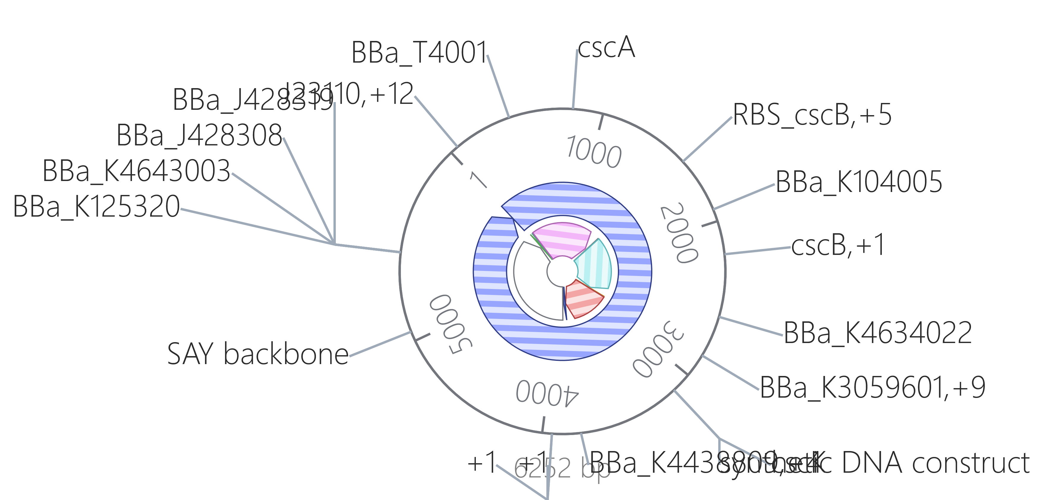
What was assembled
Built as a five-fragment Gibson assembly designed in NEBuilder v2.10.8 (overlaps ≥20 nt, Tm ≥48 °C, all PCRs with Q5 High-Fidelity polymerase), then imported into Kernel for verification.
| Fragment | Size | Origin |
|---|---|---|
| SyntheticSeq (J23110 + B0034 + leader) | 131 bp | de novo synthesis |
| cscA–cscB | 2,770 bp | PCR |
| cscK | 970 bp | PCR |
| Terminator + spacer | 142 bp | PCR |
| SAY backbone | 2,419 bp | PCR |
| Assembled | 6,252 bp |
Verification in Kernel
- Length — Kernel reports construct length 6252, matching the NEBuilder design exactly.
- Reading frames — all three CDSs are divisible by three, begin with ATG and end with a stop codon: cscA 1,434 nt, cscB 1,248 nt, cscK 915 nt.
- Internal stop codons — none in any of the three CDSs, confirmed by translation.
- Part order and orientation — the circular and linear maps confirm all three CDSs on the same strand, downstream of J23110 and upstream of the terminator, in the intended order.
- Junction integrity — the Gibson overlaps did not introduce frameshifts: each CDS translates cleanly to its expected reference length.
A finding worth recording
The GenBank file exported by NEBuilder could not be parsed as a construct on import. Inspecting it showed why: it contained no CDS, promoter, RBS or terminator features at all. Its only annotations were fragment-level gene entries naming the assembly pieces (SyntheticSeq, cscABpAVpmdhAmp, 2_cscK_pUC57, SAY) and eight primer_bind entries. It also declared one feature with coordinates crossing the origin of the circular molecule, which is a common cause of GenBank parser failure.
The file was therefore re-annotated from first principles — ORFs located by translation, regulatory elements located by sequence match — and rebuilt as a valid GenBank, which imported correctly with all features mapped.
This is a real lesson about design-tool handoff: assembly planners annotate what matters to assembly (which fragment came from where, where primers bind), not what matters to biology. A file that is complete for one purpose can be empty for another.
Design description
A single constitutive transcriptional unit carrying a three-gene catabolic operon in an episomal verification vector. The episomal stage is deliberate: the construct can be fully verified by Sanger sequencing before committing to chromosomal integration, so an assembly error is caught cheaply. The longer-term destination is chromosomal integration, removing plasmid maintenance and antibiotic dependence from the production process.
Part 4 — Optimization and quality control
Codon optimization
What I ran. Codon optimization on two of the three coding sequences: cscK (915 bp) and cscA (1,434 bp). Both runs used input sequence type DNA, expression host Escherichia coli, Asimov preset None, optimizer version SIMPLE_0_3, signal peptide algorithm off and CpG avoidance off. Restriction sites XhoI and BamHI were excluded from the output, since both must remain intact for the downstream pOSIP-CH integration step. Signal peptide handling was disabled deliberately: neither CscA nor CscK carries one, and CscB — which was not optimized — targets the membrane through internal signal-anchor sequences rather than a cleavable peptide, so the algorithm would have nothing correct to act on.
What it returned. Both runs completed in under a second and returned full-length optimized sequences. In both cases, the tool reported Asimov Codon Usage Index: N/A and RNA structure score: N/A — no quantitative scores were produced. Two independent runs on two different genes gave the same outcome, indicating a property of the tool in this configuration rather than a problem with either input.
Measuring GC content directly: the cscA input is 50.0% GC and the optimized output runs near 47%, so optimization lowered GC rather than raising it, moving the sequence toward the E. coli genomic average. The cscK input is 52.0% GC. The optimizer nonetheless rewrote a large fraction of codons — comparing the first 30 nt of cscA, input ATGACGCAATCTCGATTGCATGCGGCGCAA becomes ATGACCCAGAGCCGTCTGCATGCAGCACAG, with most positions changed.
One further consequence worth recording: the stop-codon setting replaced the native TAA with TGA. The optimized CDS is therefore not strictly synonymous with the input at the terminator, though TGA is the most common stop codon in E. coli and the substitution is expected to be neutral or mildly favorable.
What I concluded.
The interesting result here is the mismatch between how much the tool changed and how little it could demonstrate.
Codon usage matters because rare codons stall elongating ribosomes, reducing yield and promoting misfolding. But that reasoning applies to genes crossing a large phylogenetic distance, and this case is the opposite: the source is E. coli W and the host is E. coli K-12 — the same species. A 50% GC input is unremarkable for E. coli, and the optimizer moved it only slightly, which is consistent with there having been little adaptation left to do. The extensive codon rewriting reflects the algorithm substituting statistically preferred synonymous codons, not correcting a real expression liability.
Against that, two risks argue for leaving the sequences alone. Optimizing all three csc genes toward the same preferred codon set increases sequence similarity between them, raising recombination risk in a construct destined for chromosomal integration. And for cscB in particular, optimization could be actively harmful: membrane protein biogenesis depends on translation rate, and rare-codon pausing is thought to give transmembrane helices time to insert correctly, so maximizing translation speed can impair folding of a 12-transmembrane transporter rather than help it. This is why cscB was not optimized.
Without a Codon Usage Index I cannot demonstrate improvement quantitatively, only argue it. That is a genuine limitation of the analysis: the decision to retain the native sequences rests on the reasoning above and on the measured GC, not on the tool’s own scoring.
Sequence search (BLAST)
What I ran. Kernel’s Toolbox contains only three tools — Codon optimizer, Biosecurity sequence scanner and pairwise Sequence alignment — and does not include a database search. BLAST was therefore run externally at NCBI (blastn, megablast, Nucleotide collection nr/nt), first on the full 6,252 bp construct and then on the cscA coding sequence.
What it returned. ` Full construct. Query coverage 37%, with the aligned region corresponding to the vector portion rather than the operon. The top hits were cloning vectors of the pSEVA family — pSEVAb637 (MW476677.1), pSEVA633 (JX560372.1), pSEVA632 (JX560371.1) and pSEVA631 (JX560348.1), all at 99.72% identity — followed by several pSpy-series vectors at 100% identity. This identifies the SAY backbone as a pSEVA-architecture vector, which internal records had described only as a generic propagation vector. That is a useful piece of provenance: pSEVA is a documented public vector standard, so the origin and marker architecture of the backbone can be read from the literature rather than inferred.
cscA. Query length 1,434 bp, query coverage 100%, every hit in the top 100 an Escherichia coli genome. Best hit E. coli PSJ-22 (AP027634.1), score 1995 bits, E value 0.0, 91.78% identity, alignment 1,317/1,435 identities with 2 gaps (0%), aligned to positions 4,572,901–4,574,334 on the minus strand.
What I concluded.
cscA is unambiguously what it was believed to be: a natural bacterial gene, present across E. coli genomes, aligned end to end with no truncation and essentially no gaps. It is not novel, and it is not a synthetic or engineered variant.
The identity figure is worth examining rather than reporting flatly. At 91.78% with full coverage, roughly 118 positions differ across the gene — far more than the near-identity a direct genomic amplicon from the source strain would give. My first hypothesis was that the sequence had been recodified, that is, synthesized with optimized codons while preserving the protein, which would have shown up as differences concentrated in third codon positions.
Inspecting the pairwise alignment ruled that out. The mismatches are irregularly distributed and frequently fall in adjacent positions — AATGGCCTGATTTGG against AACGGCCTGATCTGG, for example — which is not the signature of systematic synonymous substitution. The pattern is instead ordinary strain-level divergence. The csc regulon is an accessory, horizontally acquired locus rather than a core gene, and accessory loci diverge considerably more between E. coli strains than core metabolic genes do. E. coli PSJ-22 is simply a different strain from the E. coli W source; the first 60 aligned bases are identical, with differences accumulating downstream.
The closest match reported here is therefore not the source organism. A filtered search restricted to higher identity would be expected to surface E. coli W itself above 99%, and that would be the more informative comparison. As run, the result confirms identity and naturalness but not provenance to the specific source strain.
Two limitations of the method are worth stating. Running BLAST on the whole construct returned only the backbone, because vector sequences are heavily represented in the database and outcompete a three-gene operon on score; per-CDS searches are the right granularity. And short synthetic regulatory parts such as J23110 and B0034 are poor BLAST queries — sequence search is built for genes. Kernel’s Part match tool, which returned J23110 against the iGEM Part Registry, was the better instrument for those.
Overall: the individual sequences are not novel. The construct is novel only in its arrangement — a natural catabolic operon decoupled from its native repressor and placed under constitutive synthetic control, in a pSEVA-architecture backbone.
Safety screen
What I ran. The SecureDNA biosecurity scanner on the full 6,252 bp construct.
What it returned. ` No flagged sequences detected
What it checks. SecureDNA screens query sequences against signatures of sequences of concern — genes from regulated pathogens and toxins — using a privacy-preserving hashing scheme, so the query is not exposed to the screening service. It is the class of screen DNA synthesis vendors apply to incoming orders.
What I expected, and how I would respond to a flag. No flags expected: all three genes are carbohydrate-metabolism enzymes from a non-pathogenic laboratory E. coli strain, and E. coli W is a standard industrial workhorse. Had something been flagged, my first question would be whether it is a domain-level homology hit rather than a functional match — MFS transporters share structural architecture with transporters found across many organisms, pathogens included, so a permease flagged on that basis would be a false positive. I would identify the triggering region, confirm the match is structural rather than to a virulence determinant, and document the reasoning rather than assume it must be spurious.
Limitations and next steps
Kernel’s genetic simulator predicts transcription and translation, not metabolic flux. It can indicate whether cscK is under-translated relative to cscA, but not the resulting sucrose uptake rate or carbon yield. That requires flux balance analysis against a genome-scale model, which is outside this assignment.
The most valuable follow-up this analysis suggests is a comparison of gene orders — cscA-cscB-cscK as built versus cscB-first — evaluated by predicted ribosome flux per ORF, given that transport is the likely rate-limiting step and is currently in the least favorable position in the polycistron.
References
- Utrilla, J. et al. (2012). E. coli JU15 strain background.
- St-Pierre, F. et al. (2013). One-step cloning and chromosomal integration of DNA. ACS Synthetic Biology.
- iGEM Registry of Standard Biological Parts: BBa_J23110, BBa_B0034.
- NCBI: Escherichia coli W (ATCC 9637) genome, accession CP002185.
- NEBuilder Assembly Tool v2.10.8, New England Biolabs.