Week 2 HW: DNA Design
Part 0: Set up in Kernel
Repository: Luisa Olivas HTBAA 2026 Folder: LuisaOlivas-SodiumAlginateAmilCP
Part 1: Define your expression target
The expressed molecule is chromoprotein — a non-fluorescent, non-enzymatic protein that folds into an intensely pigmented structure visible to the naked eye without any added substrate, UV light, or cofactor. Specifically, amilCP, derived from the coral Acropora millepora.
The coding sequence corresponds to iGEM Registry part BBa_K592009 (amilCP), a well-characterized part available in the iGEM Registry of Standard Biological Parts. It will be imported directly from the registry rather than designed de novo, since it is already codon-optimized and validated for expression in E. coli.
The host is E. coli, BSL-1 safe strain, matching the microbial biosensor approach proposed in HW1.
The product needs to go to cytoplasm. amilCP requires no secretion or membrane targeting — its color develops through intracellular folding and oxygen-dependent chromophore maturation, so the signal is visible directly within the bacterial cell body.
Part 2: Design your genetic parts
Part 3: Assemble your construct
Part 4: Optimize and quality-check
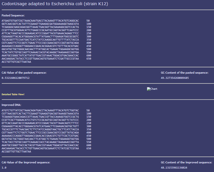
I ran codon optimization on the amilCP CDS using JCat, targeting Escherichia coli (K-12) as the expression host. The Codon Adaptation Index (CAI) improved from 0.53 in the original sequence to a perfect 1.0 after optimization, indicating that codon usage was fully adapted to E. coli’s tRNA abundance profile. GC content remained close to the E. coli genomic average (48.1% optimized vs. 50.7% genomic average, compared to 49.3% in the original sequence), confirming the optimization did not introduce an extreme GC bias that could complicate synthesis. The encoded amino acid sequence remained identical before and after optimization, confirming that only synonymous codon substitutions were made.
This screenshot shows the BLASTX (translated nucleotide-to-protein) search results for the codon-optimized amilCP coding sequence (669 bp query, RID: BPG64YRR016), run against the NCBI ClusteredNR protein database. Unlike the nucleotide-level BLASTN search — which failed to specifically identify the codon-optimized sequence due to synonymous codon substitutions — this translated search correctly identifies the encoded protein. The top-ranked cluster matches GFP-like non-fluorescent chromoprotein [Porites lutea] (XP_073233827.1) with 99% query coverage, 95.02% identity, and an E-value of 4e-157. The second-ranked cluster (NP_001310111.1) represents 89 member sequences from 23 different coral and related organisms, all annotated as the same GFP-like non-fluorescent chromoprotein, with 96.73% identity. These results confirm that, despite the nucleotide-level changes introduced by codon optimization, the CDS still encodes a bona fide member of the amilCP chromoprotein family — verifying that optimization preserved the protein’s identity and function.
Part 5: From design to build
Considering the methods covered in class, my construct can be broken down into two distinct scales of problem—and each calls for a different technique:
- Synthesis of individual fragments → Chip-based gene synthesis The longest element in my design is the amilCP CDS (669 bp). This exceeds the practical limit of a single oligo synthesized via the phosphoramidite cycle (which operates reliably up to ~150–200 nt, as the coupling error rate accumulates with each cycle, resulting in very low yields for longer oligos). Therefore, the correct approach is not to order the full gene as a single oligo, but to use chip-based gene synthesis: multiple overlapping short oligos (each made via the phosphoramidite cycle) are synthesized in parallel on a chip and then enzymatically assembled via PCR (polymerase cycling assembly) into the full double-stranded fragment. This is the standard method commercial providers (such as Twist or IDT) use to deliver gene fragments in this size range, and it is significantly cheaper than traditional column synthesis for a fragment of this length. The ProU promoter (658 bp, a natural sequence isolated from E. coli) could be obtained the same way—as a synthetic fragment—or alternatively amplified via PCR directly from E. coli genomic DNA, given that it is a native sequence available in the host organism.
- Assembly of the full construct → Gibson Assembly Once I have the individual fragments (promoter, the RBS+CDS+terminator as a synthesized cassette, and the linearized pSB1C3 backbone), the most practical way to join them into a single circular plasmid is Gibson Assembly. Fragments are designed with overlapping ends (~20–40 bp of homology between consecutive pieces), and an enzyme mix (exonuclease, polymerase, and ligase) assemblies them in a single isothermal reaction without requiring specific restriction sites. This is preferable over classical restriction-ligation cloning because it avoids being tied to the specific cleavage sites of the BioBrick standard and allows for the multi-fragment assembly of several pieces in a single step.
Part 6: What don't you know yet?
• Will the ProU promoter respond specifically to the sodium range associated with mastitis, rather than general osmotic stress? To find out, I would measure amilCP color intensity (quantified via image analysis) across a gradient of NaCl concentrations encompassing both normal levels and those associated with mastitis in breast milk, confirming that the promoter’s activation threshold falls within the clinically relevant range.
• Will the color signal be visible and sufficiently fast once the bacteria are encapsulated in the alginate hydrogel, compared to free liquid culture? To find out, I would encapsulate the transformed strain in alginate beads or patches and measure the time it takes for the color change to become visible when exposed to simulated high-sodium milk, comparing it against the response time in liquid culture.
• Will the plasmid-based construct remain stable within the hydrogel over the shelf-life of the patch without antibiotic selection pressure? To find out, I would perform a stability assay: store the encapsulated bacteria for varying periods (ranging from days to weeks) and periodically test whether the biosensor response (color change under salt stress) is maintained. This would indicate whether chromosomal integration is required before scaling up production.