I’m an aspiring synthetic biologist interested in the intersection of SynBio, phage/microbial mechanism biology, and context-aware bioengineering. Some of my current projets focus chassis’ responses to engineering conditions and simulated environmental deployment. I’m also an iGEM alum (William & Mary 2025!), and I studied anti-phage defense systems at MIT this summer.
See the nav bar on the left to look through my work for How To
Biomanufacture Almost Anything (Fall 2026)!
Subsections of Madeline Eibner-Gebhardt — HTBAA Fall 2026
Week 1 HW: Final Project Ideas
(1) What you would like to biomanufacture and why?
An often-quoted estimate predicts that antibiotic resistant infections will outpace cancer and heart disease as the global leading cause of death by 2050. Resistant bacteria are especially prevalent in hospital settings, where high antibiotic concentrations and large, diverse populations of pathogenic bacteria combine (within infected patients, in drains, etc) to drive rapid resistance evolution. Resistant bacteria are difficult to eliminate not only because 1), they, by definition, cannot be treated via traditional antibiotics, but also because, 2), they tend to form dense, sticky, and highly resilient biofilms. I want to biomanufacture something that combats antibiotic resistant pathogens – possibly via a phage-therapy based approach, taking advantage of automated and cloud-lab systems to design, engineer, and test a variety of modified phage at a large scale and create/identify promising anti-biofilm candidates against various bacterial pathogens.
(2) What living system would you use?
I would likely use Mycobacterium smegmatis as my host bacterium for phage engineering, testing, and large-scale phage production. (And I would use the large repository of Mycobacteriophages available through the HHMI SEA-PHAGES program as a starting point for engineering anti-biofilm phage therapies.) Mycobacterial species can form resilient biofilms that contribute to devastating hospital-acquired infections. M. smegmatis, as a BSL1, non-pathogenic, and relatively fast-growing Mycobacterial species, offers an easier and safer system that could discoveries relevant to treating pathogenic Mycobacteria such as M. tuberculosis and M. abscessus. (I.e., design principles that I discover apply to the anti-biofilm properties of M. smegmatis phages may help us eventually engineer therapies for M. abscessus.)
(3) How would you use automation?
I would use an Opentron liquid handling robot to automate the phage engineering and cloning-based parts of this process – so as to make it possible to engineer, reboot, and test many slightly different engineered variations of the same phage. I would likely also use a larger Cloud Lab system for the testing and measurement stages: I would need an automated and standardized way of producing biofilms with qualities of interest, potentially on surfaces designed to mimic a hospital or patient setting, and a way to measure the extent to which the biofilm was cleared by engineered phage – and then to select for and amplify/propagate more of, acquire, and sequence the effective phage.
(4) How much would you need to make, and how long would it take?
After engineering a particularly effective anti-biofilm phage via the methods above, I could expand my system to produce the relevant phage at a large scale for treatment. Phage therapy dosage can vary – one review says that they tend to be 109 - 1010 PFU (plaque forming units) per dosage for acute infections (https://pmc.ncbi.nlm.nih.gov/articles/PMC11870740/#sec10). Acute respiratory infections can require up to 4 dosages per day, so 4 x 10¹⁰ PFU /day. If the patient must be treated for 7 days, they require 28 x 1010 PFU total – aka 28 x 1010 phage. Let’s say that the typical mycobacteriophage burst size is 150 phage, and each 200 ul well in a 96 well plate holds ~107 bacterial cells, and we add inoculate each well with enough phage to kill half of the bacteria – we would end up with ~7.5 x 10^8 phage per well(?). If so, we would need to run at least ~373 infections in 200 ul wells to produce enough phage for one patient.
Timing-wise, each well-infection could run in 1-2 days and then be harvested to infect the next round. If we started with enough pure phage lysate, we could, e.g., run 96 initial parallel infections in a plate per patient in 24 hrs, then harvest the filtrate and start 2-3 new infections per harvested well in order to obtain the remaining phage in another 24 hrs.
(5) What don’t you know yet?
What is the best approach to automate engineering of mycobacteriophages? What cloning techniques would be optimal to use? How might I get started with identifying engineering modifications that would provide anti-biofilm properties? What technqiues and growth conditions can I use to grow test biofilms at large scale in a automated Cloud Lab setting?
Testing
Week 2 HW: DNA Design
What molecule are you expressing? A protein, an RNA, or an enzyme (or small set of enzymes) for a pathway. If your HW1 product is a material, food, or chemical, identify the protein(s) that produce it and design for those.
Per homework 1, I want to biomanufacture anti-biofilm phages on a large scale, engineering, testing, evolving, and producing the modified phages on a large scale to identify promising candidates for biofilm mitigation. This would likely involve CRISPR-based editing and/or assembly and rebooting of phages to stitch together modified components. For the purposes of this assignment, I will focus on designing an expression construct that overexpresses the mycobacteriophage SWU’s gp39 protein, which interferes with biofilm formation and increases the permeability of the Mycobacterial cell wall (https://www.nature.com/articles/srep28701). This protein could eventually be useful to engineer into other mycobacteriophages as a means of enhancing their ability to disrupt biofilms / infect in spite of them – but we may also want to test the effectiveness of the overexpressed protein on its own before we attempt phage engineering.
What is the sequence? Give the protein or RNA sequence, or the specific gene, you will encode, and say where it comes from (a database accession such as UniProt or NCBI, a known part, or de novo). Cite it.
What host or chassis? The living system from HW1, for example E. coli, S. cerevisiae, CHO or another mammalian line, or a cell-free system. Your host drives almost every part choice that follows.
I will express the protein in Mycobacterium smegmatis.
Where does the product need to go? Cytoplasm, secreted, membrane, or a compartment? This decides whether you need a signal peptide or targeting sequence.
Cytoplasm
Every construct is built from parts. Decide which parts your target needs, then create or import each one in Kernel with the correct part type. At minimum, choose the elements below for your host.
Tx start (Promoter):
pExTra backbone includes a tetracycline-inducible promoter - pTet
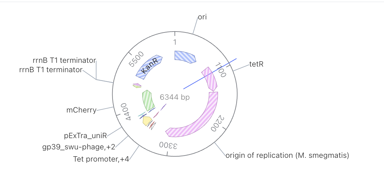
Create a new construct in your folder and add your parts in the correct 5’→3’ order for your host (for a basic expression cassette: promoter → RBS or 5’ UTR/Kozak → CDS → terminator, plus any signal peptide or tag, in the vector backbone).
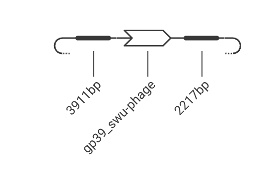
Check the assembled sequence: the reading frame is intact, the start and stop codons are where you expect, and there are no accidental internal stops. Look at the construct map Kernel renders and confirm the parts sit in the order and orientation you intended.
Brief Description: My inducible construct overexpresses gp39, a protein that the SWU mycobacteriophage uses to weaken/inhibit mycobacterial biofilm formation. I’ve selected the pExTra backbone, which the SEA-GENES program recommends for mycobacterial expression constructs.
Codon-optimize your CDS for your chosen host. Compare before and after: what changed, and why does codon usage matter for expression in this organism? Note any constraints you applied (e.g. avoiding certain restriction sites, GC content).
I have access to the IDT tool for codon optimization. IDT does not have an option to optimize for Mycobacterium smegmatis, so I selected M. tuberculosis instead for the purposes of the exercise. (If I were to do this in real life, I would likely leave it as-is and assume that a phage protein from a phage that infects M. smegmatis should be expressable in M. smeg without any special optimization.) The optimized sequence looks relatively similar to the original.
Search your sequence (BLAST / sequence search) against known sequences. Is your CDS what you think it is? Is it identical to a natural gene, or novel? What is the closest match?
I obtained the sequence from NCBI, so its top hit is the phage sequence from which I obtained it.
Run the safety screen on your construct. Summarize what it checks for and what it reports. If it flags something, explain whether it is expected for your design and how you would respond.
Returned “No flagged sequences detected”
How would you build it? Would you order it as synthetic DNA, assemble it from smaller fragments (e.g. Gibson assembly), or build it from a pool of oligos synthesized on a chip? Justify your choice given your construct’s length and complexity.
I would order the pExTra plasmid vector from SEA-GENES, then order the CDS as a gene block and insert it into the vector via either Gibson or Golden Gate Assembly. This approach is standard for assembling simple expression constructs.
What does synthesis constrain? Synthesis has error rates and struggles with certain sequences. Does your design contain anything hard to synthesize? How would error correction or fragment-based assembly change your approach?
Mycobacterial and mycobacteriophage sequences tend to have a high GC content and are harder to sythesize, assemble via Gibson, and sequence. Depending on how long it could take to troubleshoot the Gibson, Golden Gate Assembly might be preferable, as it relies on restriction enzymes – not exonucleases, which are subjected to nucleotide bond strength – to assemble the parts.
Prototype vs. scale. Mirroring HW1: the way you’d build one construct to test your idea may differ from how you’d produce DNA at the scale your product needs. Briefly describe both if they differ.
Above, I’m describing the process I would use to clone SWU gene gp39 – an phage component that intereferes with lipid synthesis and alters the cell wall through this method to prevent biofilm formation.
As in Homework #1, close by naming the edges of your design. Identify the 2–3 biggest unknowns or risks in your construct. For example: Will the promoter give the right expression level? Will the protein fold and be active in this host? Will it be secreted? Is codon optimization enough, or do you need to tune it empirically? For each, state how you would find out: the measurement, experiment, or design variant you would try next. A good answer here shows you know which decisions are still bets.
The CDS mat be too toxic to express and observe without killing the cells of interest
The promoter may be too leaky, potentially leading to bacterial cell death
Week 3 HW: Cell-Free Reaction Design
A. COMPARE CELL-FREE REACTION COMPOSITIONS (20 minutes)
Why does the concentration of potassium glutamate and magnesium glutamate differ between compositions? Which is more sensitive?
The compositions require differnt pH conditions. Varying the concentrations of potassium glutamate and magnesium glutamate helps modify the pH across compositions. It’s also stoichiometrically helpful to have a compound that delivers two glutamates vs. one.
Explain how a PEP+NTP reaction formulation is different from a Ribose+NMP reaction formulation.
A PEP+NTP reaction uses PEP to supply phosphate groups and replenish those of spent NTPs. A Ribose+NMP reaction instead uses ribose to build new nucleotides and uses pure phosphate as its phosphate source.
Choose two cell-free reaction compositions from the literature, compare and contrast the choice of reagents and the concentration of reagents.
The biggest differences between the 2004 and 2026 formulations (see below) are that the 2004 version supplies extra phosphate groups while 2026 relies on the existing compounds and that the 2004 uses only NTPs while 2026 uses a combination of NMPs (which can be converted to NTPs) and NTPs.
Note: A recommended starting point would be to review a least one published formulation, such as PANOx-SP (Jewett & Swartz 2004), a cost-optimized formulation from Olsen et al. (2026), and/or an AI-designed formulation from Smith et al. (2026).
B. DESIGN ONE REACTION COMPOSITION FOR EACH OF THE THREE PROTEINS (40 minutes)
Sign in to coopetition.htgaa.org and publish your first unique 20 µL CFPS reaction composition for each of the following (don’t worry, you can resubmit later):
sfGFP
PETase
Reteplase
Note: if you’re unable to login with your HTGAA keycloak account, please post in Zulip so that you can participate in the coopetition!
For each composition, specify the reagents you would adjust from the relevant categories, including salts, energy sources, building blocks, cofactors, and redox components.
See submission on coopetition site.
As a HTBAA research team, we will have the opportunity to suggest new reagents to add to the cell-free reagent inventory. Contribute (or upvote) at least one reagent to the “Community Reagents” section of the website and provide a short rationale. Include in your homework documentation how you decided on that specific reagent and provide links to any relevant research.
Visit the cfps-coopetition forum page to ensure you will be able to take part in the discussion. Bonus: send a note in the channel about a CFPS optimization idea that you have! If you would like clarification on aspects of the project, please message in this dedicated channel.