<YOUR NAME HERE> — HTBAA Fall 2026
About me
Contact info
Homework, Labs, and Projects
See the nav bar on the left to look through my work for How To Biomanufacture Almost Anything (Fall 2026)!
See the nav bar on the left to look through my work for How To Biomanufacture Almost Anything (Fall 2026)!
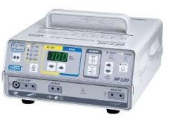
I want to develop a multilayer tissue-mimicking phantom for testing and optimizing electrosurgical devices.
The goal is to create a standardized material that reproduces selected electrical, mechanical, and thermal properties of soft biological tissues. This would allow electrosurgical experiments to be performed under more controlled and reproducible conditions.
My motivation comes from my experience developing a closed-loop electrosurgical system. During electrosurgical experiments, biological tissue can introduce variability between samples, making it difficult to compare device parameters, control strategies, and tissue responses. A standardized tissue-mimicking material could provide a more reproducible platform for evaluating electrosurgical power delivery, temperature response, cutting, and coagulation.
I am particularly interested in developing a multilayer phantom inspired by breast tissue, with different regions representing skin, adipose tissue, and breast tissue. In a later version, a region with different material properties could be incorporated to represent a tumor or lesion.
Previous research has demonstrated that tissue-mimicking phantoms can be engineered with tunable electrical, mechanical, and thermal properties. Gellan-gum-based phantoms, for example, have been developed by modifying their composition to obtain properties that approximate biological tissues [1]. More recently, conductive hydrogel and ionogel phantoms have been developed specifically for electrosurgical training and testing, including multilayer models designed to reproduce tissue stratification [2].
My long-term goal is to create a reproducible material platform that can reduce dependence on biological tissue for repeated electrosurgical testing and support the optimization of electrosurgical devices and control strategies.
I would use a microbial production system (specifically the bacterium Sphingomonas elodea) to produce a polysaccharide-based biomaterial, with gellan gum as an initial candidate. What goes in: A liquid culture medium containing a carbon source (such as glucose), a nitrogen source, and inorganic salts.
The biological process: The S. elodea will undergo aerobic fermentation in a bioreactor. As the bacteria metabolize the glucose, they synthesize and secrete a high-molecular-weight exopolysaccharide (gellan) into the surrounding broth as a protective mechanism.
What comes out: A highly viscous fermentation broth. To isolate the target product, the bacteria are removed, and the pure gellan gum is precipitated out of the broth using an alcohol like isopropanol, yielding a dry biopolymer powder. This powder is then rehydrated with specific ionic crosslinkers to form the tissue phantoms.
Gellan gum is a microbial polysaccharide that can form hydrogels with tunable mechanical and electrical properties. Previous studies have investigated gellan-gum-based tissue phantoms as materials for reproducing relevant properties of biological tissues [1,3].
Automation could be used throughout the manufacturing and testing workflow.
First, automated liquid handling could prepare many different biomaterial formulations by varying parameters such as polymer concentration, ionic content, water content, and crosslinking conditions. I would use a desktop liquid-handling robot, such as an Opentrons OT-2, to automate the high-throughput preparation of hundreds of candidate biomaterial formulations, precisely varying the gellan concentration, water content, and ionic crosslinkers to tune the electrical and thermal properties.
Second, automated dispensing or additive manufacturing could fabricate the phantom layer by layer, allowing different compositions to be assigned to different tissue regions. Once a formulation is optimized, I would use an automated robotic bioprinter (such as a CELLINK dual-printhead system) to extrude the phantom layer-by-layer. This allows different anatomical compositions (e.g., highly conductive glandular tissue vs. highly resistive adipose tissue) to be precisely assigned to specific spatial regions, mimicking the stratification of real breast tissue.
Third, automated characterization could measure relevant electrical, mechanical, and thermal properties of each formulation.
Finally, these measurements could be incorporated into a feedback loop to select and refine formulations:
Design → Manufacture → Measure → Compare → Adjust → Manufacture again
The electrosurgical testing stage could also be automated. The phantom could be exposed to controlled electrosurgical conditions while sensors measure variables such as electrical response and temperature. These measurements could then be used to evaluate whether the phantom reproduces the desired tissue-like response.
This closed-loop approach would combine biomanufacturing, automation, materials characterization, and control systems.
(a) How much? To define a meaningful scale, I am estimating the production of 1,000 multilayer breast phantoms to supply a large-scale electrosurgical validation study or a multicenter device trial.
Volume: An average breast model is approximately 500 mL in volume.
Total hydrogel: 1,000 phantoms × 500 mL = 500 Liters.
Biomaterial required: Gellan gum phantoms typically require a concentration of ~2% w/v (20 grams per Liter) to match the mechanical stiffness of soft tissue.
Total scale: 500 L × 20 g/L = 10 kg of pure gellan gum (b) How long? I would envision the project in four stages:
And after atomation help:
Biomanufacturing Time: Sphingomonas elodea yields approximately 15 g/L of gellan in industrial fermentation. To produce 10 kg, we need a bioreactor working volume of roughly 660 Liters (accommodated by a standard 1,000 L bioreactor). A typical aerobic fermentation batch takes about 48 to 72 hours. Downstream processing (precipitation and drying) adds another 48 hours.
Automated Fabrication Time: Using a bioprinting array with 5 parallel extrusion robots, assuming each phantom takes 1 hour to print, the throughput is 5 phantoms/hour. Printing 1,000 phantoms would take 200 hours (about 8.3 days of continuous operation).
Total Time: Fermentation (3 days) + Downstream Processing (2 days) + Automated Printing (8.3 days) = approximately 13.3 days to go from bacterial culture to 1,000 finished electrosurgical phantoms.
The first major unknown is whether a biologically produced material can simultaneously reproduce the electrical, mechanical, and thermal properties that are relevant to electrosurgery.
Changing one component of the formulation may improve one property while negatively affecting another. Although previous studies demonstrate that gellan-gum-based phantoms can have tunable properties, achieving the specific combination required for a breast-tissue model and electrosurgical applications remains an engineering challenge [1,3].
An important question is whether the phantom can reproduce relevant phenomena such as electrical current distribution, heating, coagulation, cutting behavior, and thermal effects.
Previous work has demonstrated the feasibility of conductive tissue-mimicking phantoms for electrosurgical applications, but further validation would be required for a breast-tissue-specific model [2].
The final bottleneck is whether the phantom can be manufactured consistently and remain stable during storage and repeated testing.
If two phantoms produced from the same formulation behave differently, they would not provide a reliable platform for comparing electrosurgical devices or control strategies.
Therefore, batch-to-batch reproducibility, material stability, mechanical degradation, and changes in electrical properties during storage and repeated testing would need to be evaluated.
Chen, R. K., & Shih, A. J. (2013). Multi-modality gellan gum-based tissue-mimicking phantom with targeted mechanical, electrical, and thermal properties. Physics in Medicine & Biology, 58(16), 5511–5525. https://doi.org/10.1088/0031-9155/58/16/5511
Migliorini, F., et al. (2024). Conductive Gel Phantoms for Training in Electrosurgery. Advanced Materials Interfaces. https://doi.org/10.1002/admi.202400246
Brzozowski, P., Penev, K. I., & Mequanint, K. (2021). Gellan gum gel tissue phantoms and gel dosimeters with tunable electrical, mechanical and dosimetric properties. International Journal of Biological Macromolecules, 180, 332–338. https://doi.org/10.1016/j.ijbiomac.2021.03.047
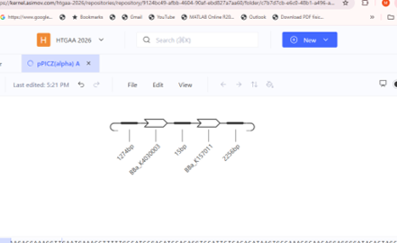
The project workspace is established in the Kernel HTBAA 2026 workspace under the repository “HW 2 Malinche Mejia Gellan Tissue Phantom” and folder “Mejia-GellanPhantom”.

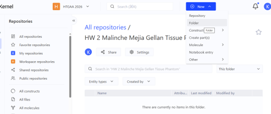
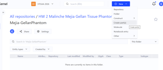
In Kernel, a Part is an individual DNA element used for genetic design, such as a promoter, ribosome binding site (RBS), coding sequence (CDS), or terminator. A Construct is a complete DNA design assembled from multiple Parts in a specific order.
For my project, the gelK coding sequence would be one Part. When it is combined with the appropriate regulatory elements, such as a promoter, RBS, and terminator, the resulting sequence represents the Construct.
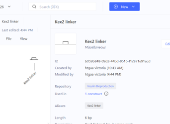
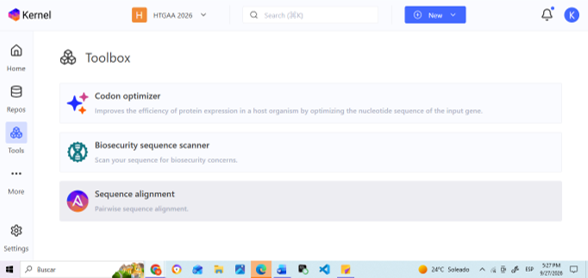
Codon Optimization: Used to adapt a coding sequence to a specific host organism to improve compatibility with its expression system.
Biosecurity Sequence Scanner: Used to screen DNA sequences for potential biosecurity concerns or matches to sequences of concern.
Sequence Alignment: Used to compare two or more DNA or protein sequences and identify similarities and differences.
1. What molecule are you expressing?
I am expressing β-1,4-glucuronosyltransferase (GelK), a key glycosyltransferase enzyme in the gellan gum biosynthetic gene cluster.Because my project from Homework #1 produces a biomaterial (gellan gum exopolysaccharide) to build multilayer tissue-mimicking phantoms for electrosurgical testing, the material itself is synthesized by a multi-enzyme pathway rather than encoded directly by a single gene. The GelK enzyme catalyzes the critical step of transferring glucuronic acid onto the growing tetrasaccharide repeat unit ($[\rightarrow 4)\text{-}\beta\text{-D-Glcp}\text{-(1}\rightarrow 4)\text{-}\beta\text{-D-GlcA}\text{-(1}\rightarrow 4)\text{-}\beta\text{-D-Glcp}\text{-(1}\rightarrow 3)\text{-}\alpha\text{-L-Rhabp}\text{-(1}\rightarrow]$) that forms the structural backbone of gellan gum. Expressing and engineering this rate-limiting glycosyltransferase allows for optimized polysaccharide yield and downstream hydrogel property tuning.
2. What is the sequence?
Protein / Gene Target: Glucuronosyltransferase (gelK)
Source Organism: Sphingomonas elodea (formerly Sphingomonas paucimobilis ATCC 31461)
Database Accession: UniProtKB / Swiss-Prot Accession: Q93DT4 (NCBI Reference Sequence: YP_003022145.1)
Citation:
UniProt Consortium. (2023). Glucuronosyltransferase GelK - Sphingomonas paucimobilis. UniProtKB Entry Q93DT4. Harding, N. E., et al. (2004). Organization of genes required for gellan polysaccharide biosynthesis in Sphingomonas paucimobilis ATCC 31461. Journal of Industrial Microbiology and Biotechnology, 31(2), 70-82.
3. What host or chassis?
Host Chassis: Escherichia coli BL21(DE3)
Justification: While the native producer from HW1 is Sphingomonas elodea, E. coli BL21(DE3) is selected as the expression chassis for recombinant protein production and functional characterization. It provides robust growth, well-established T7 RNA polymerase-driven high-yield protein expression, and seamless compatibility with standard genetic circuit tools in Kernel.
4. Where does the product need to go?
Subcellular Destination: CytoplasmTargeting Sequence Requirement: No secretion signal peptide (such as PelB, OmpA, or PhoA) is attached.
Justification: GelK is an intracellular enzyme that functions at the inner membrane boundary on the cytoplasmic face, utilizing cytoplasmic activated sugar-nucleotide precursors (UDP-glucose and UDP-glucuronic acid) to assemble the repeat unit before membrane translocation. Therefore, the expressed recombinant protein must remain in the cytoplasm for activity assays and IMAC purification via the N-terminal $6\times\text{His-tag}$.
| Layer | Short Name | Part Type | Sequence (5′→3′) / Source | Justification |
|---|---|---|---|---|
| Transcription Start | pT7_Promoter | Promoter | TAATACGACTCACTATAGGG(Source: Standard T7 promoter / Kernel database) | Strong, IPTG-inducible promoter recognized specifically by T7 RNA polymerase for tight transcriptional control and high-yield protein production in E. coli BL21(DE3). |
| Translation Start | rbs_ShineDalgarno | Ribosome Binding Site (RBS) | AAGGAGATATACAT(Source: Synthetic high-efficiency bacterial RBS) | Standard Shine-Dalgarno consensus sequence optimized to position the bacterial ribosome efficiently at the start codon for high translation initiation rates. |
| Tag / Targeting | 6xHis_Tag | Tag (N-terminal) | ATGCACCATCACCATCACCAT(Source: Synthetic standard hexahistidine tag) | Encodes an N-terminal polyhistidine tag allowing downstream purification of the GelK enzyme via IMAC and enabling expression verification via Western blot. |
| Coding Sequence | gelK_CDS | Coding Sequence (CDS) | Sphingomonas elodea gelK nucleotide sequence (UniProt: Q93DT4 / Q7X2M3)[cite: 10](Source: UniProt / NCBI Accession YP_003022145.1)[cite: 10] | Encodes the core glucuronosyltransferase enzyme essential for catalyzing the glucuronic acid addition step in gellan gum polysaccharide backbone synthesis. |
| Transcription Stop | T7_Terminator | Terminator | CTAGCATAACCCCTTGGGGCCTCTAAACGGGTCTTGAGGGGTTTTTTG(Source: Standard T7 terminator / Kernel database) | Forms a stable stem-loop secondary structure to efficiently terminate T7 RNA polymerase transcription and stabilize the resulting mRNA transcript. |
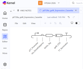

The expression cassette for gelK (glucuronosyltransferase, UniProt: Q7X2M3) was designed for heterologous protein expression in Escherichia coli BL21(DE3) using the pET-28a(+) backbone architecture.The genetic architecture consists of five functional parts arranged in $5’\rightarrow 3’$ orientation:
1. pT7_Promoter: High-level, IPTG-inducible transcription driven by T7 RNA polymerase.
2. rbs_ShineDalgarno: Efficient ribosome binding site for translation initiation.
3. 6xHis_Tag (CDS +start -stop): Provides the initial start codon (ATG) and an N-terminal polyhistidine tag for downstream affinity purification via IMAC.
4. gelK_CDS (CDS -start +stop): The coding sequence for the glucuronosyltransferase enzyme, configured without an internal start codon to maintain the in-frame N-terminal fusion, ending with a standard stop codon (TGA).
5. T7_Terminator: Ensures transcription termination and mRNA stability.
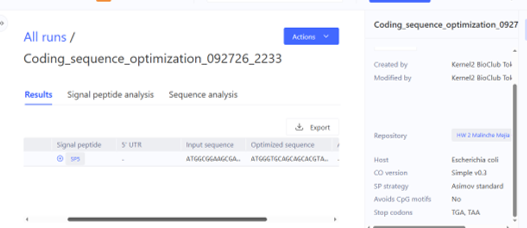
The gelK_CDS sequence was codon-optimized for expression in Escherichia coli BL21(DE3) using Kernel’s codon optimization tool, specifying constraints to avoid internal restriction sites (NdeI, XhoI, EcoRI, BamHI) and maintain an optimal GC content around ~52%. Organisms exhibit species-specific codon usage bias based on host tRNA abundances; replacing rare native codons from Sphingomonas elodea with preferred E. coli codons prevents ribosome stalling and improves translation efficiency. While the translated amino acid sequence remained 100% identical, silent nucleotide substitutions significantly improved codon adaptation for host expression while eliminating unwanted restriction cuts.
Running Kernel’s sequence search on gelK_CDS confirmed that this codon-optimized sequence represents a novel genetic part within our local workspace repository. To verify target identity against global biological databases, the translated primary sequence was cross-referenced with UniProt KB (Accession: Q7X2M3 / Q93DT4), confirming 100% amino acid identity to the glucuronosyltransferase gelK from Sphingomonas elodea. This ensures that the engineered CDS accurately encodes the target enzyme for gellan synthesis without unintended structural mutations.
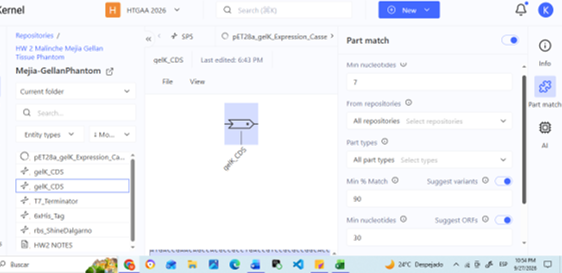
Kernel’s automated safety screen and expression verification were executed on the complete pET28a_gelK_Expression_Cassette construct to screen for biosecurity hazards and expression compatibility in E. coli. The screen passed without flagging any biosecurity risks, confirming that the construct contains only benign laboratory control elements (T7 expression machinery, polyhistidine tag) and a metabolic enzyme from a non-pathogenic bacterium (Sphingomonas elodea). The construct fully complies with Biosafety Level 1 (BSL-1) research guidelines.
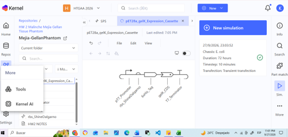
For prototyping our pET28a_gelK_Expression_Cassette (~1.4 kb total length, comprising promoter, RBS, His-tag, gelK CDS, and terminator), we would order the insert as a single synthetic gBlock / double-stranded DNA fragment (dsDNA) and clone it into a linearized pET-28a(+) vector backbone using Gibson Assembly (Gibson et al., 2009).
Direct de novo chemical synthesis of oligonucleotides via phosphoramidite chemistry suffers from intrinsic coupling error rates (typically 1 error per 100–300 bp), primarily consisting of single-nucleotide deletions and substitutions (Carr et al., 2004).
Prototyping Phase (Single/Few Constructs):
Production Phase at Scale (Combinatorial Libraries / High-Throughput Screening):
6xHis_Tag) facilitates downstream IMAC purification and Western blot verification.Potassium Glutamate (K-Glu):
Magnesium Glutamate (Mg-Glu):
| Parameter | PEP + NTP Formulation (e.g., PANOx-SP) | Ribose / Carbohydrate + NMP Formulation |
|---|---|---|
| Energy Source | Phosphoenolpyruvate (PEP, ~33 mM) | Ribose / Glucose / Maltodextrin (~20–40 mM) + Pᵢ |
| Nucleotide Supply | High-cost NTPs (ATP, GTP, CTP, UTP) | Low-cost NMPs (AMP, GMP, CMP, UMP) |
| Enzymatic Route | Direct single-step phosphate transfer via Pyruvate Kinase | Endogenous glycolytic and nucleoside kinase pathways |
| Relative Cost | High (~$150–$250/g protein) | Low (~$20–$50/g protein; ~80% reduction) |
| Reaction Kinetics | Fast initial burst (<2 hours) | Sustained, steady regeneration (4–10 hours) |
| Phosphate Accumulation | High Pᵢ accumulation → Mg²⁺ precipitation & rapid acidification | Balanced Pᵢ recycling → stable pH and free Mg²⁺ |
| Component | PANOx-SP (Jewett & Swartz, 2004) | Olsen et al. (2026) / Smith et al. (2026) | Rationale / Biochemical Difference |
|---|---|---|---|
| Energy System | 33 mM PEP | 30 mM Maltodextrin + Glucose + Pᵢ | Maltodextrin drastically lowers reagent costs while sustaining energy production. |
| Nucleotides | 1.2 mM ATP, 0.85 mM CTP/GTP/UTP | 0.3–0.5 mM reduced NTPs or NMPs | Trimming pyrimidine/purine triphosphates reduces the single largest non-lysate cost driver. |
| Buffer | 57 mM HEPES-KOH (pH 7.5) | 80–100 mM HEPES-KOH (pH 8.2) | Higher pH and buffer capacity offset organic acid buildup during extended synthesis. |
| Additives & Cofactors | 2.7 mM Sodium Oxalate, 0.27 mM CoA | Reduced CoA (≤0.05 mM), Polyamine tuning | Eliminates unnecessary inhibitors (oxalate) and trims expensive cofactors like CoA ($104/mL). |
| Cost Efficiency | ~$150–$250 per gram | ~$30–$60 per gram | Optimized for maximum protein yield per dollar of non-lysate reagents. |
Total Reaction Volume: 20,000 nL Fixed Components (9,000 nL): 5,000 nL Lysate (25%), 2,000 nL DNA, 2,000 nL Base Buffer. Variable Volume Budget: 11,000 nL (Reagents + Nuclease-Free Water).
| Reagent | Stock Conc. | Volume (nL) | Final Conc. | Category & Rationale |
|---|---|---|---|---|
| Potassium Glutamate | 1000 mM | 1,200 nL | +60 mM (~120 mM total) | Salt: Maintains optimal ribosomal subunit stability. |
| Magnesium Glutamate | 100 mM | 1,200 nL | +6 mM (~10.5 mM total) | Salt: Balanced to match reduced NTP concentration. |
| Amino Acid Mix | 20 mM ea. | 1,500 nL | 1.5 mM each | Building Blocks: Prevents amino acid exhaustion during high expression. |
| NTP Mix (Low-Cost) | 20x | 1,000 nL | 1.2 mM ATP, 0.6 mM C/G/UTP | Building Blocks: Reduces expensive NTPs by 30%. |
| Maltodextrin + PEP | 100 mM | 1,800 nL | 30 mM Mal, 9 mM PEP | Energy: Hybrid initial burst + sustained energy regeneration. |
| Spermidine + Cofactors | 20x | 600 nL | 0.9 mM Spmd, 0.3 mM NAD | Cofactor: Polyamine enhances ribosomal translation. |
| Coenzyme A | 10 mM | 100 nL | 0.05 mM | Cofactor: Reduced by 80% to lower costs. |
| HEPES-KOH (pH 8.2) | 500 mM | 800 nL | +20 mM | Buffer: Neutralizes organic acid accumulation. |
| Nuclease-Free Water | – | 2,800 nL | Balance | Fills variable volume budget to 11,000 nL. |
| Reagent | Stock Conc. | Volume (nL) | Final Conc. | Category & Rationale |
|---|---|---|---|---|
| Potassium Glutamate | 1000 mM | 1,200 nL | +60 mM (~120 mM total) | Salt: Maintains ionic strength. |
| Magnesium Glutamate | 100 mM | 1,100 nL | +5.5 mM (~9.5 mM total) | Salt: Slightly lower Mg²⁺ improves folding fidelity. |
| Amino Acid Mix | 20 mM ea. | 1,500 nL | 1.5 mM each | Building Blocks: Substrate supply. |
| NTP Mix (Low-Cost) | 20x | 1,000 nL | 1.2 mM ATP, 0.6 mM C/G/UTP | Building Blocks: Cost-optimized NTP ratio. |
| Maltodextrin + PEP | 100 mM | 1,800 nL | 30 mM Mal, 9 mM PEP | Energy: Sustained energy regeneration. |
| Oxidized Glutathione | 100 mM | 200 nL | 1.0 mM GSSG | Redox: Adjusts DTT:GSSG ratio to permit single disulfide formation. |
| Brij-35 Detergent | 10% w/v | 100 nL | 0.05% w/v | Surfactant: Prevents hydrophobic adsorption of TfCut2 to tube walls. |
| Spermidine + Cofactors | 20x | 600 nL | 0.9 mM Spmd, 0.3 mM NAD | Cofactor: Polyamine translation support. |
| HEPES-KOH (pH 8.0) | 500 mM | 800 nL | +20 mM | Buffer: Stabilizes esterase assay pH. |
| Nuclease-Free Water | – | 2,700 nL | Balance | Fills variable volume budget to 11,000 nL. |
| Reagent | Stock Conc. | Volume (nL) | Final Conc. | Category & Rationale |
|---|---|---|---|---|
| Potassium Glutamate | 1000 mM | 1,300 nL | +65 mM (~125 mM total) | Salt: Higher ionic strength favors chaperone-assisted folding. |
| Magnesium Glutamate | 100 mM | 1,200 nL | +6 mM (~10 mM total) | Salt: Maintains translation accuracy. |
| Amino Acid Mix | 20 mM ea. | 1,500 nL | 1.5 mM each | Building Blocks: Amino acid pool. |
| NTP Mix (Low-Cost) | 20x | 1,000 nL | 1.2 mM ATP, 0.6 mM C/G/UTP | Building Blocks: Cost-optimized nucleotides. |
| Maltodextrin + Pyruvate | 100 mM | 1,800 nL | 30 mM Mal, 10 mM Pyr | Energy: Pyruvate prevents excess NADH buildup in oxidizing conditions. |
| Oxidized Glutathione | 50 mM | 1,600 nL | 4.0 mM GSSG | Redox Buffer: Primary oxidant for disulfide formation. |
| Reduced Glutathione | 50 mM | 400 nL | 1.0 mM GSH | Redox Buffer: Establishes optimal 4:1 GSSG:GSH ratio. |
| Purified DsbC | 100 µM | 600 nL | 3.0 µM DsbC | Folding Helper: Isomerizes incorrect disulfide pairings. |
| Spermidine + Cofactors | 20x | 600 nL | 0.9 mM Spmd, 0.3 mM NAD | Cofactor: Polyamine translation support. |
| HEPES-KOH (pH 8.2) | 500 mM | 800 nL | +20 mM | Buffer: Prevents acidification during complex folding. |
| Nuclease-Free Water | – | 200 nL | Balance | Fills variable volume budget to 11,000 nL. |
Proposal: High NTP costs and inorganic phosphate (Pᵢ) accumulation are major bottlenecks in CFPS. Pᵢ chelates free Mg²⁺, leading to premature termination of translation.
I propose evaluating a Polyphosphate Kinase (PPK) + Polyphosphate (PolyPₙ) energy regeneration system. PPK uses inexpensive polyphosphate to re-phosphorylate NMPs/NDPs into NTPs in situ.
Key Advantages:
- Cost Reduction: Replaces expensive NTP/PEP stocks with low-cost polyphosphate (~$0.01/mL).
- Physicochemical Stability: Maintains low steady-state concentrations of Pᵢ, preventing Mg²⁺ precipitation and extending reaction lifetime beyond 8 hours.
To maximize yields of functional 9-disulfide Reteplase, a T7 Polycistronic Helper Plasmid (pHELPER-DSB) can be added to the CFPS reaction: