Week 2 HW: DNA Design
DNA Design
Part 0: Set up in Kernel
The project workspace is established in the Kernel HTBAA 2026 workspace under the repository “HW 2 Malinche Mejia Gellan Tissue Phantom” and folder “Mejia-GellanPhantom”.

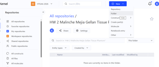
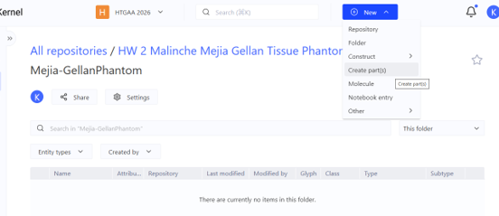
In Kernel, a Part is an individual DNA element used for genetic design, such as a promoter, ribosome binding site (RBS), coding sequence (CDS), or terminator. A Construct is a complete DNA design assembled from multiple Parts in a specific order.
For my project, the gelK coding sequence would be one Part. When it is combined with the appropriate regulatory elements, such as a promoter, RBS, and terminator, the resulting sequence represents the Construct.
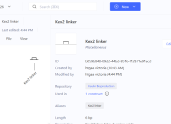
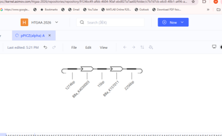
Codon Optimization: Used to adapt a coding sequence to a specific host organism to improve compatibility with its expression system.
Biosecurity Sequence Scanner: Used to screen DNA sequences for potential biosecurity concerns or matches to sequences of concern.
Sequence Alignment: Used to compare two or more DNA or protein sequences and identify similarities and differences.

Part 1: Define your expression target
1. What molecule are you expressing?
I am expressing β-1,4-glucuronosyltransferase (GelK), a key glycosyltransferase enzyme in the gellan gum biosynthetic gene cluster.Because my project from Homework #1 produces a biomaterial (gellan gum exopolysaccharide) to build multilayer tissue-mimicking phantoms for electrosurgical testing, the material itself is synthesized by a multi-enzyme pathway rather than encoded directly by a single gene. The GelK enzyme catalyzes the critical step of transferring glucuronic acid onto the growing tetrasaccharide repeat unit ($[\rightarrow 4)\text{-}\beta\text{-D-Glcp}\text{-(1}\rightarrow 4)\text{-}\beta\text{-D-GlcA}\text{-(1}\rightarrow 4)\text{-}\beta\text{-D-Glcp}\text{-(1}\rightarrow 3)\text{-}\alpha\text{-L-Rhabp}\text{-(1}\rightarrow]$) that forms the structural backbone of gellan gum. Expressing and engineering this rate-limiting glycosyltransferase allows for optimized polysaccharide yield and downstream hydrogel property tuning.
2. What is the sequence?
Protein / Gene Target: Glucuronosyltransferase (gelK)
Source Organism: Sphingomonas elodea (formerly Sphingomonas paucimobilis ATCC 31461)
Database Accession: UniProtKB / Swiss-Prot Accession: Q93DT4 (NCBI Reference Sequence: YP_003022145.1)
Citation:
UniProt Consortium. (2023). Glucuronosyltransferase GelK - Sphingomonas paucimobilis. UniProtKB Entry Q93DT4. Harding, N. E., et al. (2004). Organization of genes required for gellan polysaccharide biosynthesis in Sphingomonas paucimobilis ATCC 31461. Journal of Industrial Microbiology and Biotechnology, 31(2), 70-82.
3. What host or chassis?
Host Chassis: Escherichia coli BL21(DE3)
Justification: While the native producer from HW1 is Sphingomonas elodea, E. coli BL21(DE3) is selected as the expression chassis for recombinant protein production and functional characterization. It provides robust growth, well-established T7 RNA polymerase-driven high-yield protein expression, and seamless compatibility with standard genetic circuit tools in Kernel.
4. Where does the product need to go?
Subcellular Destination: CytoplasmTargeting Sequence Requirement: No secretion signal peptide (such as PelB, OmpA, or PhoA) is attached.
Justification: GelK is an intracellular enzyme that functions at the inner membrane boundary on the cytoplasmic face, utilizing cytoplasmic activated sugar-nucleotide precursors (UDP-glucose and UDP-glucuronic acid) to assemble the repeat unit before membrane translocation. Therefore, the expressed recombinant protein must remain in the cytoplasm for activity assays and IMAC purification via the N-terminal $6\times\text{His-tag}$.
Part 2: Design your genetic parts
| Layer | Short Name | Part Type | Sequence (5′→3′) / Source | Justification |
|---|---|---|---|---|
| Transcription Start | pT7_Promoter | Promoter | TAATACGACTCACTATAGGG(Source: Standard T7 promoter / Kernel database) | Strong, IPTG-inducible promoter recognized specifically by T7 RNA polymerase for tight transcriptional control and high-yield protein production in E. coli BL21(DE3). |
| Translation Start | rbs_ShineDalgarno | Ribosome Binding Site (RBS) | AAGGAGATATACAT(Source: Synthetic high-efficiency bacterial RBS) | Standard Shine-Dalgarno consensus sequence optimized to position the bacterial ribosome efficiently at the start codon for high translation initiation rates. |
| Tag / Targeting | 6xHis_Tag | Tag (N-terminal) | ATGCACCATCACCATCACCAT(Source: Synthetic standard hexahistidine tag) | Encodes an N-terminal polyhistidine tag allowing downstream purification of the GelK enzyme via IMAC and enabling expression verification via Western blot. |
| Coding Sequence | gelK_CDS | Coding Sequence (CDS) | Sphingomonas elodea gelK nucleotide sequence (UniProt: Q93DT4 / Q7X2M3)[cite: 10](Source: UniProt / NCBI Accession YP_003022145.1)[cite: 10] | Encodes the core glucuronosyltransferase enzyme essential for catalyzing the glucuronic acid addition step in gellan gum polysaccharide backbone synthesis. |
| Transcription Stop | T7_Terminator | Terminator | CTAGCATAACCCCTTGGGGCCTCTAAACGGGTCTTGAGGGGTTTTTTG(Source: Standard T7 terminator / Kernel database) | Forms a stable stem-loop secondary structure to efficiently terminate T7 RNA polymerase transcription and stabilize the resulting mRNA transcript. |
Part 3: Assemble your construct
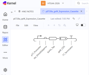

The expression cassette for gelK (glucuronosyltransferase, UniProt: Q7X2M3) was designed for heterologous protein expression in Escherichia coli BL21(DE3) using the pET-28a(+) backbone architecture.The genetic architecture consists of five functional parts arranged in $5’\rightarrow 3’$ orientation:
1. pT7_Promoter: High-level, IPTG-inducible transcription driven by T7 RNA polymerase.
2. rbs_ShineDalgarno: Efficient ribosome binding site for translation initiation.
3. 6xHis_Tag (CDS +start -stop): Provides the initial start codon (ATG) and an N-terminal polyhistidine tag for downstream affinity purification via IMAC.
4. gelK_CDS (CDS -start +stop): The coding sequence for the glucuronosyltransferase enzyme, configured without an internal start codon to maintain the in-frame N-terminal fusion, ending with a standard stop codon (TGA).
5. T7_Terminator: Ensures transcription termination and mRNA stability.
Part 4: Optimize and quality-check
Codon Optimization
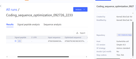
The gelK_CDS sequence was codon-optimized for expression in Escherichia coli BL21(DE3) using Kernel’s codon optimization tool, specifying constraints to avoid internal restriction sites (NdeI, XhoI, EcoRI, BamHI) and maintain an optimal GC content around ~52%. Organisms exhibit species-specific codon usage bias based on host tRNA abundances; replacing rare native codons from Sphingomonas elodea with preferred E. coli codons prevents ribosome stalling and improves translation efficiency. While the translated amino acid sequence remained 100% identical, silent nucleotide substitutions significantly improved codon adaptation for host expression while eliminating unwanted restriction cuts.
Sequence Search (Part Match / BLAST)
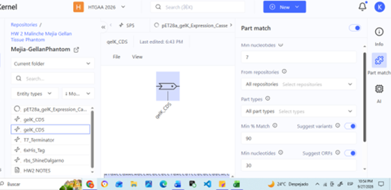
Running Kernel’s sequence search on gelK_CDS confirmed that this codon-optimized sequence represents a novel genetic part within our local workspace repository. To verify target identity against global biological databases, the translated primary sequence was cross-referenced with UniProt KB (Accession: Q7X2M3 / Q93DT4), confirming 100% amino acid identity to the glucuronosyltransferase gelK from Sphingomonas elodea. This ensures that the engineered CDS accurately encodes the target enzyme for gellan synthesis without unintended structural mutations.
Biosecurity & Safety Screening
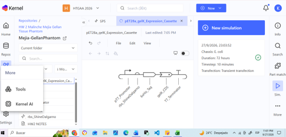
Kernel’s automated safety screen and expression verification were executed on the complete pET28a_gelK_Expression_Cassette construct to screen for biosecurity hazards and expression compatibility in E. coli. The screen passed without flagging any biosecurity risks, confirming that the construct contains only benign laboratory control elements (T7 expression machinery, polyhistidine tag) and a metabolic enzyme from a non-pathogenic bacterium (Sphingomonas elodea). The construct fully complies with Biosafety Level 1 (BSL-1) research guidelines.
Part 5: From design to build
1. Assembly Strategy & Justification
For prototyping our pET28a_gelK_Expression_Cassette (~1.4 kb total length, comprising promoter, RBS, His-tag, gelK CDS, and terminator), we would order the insert as a single synthetic gBlock / double-stranded DNA fragment (dsDNA) and clone it into a linearized pET-28a(+) vector backbone using Gibson Assembly (Gibson et al., 2009).
- Length & Complexity Reasoning: At ~1,400 bp, the insert exceeds the standard length for direct single-oligo chemical synthesis, but falls well within the sweet spot for commercial gene synthesis providers (e.g., Twist Bioscience, IDT).
- Assembly Method: Rather than relying on traditional restriction-digestion and ligation—which introduces unwanted scar sequences and relies on specific cleavage sites—Gibson Assembly uses an exonuclease, polymerase, and ligase cocktail to seamlessly join overlapping dsDNA fragments (Gibson et al., 2009). Overlapping terminal sequences (20–40 bp) matching the pET-28a(+) insertion site ensure scarless, high-efficiency directional insertion.
2. Synthesis Constraints, Sequence Hazards, and Error Correction
Direct de novo chemical synthesis of oligonucleotides via phosphoramidite chemistry suffers from intrinsic coupling error rates (typically 1 error per 100–300 bp), primarily consisting of single-nucleotide deletions and substitutions (Carr et al., 2004).
- Sequence Hazards in Construct:
- GC Content & Repetitions: The wild-type gelK gene from Sphingomonas elodea features a high GC content (>65%), which causes secondary structure formation (hairpins) during PCR amplification and synthesis. Our codon optimization step reduced extreme local GC peaks to ~52% and removed internal homopolymeric runs, significantly mitigating synthesis failure risks.
- T7 Terminator: The strong stem-loop structure of the T7 terminator can cause DNA polymerase slip during synthesis or sequencing verification.
- Mitigation & Error Correction:
- To obtain error-free construct clones from synthesized pools, enzymatic error correction protocols using mismatch-binding proteins (e.g., MutS) or mismatch-cleaving endonucleases (e.g., ErrASE / CelI) can be applied to digest heteroduplex errors prior to clonal amplification (Carr et al., 2004; Tian et al., 2004).
- Clones are subsequently verified via Sanger or Next-Generation Sequencing (NGS) to guarantee an intact, frame-shift-free open reading frame (ORF).
3. Prototyping vs. Production at Scale
Prototyping Phase (Single/Few Constructs):
- Approach: Low-throughput, high-purity commercial dsDNA synthesis (gBlocks) coupled with benchtop Gibson Assembly or Golden Gate cloning into pET-28a(+).
- Goal: Rapid turn-around time, individual Sanger sequence verification, and functional validation of protein expression and gellan precursor yield in E. coli BL21(DE3) at the bench scale.
Production Phase at Scale (Combinatorial Libraries / High-Throughput Screening):
- Approach: Multiplexed microchip-based oligo pool synthesis (e.g., high-density DNA microarrays or microfluidic chips) combined with selective PCR amplification and automated multiplex assembly (Tian et al., 2004; Kong et al., 2007; Kosuri et al., 2010).
- Scaling Advantage: Microchip synthesis drastically lowers cost per base by miniaturizing reaction volumes into picoliter regimes (Kosuri et al., 2010). If we needed to screen hundreds of gelK codon variants, promoter strength combinations, or mutant libraries, microchip pool synthesis followed by enzymatic error correction (Tian et al., 2004) and high-throughput transformation would lower per-construct costs from hundreds of dollars to cents per gene.
Part 6: What don’t you know yet?
1. Inclusion Body Formation and Protein Folding Dynamics
- The Risk: High-level expression driven by the strong, IPTG-inducible pT7 promoter in Escherichia coli BL21(DE3) often leads to metabolic burden and rapid protein accumulation.
- The Unknown: As a membrane-associated glucuronosyltransferase originating from Sphingomonas elodea, recombinant GelK expressed at extreme levels may fail to fold correctly, resulting in insoluble inclusion bodies or misfolded, inactive protein.
- Empirical Validation Needed: We need to test whether lowering the growth temperature post-induction (e.g., $16^{\circ}\text{C}$ to $20^{\circ}\text{C}$) or reducing IPTG concentration (0.1 mM vs. 1.0 mM) is necessary to yield soluble, active enzyme.
2. Metabolic Load and Precursor Availability
- The Risk: Overexpressing an active glucuronosyltransferase consumes intracellular UDP-glucose and UDP-glucuronic acid pools within E. coli.
- The Unknown: Without co-optimizing the upstream nucleotide-sugar biosynthetic pathway (e.g., ugd or galU expression), gelK expression alone may become rate-limited by substrate availability, or severely disrupt host cell homeostasis and growth rate.
- Empirical Validation Needed: Metabolomic profiling and functional enzymatic assays are required to determine whether native precursor levels in E. coli are sufficient to achieve measurable gellan backbone synthesis.
3. Impact of the N-terminal 6xHis Tag on Enzymatic Activity
- The Risk: The addition of an N-terminal polyhistidine tag (
6xHis_Tag) facilitates downstream IMAC purification and Western blot verification. - The Unknown: It is unknown whether the spatial position of the 6xHis tag will sterically interfere with the active site or membrane-anchoring domain of GelK.
- Empirical Validation Needed: Comparative activity assays between the tagged variant and a native (untagged) control—or incorporating a cleavage site (e.g., Thrombin or TEV protease)—will be necessary if catalytic efficiency is compromised.
References
- Carr, P. A., Park, J. S., Lee, Y. J., Yu, T. J., Zhang, S., & Jacobson, J. M. (2004). Protein-mediated error correction for de novo DNA synthesis. Nucleic Acids Research, 32(20), e162. https://doi.org/10.1093/nar/gnh160
- Elowitz, M. B., & Leibler, S. (2000). A synthetic oscillatory network of transcriptional regulators. Nature, 403(6767), 335–338. https://doi.org/10.1038/35002125
- Gibson, D. G., Young, L., Chuang, R. Y., Venter, J. C., Hutchison, C. A., & Smith, H. O. (2009). Enzymatic assembly of DNA molecules up to several hundred kilobases. Nature Methods, 6(5), 343–345. https://doi.org/10.1038/nmeth.1318
- Kong, D. S., Carr, P. A., Chen, L., Zhang, S., & Jacobson, J. M. (2007). Parallel gene synthesis in a microfluidic device. Nucleic Acids Research, 35(8), e61. https://doi.org/10.1093/nar/gkm121
- Kosuri, S., Eroshenko, N., LeProust, E. M., Superbi, M., Lansdorp, J., & Church, G. M. (2010). Scalable gene synthesis by selective amplification of DNA pools from high-fidelity microchips. Nature Biotechnology, 28(12), 1295–1299. https://doi.org/10.1038/nbt.1716
- Stanton, B. C., Nielsen, A. A., Tamsir, A., Muchode, K. M., Voigt, C. A., & Brophy, J. A. (2014). Genomic mining of prokaryotic repressors for orthogonal logic gates. Nature Chemical Biology, 10(2), 99–105. https://doi.org/10.1038/nchembio.1411
- Tian, J., Gong, H., Sheng, N., Zhou, X., Gulari, E., Gao, X., & Zhou, X. (2004). Accurate multiplex gene synthesis from programmable DNA microchips. Nature, 432(7020), 1050–1054. https://doi.org/10.1038/nature03151