Week 02: DNA design
HTBAA Homework #2 — AntiCTnI_Fab_ExpressionPlasmid
Part 0: Setup in Kernel
Repository: HTBAA26_WilliamandMary. Folder: Marcela Mejia.
Part 1: Expression Target
Molecule: A recombinant Fab (antibody fragment) directed against human cardiac troponin I (cTnI). A Fab is a protein made of two separate chains — a heavy chain fragment (VH-CH1) and a light chain (VL-CL) — that associate after folding to form one functional antigen-binding unit. A Fab is chosen over a full antibody because it is smaller, simpler to express in bacteria, and sufficient for the diagnostic/binding function needed.
Sequence and source: The construct is a chimeric Fab. The variable domains (VH and VL) come from PDB 8DZV, a chicken anti-cardiac Troponin I antibody–peptide complex structure (Islam et al., 2023). These are fused to human constant domains: CH1 from human IgG1, UniProt P01857 (IGHG1), for the heavy chain, and the human IGLC (lambda) constant region for the light chain. Each chain’s protein sequence was reverse-translated to DNA and codon-optimized for E. coli, then verified by back-translation to confirm an exact match to the source protein sequence before being built as Kernel CDS parts. I chose this chimeric design because chicken antibodies are commonly raised against highly conserved mammalian proteins like cardiac troponin. Human cTnI looks too similar to a mammal’s own troponin to trigger a strong immune response, so mammals make weak antibodies against it. Chickens are different enough from mammals that their immune system reacts strongly to human cTnI, producing good, high-affinity antibodies instead. So if we take the binding part of that chicken antibody (VH/VL) and attach it to a standard human antibody “body” (CH1/CL), the product has stronger binding.
Host/chassis: Escherichia coli, generic strain (no DE3/T7 RNA polymerase available), which ruled out T7-driven expression and drove the choice of a Lac promoter recognized by native E. coli RNA polymerase.
Product localization: Periplasm. Proper Fab assembly requires correct inter-chain disulfide bond formation, which is far more reliable in the oxidizing environment of the periplasm than in the reducing cytoplasm. This is why the design includes PelB (heavy chain) and OmpA (light chain) signal peptides, each fused in-frame to its respective chain to direct Sec-dependent periplasmic translocation.
Part 2: Genetic Parts
| Layer | Part | Length | Source | Justification |
|---|---|---|---|---|
| Transcription start | Lac promoter | 31 bp | Kernel part | Recognized directly by native E. coli RNA polymerase (no DE3/T7 polymerase available in this host), while still IPTG-inducible for controlled expression timing. |
| Translation start (chain 1) | RBS1 (BBa_B0034) | ~12 bp | Kernel part (iGEM registry) | Strong, well-characterized ribosome binding site placed immediately upstream of PelB to initiate translation of the heavy-chain cistron. |
| Targeting (heavy chain) | PelB signal peptide (BBa_K1507017) | 66 bp | Kernel part | Canonical E. coli periplasmic leader sequence (MKYLLPTAAAGLLLLAAQPAMA), fused in-frame to VH-CH1 to direct Sec-dependent export to the periplasm, needed for correct disulfide-bond formation. |
| Coding sequence (heavy) | VH-CH1_HeavyChain | 672 bp | VH from PDB 8DZV + CH1 from human IgG1 (UniProt P01857); codon-optimized for E. coli | The antigen-binding and first constant domain of the heavy chain; the actual gene of interest for this product. |
| Translation start (chain 2) | RBS2 (BBa_B0034) | ~12 bp | Kernel part (iGEM registry) | Second copy of the same RBS, placed before OmpA to independently initiate translation of the light-chain cistron in this bicistronic design. |
| Targeting (light chain) | OmpA signal peptide | 63 bp | Kernel part | Canonical E. coli periplasmic leader, fused in-frame to VL-CL to direct the light chain to the periplasm alongside the heavy chain. |
| Coding sequence (light) + tag | VL-CL_LightChain + 6xHis_Tag | 651 bp + tag | VL from PDB 8DZV + human IGLC (lambda); codon-optimized for E. coli | Antigen-binding and constant domain of the light chain; the C-terminal 6xHis tag enables downstream Ni-affinity purification. |
| Transcription stop | Terminator (BBa_B0015) | 129 bp | Kernel part (iGEM registry, double terminator) | Strong, reliable transcriptional termination compatible with native E. coli RNA polymerase. |
| Vector context | pUC-AmpR v3 backbone | 2077 bp | Kernel part (reused, not hand-designed) | Provides the pUC origin of replication (high copy number in E. coli) and an ampicillin resistance marker for plasmid selection. |
Both signal-peptide fusions were verified translation-frame-correct in Kernel’s sequence view: each chain’s cistron reads start-to-stop with no premature stop codons at the fusion junctions.
Part 3: Assembled Construct
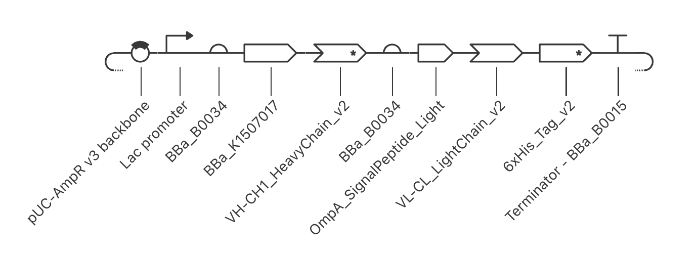
AntiCTnI_Fab_ExpressionPlasmid is a periplasmic E. coli expression construct for an anti-cardiac Troponin I (cTnI) Fab fragment, driven by a single Lac promoter with the heavy chain (PelB-VH-CH1) and light chain (OmpA-VL-CL) translated independently from separate RBSs as a bicistronic operon. Both signal peptides direct their chains to the periplasm for proper folding and disulfide bond formation; the light chain carries a C-terminal 6xHis tag for Ni-affinity purification, and the construct is closed out with a BBa_B0015 terminator and a pUC-AmpR v3 backbone for high-copy propagation and ampicillin selection in E. coli.
5’→3’ order (as built): pUC-AmpR v3 backbone → Lac promoter → RBS1 → PelB → VH-CH1 → RBS2 → OmpA → VL-CL → 6xHis tag → BBa_B0015 terminator → back into the backbone.
Verification performed: The assembled sequence was checked base-by-base in Kernel’s translation view. Both signal-peptide junctions (PelB→VH-CH1 and OmpA→VL-CL) read as continuous, in-frame fusions with no stop codons at the junction, confirmed by the correct canonical leader-sequence amino acids (PelB: MKYLLPTAAAGLLLLAAQPAMA; OmpA: MKKTAIAIAVALAGFATVAQA) running straight into each chain’s variable-domain sequence. VH-CH1 terminates with a clean double stop codon (TGA, TAA) before RBS2, and VL-CL + 6xHis terminates with a clean double stop before the BBa_B0015 terminator. Total construct length: 3740 bp, consistent with the sum of all individual part lengths.
Part 4: Optimize and Quality-Check
Codon optimization. As a second, independent pass, VH-CH1 and VL-CL were each re-run through Kernel’s Coding Sequence Optimization tool. The protein sequence output was identical before and after (codon optimization is synonymous), but individual codons changed — for example GCG→GCA (Ala), GTG→GTT (Val), GGC→GGT (Gly), and TGC→TGT (Cys). Codon usage matters because E. coli’s tRNA pool is unevenly abundant across synonymous codons; a codon that is common in the human/chicken source sequences can be rare in E. coli, causing ribosomal stalling and reduced yield, while recoding to E. coli-preferred codons keeps translation efficient without altering the protein. No restriction-site avoidance or GC-content constraint was applied in this run.
Part 5: From Design to Build
How I would build it. At 3740 bp total, with a ~1.6 kb insert (promoter through terminator) cloned into a 2077 bp backbone, this construct is well within the size range for commercial whole-gene synthesis. I would order the insert as synthetic DNA from a vendor and join it to the linearized pUC-AmpR backbone using Gibson assembly. Gibson et al. (2009) describe a one-pot isothermal method that joins DNA fragments sharing short (~20–40 bp) overlapping ends using an exonuclease, polymerase, and ligase acting together, without restriction sites — well suited here since the construct combines several registry parts (promoter, RBS, signal peptides, terminator) with two custom CDS inserts and a reused backbone, and Gibson assembly avoids needing compatible restriction sites across all of them.
What synthesis constrains. Gene synthesis has non-trivial per-base error rates, and struggles with highly repetitive sequences, long homopolymer runs, and extreme GC content — none of which this construct has in excess, but the repeated use of the same RBS (BBa_B0034) twice and two independent signal-peptide/CDS fusions are worth flagging as regions to sequence-verify post-synthesis carefully. Carr et al. (2004) describe protein-mediated error correction (using mismatch-binding proteins like MutS to remove error-containing strands from a synthesis pool before amplification), which becomes especially relevant if any part of this construct were instead built from chip-synthesized oligo pools rather than vendor gene synthesis — chip-based methods (Tian et al. 2004; Kosuri et al. 2010; Kong et al. 2007) have substantially higher per-base error rates than column-based vendor synthesis and would need that kind of error correction step before assembly.
Prototype vs. scale. For an initial prototype — testing whether the Fab expresses and binds cTnI at all — ordering one synthesized insert and assembling a handful of clones by Gibson assembly is sufficient and fast. If this moved toward production scale, I would shift toward chip-based parallel oligo synthesis (Tian et al. 2004; Kosuri et al. 2010), which produces many DNA sequences in parallel far more cheaply per base than individual gene synthesis orders, paired with the error-correction step above to manage its higher error rate.
Part 6: What Don’t You Know Yet?
Will the chimeric chicken-VH/VL + human-constant-domain fusion fold and assemble correctly? This is a chimeric Fab, not a natural sequence, so the VH/CH1 and VL/CL domain interfaces have not been validated as compatible. How I’d find out: express the construct, extract the periplasmic fraction, and run non-reducing SDS-PAGE to check for a disulfide-linked heavy/light complex, followed by an ELISA against cTnI to confirm the assembled Fab actually binds antigen.
Will the two chains express in a balanced enough ratio for proper 1:1 Fab assembly? A single promoter driving two independently initiated cistrons (via RBS1 and RBS2) does not guarantee equal translation rates for the heavy and light chains. How I’d find out: run a Western blot post-induction distinguishing heavy and light chain bands (by size or epitope tag) to quantify relative expression levels, and if imbalanced, swap in a different-strength RBS on one cistron to rebalance.
Is codon optimization alone sufficient, or will mRNA secondary structure near the RBS/start codons hinder translation initiation? Two independent RBS-CDS pairs in one transcript raise the chance of unintended secondary structure forming between the two cistrons. Kernel’s RNA structure score tool returned N/A on this run, so this remains unverified in software. How I’d find out: test expression via an IPTG induction time-course and compare yield to a single-cistron control; if expression is lower than expected, use Kernel’s structure-scoring tool on the 5’ region around each RBS to check for the problem area.