Matt Woslum is the founder and Lab Director of BioOlympia, an independent [Bio]Punk laboratory in Olympia, Washington, that works with mammalian cell biology, CRISPR gene editing, and adipose-derived stem cell (ASC) research. At BioOlympia, Matt and his team have built out lab infrastructure from the ground up by restoring and hacking lab equipment in order to conduct open science experiments. The purpose of BioOlympia is to disseminate information freely and without IP or paywalls. His team recently developed the “eDNA Sniffer”, an open-source, 3D-printed airborne environmental DNA sampling device that was then presented at DEF CON 34’s Biohacking Village in August 2026. Matt is also a co-author of The [Bio]Punk Cookbook, a technically grounded DIY biology guide written for the biohacker and citizen-scientist community. When he’s not in the lab, Matt plays 7-string guitar and sings/screams for the experimental hardcore rock band Electric Sweats.
See the nav bar on the left to look through my work for How To
Biomanufacture Almost Anything (Fall 2026)!
Subsections of <Matthew Woslum> — HTBAA Fall 2026
Week 1 HW: Final Project Ideas
HTBAA Homework #1
Biomanufacturing Proposal: 3D-Bioprinted Bone Artifacts from Adipose-Derived Stem Cells
Matthew Woslum - September 22, 2026
(1) What I would like to biomanufacture, and why
Target output
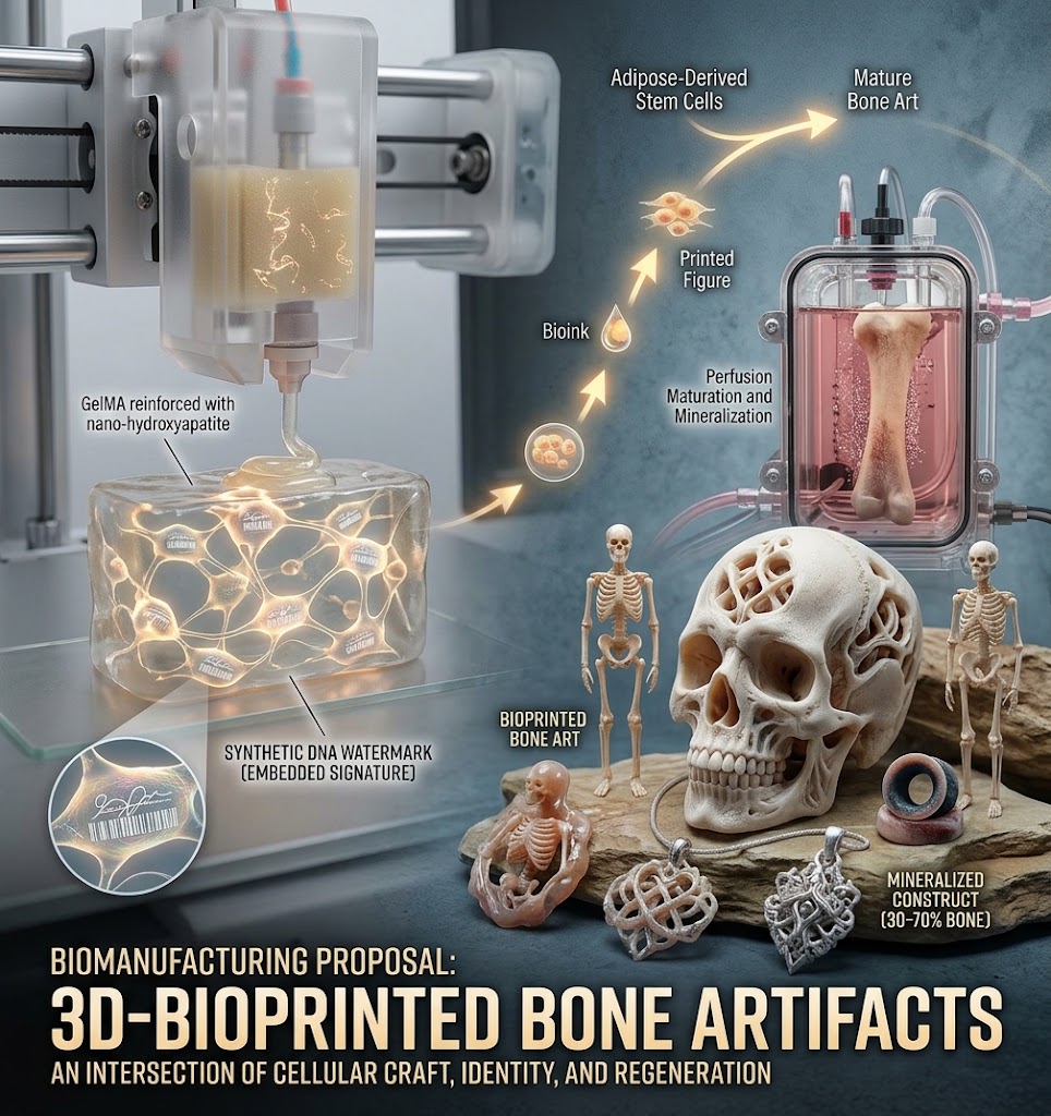
3D-bioprinted figurines made from living osteoblasts embedded in a degradable, mineralizing scaffold. The osteoblasts would be derived from adipose-derived stromal/stem cells (ASCs) expanded in serum-free or low-cost alternatively supplemented media, printed into a cell-adherent composite bioink, and matured in a perfusion bioreactor until the construct mineralizes into bone-like tissue.
Motivation: utilitarian and artistic
A streamlined, inexpensive process for bioprinting client-specific osteoblast–scaffold constructs would open tissue culture as an artistic medium. Miniature skeletons, jewelry grown from a client’s own cells, gauged ear plugs made from your own (or your partner’s) bone. The project would give me hands-on experience with low-cost mammalian cell culture, osteogenic differentiation, and CRISPR knock-in editing. Each artwork’s cells would carry a synthetic DNA watermark, a signature embedded in every cell of the piece.
Who is it for? Initially, my own lab skills practice. But with refinement, commissioned art or utilitarian bone objects could replace many plastic household objects. I find it interesting because it sits at the intersection of tissue engineering, craft, and identity: what does it mean to make (and own) an object literally grown from yourself or someone you love?
(2) What living system would I use?
The chassis will be primary human ASCs. Adipose tissue is clinically abundant and easy to harvest, and ASCs are well characterized for expansion and osteogenic differentiation.
Workflow:
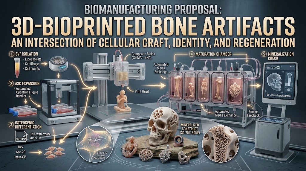
Harvest lipoaspirate → isolate the stromal vascular fraction (SVF) by collagenase digestion and centrifugation.
Select adherent ASCs and expand them in serum-free (or dilute human platelet-lysate) media to reduce cost and lot-to-lot variability relative to fetal bovine serum.
Early in expansion, optionally knock in a synthetic DNA watermark sequence (CRISPR/Cas9) so that every cell in the final piece carries the maker’s signature + reporter/selection marker.
Drive osteogenic differentiation with dexamethasone, ascorbate-2-phosphate, and β-glycerophosphate, applied during/after printing.
Print the ASC-laden bioink into the target geometry, crosslink, and mature the construct in a perfusion bioreactor.
Inputs: lipoaspirate, culture media and growth factors, and a composite bioink. Process: ASC expansion, osteogenic differentiation, and extracellular matrix mineralization. Output: a partially mineralized (~30–70%), cell-laden bone-like construct.
(3) How would I use automation?
Expansion: an Opentrons liquid-handling robot would automate feeding, passaging, and plating across parallel flasks on a fixed schedule. Media changes are the largest recurring labor and contamination risk in mammalian culture, so this is the best step to automate.
Fabrication: a desktop extrusion bioprinter deposits the ASC-laden bioink in the figure geometry with reproducible cell density. The resolution would be dependent on the bioprinter.
Maturation: a perfusion bioreactor automates media exchange and flows medium through the printed construct. This is essential rather than optional: static culture would not mineralize a construct this thick (diffusion limits) and would cause a cause a necrotic core, whereas perfusion enables greater scale[6].
If the process were ever scaled up, the same biology would run in multiple, automated perfusion chambers with sensor feedback rather than manually tended flasks. The prototyping stack (Opentrons + bioreactor) differs from the manufacturing stack (parallel reactors) mainly in reactor count and integration.
(4) How much would I need to make, and how long would it take?
(a) How much?
A 15 cm figure-scale object has a bulk volume of roughly 100–150 cm³ as a mostly solid bone construct, that is ~80–100 cm³ of bone/scaffold composite.
Cells: Bone tissue engineering typically seeds scaffolds at 5 × 10⁶ cells/cm³ [1]. 3D-bioprinted bone constructs commonly use 5–10 × 10⁶ cells/mL of bioink [2]. Using 5 × 10⁶ cells/cm³:
80–100 cm³ × 5 × 10⁶ cells/cm³ = 4–5 × 10⁸ cells. Buffering ~2× for cell death and incomplete differentiation gives a production target of ~10⁹ ASCs.
Lipoaspirate: 1 g of lipoaspirate yields ~2–5 × 10⁵ nucleated SVF cells [3]. About 1% are true adherent, colony-forming ASCs, therefore, ~2–5 × 10³ ASCs per gram [3].
Starting with 6 g of fat: 6 g × (2–5 × 10³ ASCs/g) = 1.2–3 × 10⁴ ASCs. Required expansion: 10⁹ / 2 × 10⁴ ≈ 5 × 10⁴-fold ≈ 15–16 doublings (log₂(5 × 10⁴) ≈ 15.6). ASCs can undergo ~15–20 doublings before senescence [4], so this is feasible but near the conservative limit. Hence, starting with 6 g rather than 2 g.
Scaffold: ~80–100 cm³ of degradable, osteoconductive bioink: gelatin methacrylate (GelMA) — which carries native cell-adhesive RGD motifs and is enzymatically degradable — reinforced with nano-hydroxyapatite (~10–20 wt%) for osteoconductivity and printability [2]. I wish to avoid non-degradable PLA so that the finished artifact is as close to living tissue as possible.
Mass: Fully mineralized cortical bone is 1.8–1.9 g/cm³ [5]. Because I expect only ~30–70% mineralization, the finished piece would weigh roughly 45–135 g (80–100 cm³ × 1.8–1.9 g/cm³ × 0.3–0.7). A 100% mineralized upper bound would be ~145–190 g.
(b) How long?
SVF isolation and ASC selection: ~1 week.
Expansion to ~10⁹ cells: ASC doubling time ~2–3 days [4]; 15–16 doublings ≈ 7 weeks of pure growth, or ~8–10 weeks in practice with passaging roughly once per week (~3 doublings per passage, ~5–6 passages). Automated feeding/passaging (OT-2) keeps this schedule unattended and parallelizable.
Bioprinting and onset of differentiation: ~1–2 weeks.
Maturation and mineralization in the perfusion bioreactor: ~8–12 weeks, organized matrix (rather than a brittle mineral crust) is desired.
Total: approximately 4–6 months end-to-end per figure. Automation throughput matters mainly in the expansion phase, where one liquid handler can tend far more parallel culture than a person can; the maturation phase is throughput-limited by perfusion-chamber footprint rather than labor.
(5) What I don’t know yet
Lipoaspirate sourcing and donor variability: access to clinical-grade tissue, how much ASC yield and potency vary between donors, and whether xeno-free media match serum-based performance at acceptable cost.
Scaffold mechanics versus biology: can a fully degradable GelMA/nano-hydroxyapatite ink reach the stiffness and compressive strength to stand as a free-standing 15 cm object while still supporting ASC osteogenesis? Substrate stiffness is itself a key osteogenic cue, so the design is a genuine trade-off.
Differentiation efficiency in 3D: what fraction of printed ASCs actually deposit mineralized matrix inside a thick construct, and how much the DNA-watermark knock-in reduces expansion capacity or osteogenic potential.
References
[1] Holy, C. E., Shoichet, M. S., & Davies, J. E. (2000). Engineering three-dimensional bone tissue in vitro using biodegradable scaffolds: investigating initial seeding density and culture period. Journal of Biomedical Materials Research, 51(3), 376–382.
[2] Gungor-Ozkerim, P. S., Inci, I., Zhang, Y. S., Khademhosseini, A., & Dokmeci, M. R. (2018). Bioinks for 3D bioprinting: an overview. Biomaterials Science, 6(5), 915–946.
[3] Yoshimura, K., et al. (2006). Characterization of freshly isolated and cultured cells derived from the fatty and fluid portions of liposuction aspirates. Journal of Cellular Physiology, 208(1), 64–76.
[4] Pittenger, M. F., et al. (1999). Multilineage potential of adult human mesenchymal stem cells. Science, 284(5411), 143–147; see also Bonab, M. M., et al. (2006). Aging of mesenchymal stem cell in vitro. BMC Cell Biology, 7, 14.
[5] Rho, J.-Y., Kuhn-Spearing, L., & Zioupos, P. (1998). Mechanical properties and the hierarchical structure of bone. Medical Engineering & Physics, 20(2), 92–102.
[6] Grayson, W. L., et al. (2008). Engineering anatomically shaped human bone grafts. Proceedings of the National Academy of Sciences, 105(37), 13454–13459.
AI assistants (Kimi - Moonshot AI, Gemini - Google) were used to sanity-check the arithmetic and units in the back-of-the-envelope estimates and genereate images. Prompts: “Check the math on cell counts for SVF sufficient for enough ADSCs to seed a 15 cm tall figure comprised of bone-like mineralized tissue and scaffold”, “Use this project proposal to generate an introductory image and a workflow image” All biological parameters were verified against the primary literature cited above.
Biosecurity, Biosafety & Risk Assessment Matrix
Evaluated Options:
Option 1 (Baseline / Manual Culture): Manual primary cell isolation and expansion in standard open flasks, manual media changes, static differentiation, standard laboratory biosafety level (BSL-2).
Yes (enclosed fluidics allow easy neutralization of run media)
Other considerations
• Minimizing costs and burdens
High labor footprint (lipoaspirate collection is labor-intensive)
Same
• Feasibility?
High (established protocols)
High (validated open-hardware & frugal chemistry)
• Not impede research
Yes
Yes (fully open hardware and accessible wetware)
• Promote constructive applications
Yes
Yes (democratizes thick-tissue engineering and accessible bioreactor designs)
Week 2 HW: DNA Design
HTBAA Homework #2 — Writeup
Matthew Woslum · September 29, 2026 Project: DNA Watermarking of Biomanufactured Bone (HW1 follow-up)
Design framing
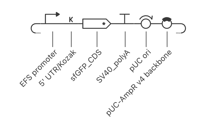
My construct is a minimal mammalian expression plasmid for HEK cells that expresses sfGFP as a fluorescent confirmation of successful delivery, and carries a unique, non-coding synthetic watermark sequence detectable by PCR/sequencing.
Important note for Part 6:
A transient plasmid watermark is diluted ~2-fold per cell division and is effectively gone within days-to-weeks of culture — long before mineralization completes. Therefore this plasmid is a prototype / ** proof of concept** that validates the editing step, not the final authentication mechanism for the final product. Permanent product authentication requires the watermark to be integrated into the host genome (CRISPR knock-in). An integrated watermark in cells embedded in mineral should persist essentially as long as the material itself: hydroxyapatite in bone binds DNA strongly. This construct is the simple, first version. Part 6 talks about the knock-in step as the key follow-up.
Part 0: Set up in Kernel
Logged in via the Biopunk node login. Repository: Woslum-Watermarking, folder: DNA-Watermarking.
Part 1: Define your expression target
Molecule(s): one protein (sfGFP, reporter) plus one non-coding DNA element (synthetic watermark). The watermark is DNA, not RNA/protein — it only needs to be present and PCR-detectable; it does not need to be expressed. Placing it outside the transcription unit avoids any effect on sfGFP expression.
Sequences:
sfGFP: engineered variant of Aequorea victoria GFP, 238 aa, from Pedelacq et al., Nat. Biotechnol. 24, 79–88 (2006). Sequence obtained from Kernel.
Watermark: de novo, ~300 bp synthetic sequence. Designed to be unique: no significant BLAST hit against the human genome (RefSeq)(see Part 4), balanced GC, no long homopolymers/repeats, and free of the restriction sites used for cloning. Generated randomly, then filtered with these requirements.
De novo watermark sequence: GTAGCGTGCTGTGCGTTCCCACGAATACGGTCCTTGATGATGTGATCAGCTTGATGTGATTCATTATCGGCTGTGCAAAACAAACCTAAGAGAGATACCAGCCCATGCACGTGATCGCACTGACAGTTAGATTAGAGGACGGAAGATGCAACGCGATTCTGATACGTCCTAGGGCGCGAAATCAAAGGCTAGCGTTGCTCGCTTTTAATCTACCCTCGGCTCAACTATTTCTAGCTCTCTTTTAACGCCAGCGGAAGCATGGAATGAGCGGGCCCACTCACGTTTGCTAAGCATAATTCG
Chassis: HEK293 cells. Mammalian host drives promoter choice (CMV), Kozak context, and a poly(A) signal.
Product localization: sfGFP is a small (~27 kDa) cytoplasmic protein. It does not need a signal peptide (not secreted) or an NLS. This plain cytoplasmic reporter is simple and sufficient for confirming delivery.
Part 2: Genetic parts
Order
Part (type)
Kernel Part
Justification
1
hEF1a (constitutive)
EFS Promoter
Human Elongation Factor 1 alpha. Long-term transcriptional activity. (CMV is sometimes epigenetically silenced.)
2
5’ UTR + Kozak (GCCACCAUGG)
5’ UTR + Kozak
Efficient translation initiation in mammalian cells
3
sfGFP CDS + start/stop
sfGFP CDS
Bright, fast-folding, low-aggregation reporter
4
SV40 poly(A) signal
SV40_poly(A)
Clean 3’ end, stable mRNA in mammalian cells
5
Watermark (non-coding DNA)
De novo (this work)
Unique, non-expressed authentication signature; placed downstream of poly(A), outside the transcript
6
Backbone: pUC ori + AmpR
Standard cloning vector
E. coli propagation for plasmid production; no mammalian selection marker, to keep the construct minimal
Reading frame check: CDS begins at the Kozak ATG and ends with a single stop codon; no internal stops in the assembled sequence.
Construct map in Kernel confirms part order and orientation.
Part 4: Optimize and quality-check
Codon optimization: the sfGFP CDS was human-codon-optimized in Kernel (sfGFP is from a cnidarian; human codon usage improves translation in HEK cells). Constraints applied: avoid the restriction sites used in cloning; keep GC 40–60%. No codons were changed.
BLAST/sequence search: sfGFP matches A. victoria GFP-derived reporters (expected — confirms identity); the watermark has no significant hits, confirming it is novel and suitable as an authentication signature.
Safety screen: ran Kernel’s Biosecurity scanner on the full construct; it checks for pathogen sequences, toxins, and other regulated elements.
Results: No flagged sequences detected
Part 5: From design to build
Prototype (one construct to test): the plasmid is ~4 kb. Simplest route is ordering the whole expression cassette as one synthetic double-stranded DNA plasmid directly from a synthesis vendor.
Synthesis constraints: current synthesis is reliable to ~1–2 kb per fragment with low error rates; long homopolymers, extreme GC, and repeats are difficult. Our design avoids these (balanced GC, no homopolymers, watermark filtered for repeats), so a single-fragment order is feasible.
Scale (DNA at product scale): if every production run needs unique watermarked plasmid at scale, chip-based oligo-pool synthesis with selective amplification is far cheaper per base than one-at-a-time gene synthesis: Kosuri et al. (Nat. Biotechnol. 28, 1295–1299, 2010) amplified complex chip-derived oligo pools for gene assembly, and Tian et al. (Nature 432, 1050–1054, 2004) showed accurate multiplex gene synthesis from programmable microchips. Fragment-based Gibson assembly of pooled amplicons also bridges prototype to scale.
Part 6: What I don’t know yet
Transient vs. integrated watermark — the biggest unknown, and it decides whether the product itself can be authenticated.
A transient plasmid does not replicate with the host genome and is diluted ~2-fold per division; it is effectively undetectable within days-to-weeks, long before mineralization completes. So this construct can only validate the editing step. An integrated watermark is a different story, though. Genomic DNA encapsulated in hydroxyapatite should persist as long as the material. Bone mineral binds DNA strongly and shields it from nucleases and oxidation. For long term authentication, I would need to integrate this watermark into the osteocyte genome using CRISPR or some other gene editing tool.
How I would find out: build an integrating version (CRISPR knock-in of the watermark cassette) alongside this transient plasmid; mineralize both; extract DNA at intervals over the culture; quantify watermark copies by qPCR; and confirm detection sensitivity by spiking known cell numbers into mineralized bone. Then observe which delivery gives a PCR-detectable signature in the final product, and for how long.
Will sfGFP signal survive processing? GFP fluorescence is lost in cross-linked/ mineralized material, so it validates editing during development, but not the final product. DNA-level watermark is the real end-product signature (see #1). Next step: confirm watermark detectability by PCR/qPCR from mineralized samples.
Is expression level/toxicity OK? Constitutive CMV expression of GFP is usually benign in HEK, but expression burden could matter at production scale. Next step: measure fluorescence across time and passage, and compare CMV vs. a weaker promoter (hPGK) as a tunable variant.
AI use
AI (Kernel AI / Kimi) was used to critique and simplify the initial draft design, design a safe non-coding watermark sequence, and to reason about watermark stability in mineralized matrix. All design choices were verified and re-derived by me.
Week 3 HW: CFPS Design
HTBAA Week 3: Designing a Cell-Free Reaction Composition
Iterative experimental design
Matthew Woslum - September 22, 2026
Part A: Compare Cell-Free Reaction Compositions
Potassium Glutamate vs. Magnesium Glutamate:
Potassium maintains ionic strength and protein synthesis, while magnesium is essential for transcription and translation.
PEP+NTP vs. Ribose+NMP Formulation:
PEP directly regenerates ATP, while ribose and NMPs require cellular machinery to synthesize NTPs.