<Dr. Michellie Hernandez> — HTBAA Fall 2026

cover image cover image

About me

I am a medical doctor in the Dominican Republic. I ❤️ learning and consider myself a lifelong learner. I have a blog called MD Biomimicry integrating medicine, AI and Biomimicry where I edit AI generated content and add my own ideas and research suggestions. In 2020 I came up with the HOPE method; reverse engineering antibodies from cryo-EM and mass spectrometry data with de novo synthesis and published a more detailed paper in 2023 with a preprint in 2022. Since then I have a strong interest to transition into the field. I suffer from an immunodeficiency thus I know the HOPE monoclonal antibodies can bring to patients, hence why I would love to help build a biomanufacturing factory dedicated to producing monoclonal antibodies and mRNA vaccines.

Contact info

Homework, Labs, and Projects

See the nav bar on the left to look through my work for How To Biomanufacture Almost Anything (Fall 2026)!

Subsections of <Dr. Michellie Hernandez> — HTBAA Fall 2026

Week 1 HW: Final Project Ideas

cover image cover image

Author: Dr. Michellie Hernandez, MD

Assisted by Chat-GPT for content clarity and Gemini for estimated feasibility calculations. Content draft and conception was all done by Dr. Michellie Hernandez, MD

Released September 20, 2026

(1) What you would like to biomanufacture and why?

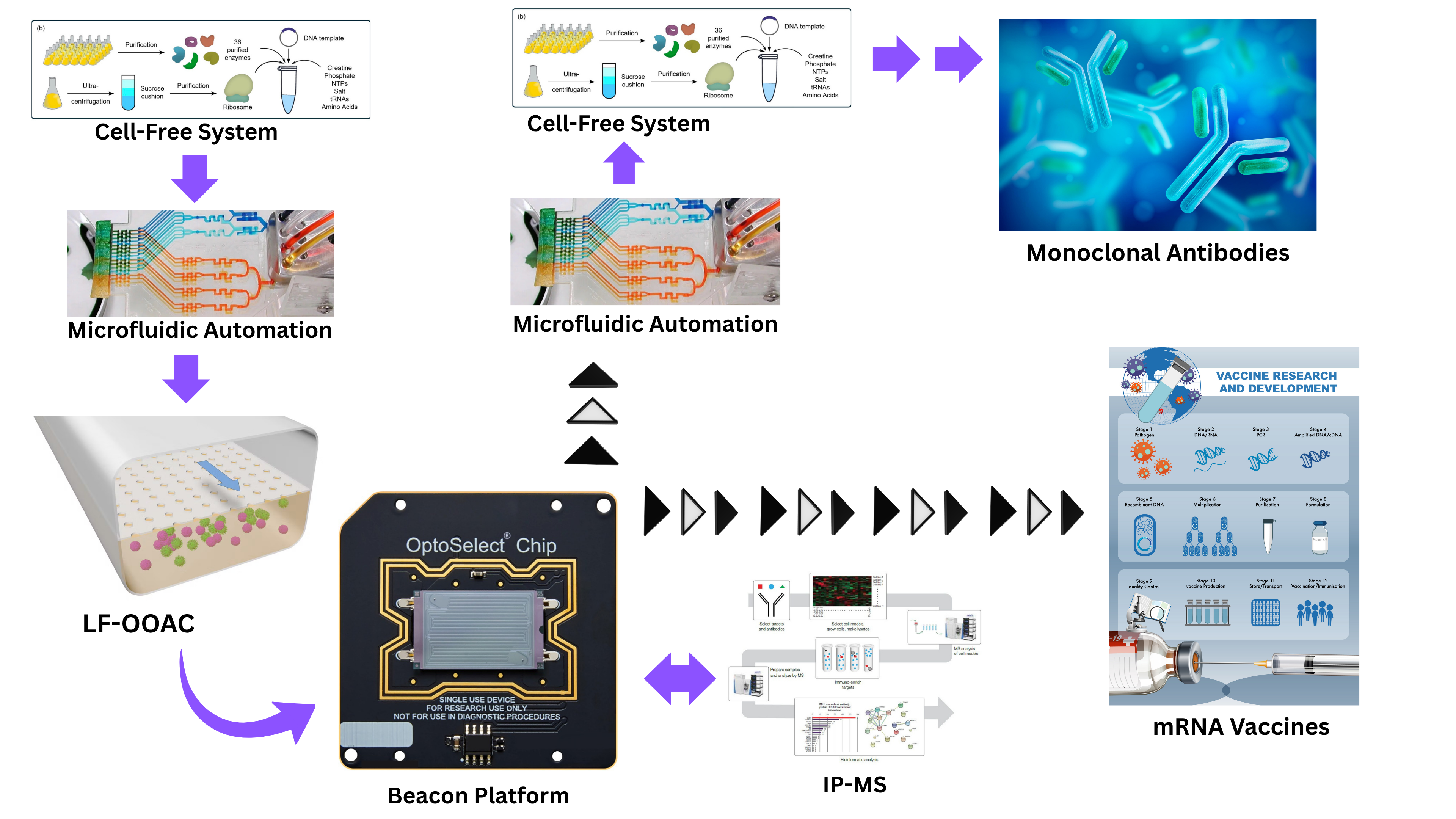
I would love to help build a monoclonal antibody and mRNA biomanufacturing factory that I would like to call the “HOPE” Factory.

(a) Lymphoid follicles organ-on-a-chip (LF-OOAC) to recreate 3D germinal-center microenvironments in microfluidics can be introduced to the antibody discovery, diagnostic and therapeutic monoclonal antibody development and vaccine development.

(b) A cell-free system to help produce the substrates for the LF-OOAC and automation to help regulate the substrates.

(c) Microfluidic automation to regulate and transfer content from LF-OOAC into Beacon Opto plasma B cell discovery and another microfluidic platform that helps extract the antibodies for analysis in Immunoprecipitation Mass spectroscopy scaling antibody affinity to target antigen of interest (preferably a constant domain for viral pathogens).

(d) Cell-free system and automation to help produce regulate the needed substrates respectively, for monoclonal antibody production.

(e) De novo protein synthesis of the epitope selected antibodies bind to, to predict the epitope’s mRNA for the development of mRNA vaccines.

Current monoclonal antibody production utilizing the Opto Plasma B Discovery Beacon platform enables the isolation and selection of plasma cells that produce antibodies specific to a pathogen or antigen of interest. Using opto-electro positioning (OEP) technology, individual plasma cells can be directed into nano-pen chambers, where they are clonally isolated and their secreted antibodies are analyzed. During the selection process, immunofluorescence-based assays are used to determine whether the antibodies produced by the sampled B cells recognize the antigen of interest. However, the current selection workflow evaluates antibody specificity rather than directly measuring antibody affinity. Plasma B cells are difficult to sample from human blood samples as they tend to be located mainly in the bone marrow. Memory B cells on the contrary can be found in human blood samples.

This distinction becomes particularly important when applying the platform to memory B cells. Memory B cells generally do not constitutively secrete antibodies; therefore, they must first be differentiated into antibody-secreting cells before their antigen specificity can be evaluated using the Beacon platform. Vogt et al. (2020), for example, addressed this requirement by differentiating memory B cells into memory B cell–derived lymphoblastoid cell lines (LCLs). Following Epstein–Barr virus exposure in vitro, these cells were capable of secreting antibodies, allowing individual clones to be evaluated within the Beacon platform using immunofluorescence assays to identify antibodies specific to the antigen of interest.

I propose modifying this approach by differentiating antigen-specific memory B cells into long-lived plasma cells (LLPCs) rather than LCLs and incorporating an antibody-affinity assessment into the selection workflow before mRNA extraction. The objective would be to identify individual B-cell clones that not only recognize the target antigen but also produce high-affinity antibodies. I propose developing a lymphoid follicles organ-on-a-chip (LF-OOAC) in vitro 3D germinal-center microenvironment that facilitates the differentiation of memory B cells to LLPCs, which will be attached to a cell-free system for LF-OOAC maintenance. The human blood samples containing memory B cells will be placed into these LF-OOAC and the differentiation of LLPCs are confirmed which can last up to 3 days in culture for differentiation to occur. Afterwards, the sample can be placed onto the Beacon platform for further isolation and selection analysis.

This proposal is based on several biological questions. During a natural immune response, germinal-center reactions promote the selection and maturation of B-cell clones with progressively increased affinity for their cognate antigen. As B cells mature, high-affinity clones are selected to continue differentiation while low-affinity clones are eliminated, thus differentiating to LLPCs mostly with high-affinity antibody production. Therefore, an important question is whether an in vitro system could reproduce sufficient aspects of this natural differentiation and selection process to generate LLPCs that secrete high-affinity antibodies. A second question concerns the relationship between the cell type used for antibody discovery and the properties of the resulting antibody. Specifically, do memory B cell–derived LCLs generate antibodies with affinity characteristics comparable to those produced by LLPCs derived from the same antigen-specific B-cell population? Which of the two; LCLs or LLPCs produce more high-affinity antibodies?

I therefore propose investigating whether memory B cells can be differentiated into LLPCs before the isolation and the selection process to assure that all specific memory B cells are evaluated during the process. This could potentially be explored through two complementary approaches: (1) specialized in vitro culture conditions designed to promote plasma-cell differentiation and maturation, and (2) bioengineered or bioprinted tissue models that reproduce key structural and signaling features of germinal-center microenvironments. A LF-OOAC 3D germinal-center-like microenvironment could be particularly interesting because antibody affinity maturation is not determined solely by soluble factors; it also depends on the spatial and cellular organization of the germinal-center reaction, including interactions among B cells, T follicular helper cells, follicular dendritic cells, antigen, and associated signaling molecules. Recreating selected aspects of this microenvironment could therefore provide a platform for studying whether memory B cells can be driven toward highly functional plasma-cell states in vitro.

The proposed workflow would consequently extend the current Beacon strategy from simple identification of antigen-specific antibody-producing cells toward the identification of clones with both antigen specificity and high functional affinity: Memory B cell → plasma-cell differentiation → individual-cell isolation in Beacon nano-pens → antibody specificity screening → affinity assessment → selection of high-affinity clone → mRNA extraction → cell-free system antibody production → de novo protein synthesis of the epitope that binds to the selected antibody → mRNA vaccine production. The overarching hypothesis is that combining controlled plasma-cell differentiation, affinity-based selection, and single-cell antibody recovery could improve the ability to identify naturally occurring high-affinity antibodies. Incorporating a LF-OOAC with a germinal-center-inspired bioengineered microenvironment could further provide a means of investigating whether aspects of the natural affinity-maturation process can be reproduced outside the human body.

(2) What living system would you use?

  1. Cell-free system producing the favorable substrates for B cell differentiation
  2. Microfluidics automation sampling and measuring to regulate substrates
  3. Lymphoid follicle Organ-on-a-Chip (LF-OOAC) to differentiate memory B cells to long-lived plasma cells
  4. Immunoprecipitation-mass spectrometry (IP-MS) to assess antibody affinity
  5. Microfluidics automation for sampling and measuring to regulate substrates necessary for monoclonal antibodies production
  6. Cell-free system to produce monoclonal antibodies from the selected plasma cell DNA or mRNA
  7. Isolation of the Epitope of the selected antibody and de novo synthesis of its mRNA for mRNA vaccine production.

The LF-OOAC 3D germinal center extracellular matrix microenvironment can be maintained by cell-free systems that supply a rich in favorable cytokines (IL-2, IL-4 and IL-21), antigen of interest, and other media to favor differentiation of memory B cells to long lived plasma B cells. For this research proposal I would also like to develop a cell-free system that biomanufactures the monoclonal antibodies consisting of ribosomes, golgi apparatus, and programmed plasmid replicating the mRNA genome that is extracted from the plasma cells isolated from the Beacon platform.

(3) How would you use automation? The LF-OOAC, a 3D germinal center microenvironment in microfluidics can utilize automation to maintain and regulate the favorable conditions in which the microenvironment stimulates the memory B cells of the human blood samples to initiate memory B cell differentiation to LLPCs. The cell-free system maintaining the LF-OOAC can utilize automation to provide a continuous source of substrates, such as CD40 ligands, glucose and cytokines for the differentiation of memory B cell system. While the cell-free system for the development of monoclonal antibodies can utilize automation to provide a continuous source of substrates for monoclonal antibodies production such as mRNA, amino acids and glucose. Regulation of the substrates provided to both cell-free systems will require screening, sampling and measurement of the substrates to determine the quantity of the substrates that need to be added to the cell-free system for its maintenance.

(4) How much would you need to make, and how long would it take? Ideally the LF-OOAC will allow for complete removal of the human sample and sterilization of the biological sample without destroying LF-OOAC, thus allowing it to be reused multiple times with multiple different samples. The need for additional cells not from the human blood sample, like follicular T helper cells or dendritic cells will be determined if the cell-free system connected to the LF-OOAC and providing the substrates for memory B cell differentiation is insufficient to enhance the maturation of LLPCs. A continuous source of antigens of interest, cytokines and CD-40 would be needed for the cell-free system differentiation of memory B cells to LLPCs. A continuous source of amino acids, organelles and glucose will be needed for the cell-free system making monoclonal antibodies. The complete manufacturing process of monoclonal antibody production with prior memory B cell differentiation will add roughly 3 to 5 days for differentiation, 1 day for immunoprecipitation mass spectrometry affinity test, 1 day for Beacon platform selection, and 1-2 weeks downstream processing and verification which can be reduced pending the development of cell-free system for monoclonal antibodies production as isolation and purification cost and duration will be reduced as a result of the cell-free system The average cost of monoclonal antibody production estimation will roughly be $30,000 to $100,000 with this research proposal may increase production cost, but should assure high-affinity monoclonal antibodies potentially assuring good quality immune responses and provide mutation-resistant monoclonal antibodies and vaccines if constant domain antigens are presented during differentiation phase to assure memory B cells specific against constant domain antigens move forward along the development of monoclonal antibodies.

(5.) What don’t you know yet?

Goyal et. al 2019 provided proof of concept that high-affinity antibodies production is obtainable within in vitro in a LF-OOAC from samples and exposure to antigen and favorable conditions. I believe the major bottlenecks on the research proposal would be obtaining the continuous sources of substrates needed for the maintenance of cell-free systems and LF-OOAC, as well as the knowledge gap in the germinal center’s microenvironment that enhances the development of LLPCs. Experimental trials would determine whether or not exposing the memory B cells to favorable conditions will be sufficient to enhance differentiation.

AI used for this homework:

ChatGPT v.5.6 prompt:

  • Rephrase to explain the following more clearly: The Opto plasma B discovery Beacon platform can isolate and select plasma cells specific to a pathogen of interest, with its opto-electro positioning (OEP) technology guiding plasma cells into nano-pen chambers where plasma cloning and antibody production takes place. Immunofluorescent assays are done during the selection process and tests the antibodies specificity to an antigen of interest, but the current model does not test for antibody affinity as of yet. However, because the selection process tests antibody specificity to an antigen of interest, memory B cells must first be differentiated into plasma cells, as memory B cells do not produce antibodies and require differentiation to do so. Vogt et al. (2020) differentiated the memory B cells into memory B cell–derived lymphoblastoid cell lines (LCLs) that secrete antibodies upon Epstein-Barr virus exposure in vitro to facilitate their selection with immunofluorescent assays within the Beacon platform (2) to confirm specificity against the antigen of interest. I propose differentiation to long-lived plasma cells instead of LCLs, as well as an affinity evaluation to be done prior to the mRNA extraction within the Beacon B cell discovery platform. Behind my reasoning are these daunting questions: Can we emulate the antibodies of long-lived plasma cells that naturally produce high-affinity antibodies towards the antigen and produce a potent immune response? In the same way, can high-affinity monoclonal antibodies more efficiently produce a potent immune response? Do LCLs have the same affinity for the antigen of interest than long-lived plasmatic B cells or do they produce low affinity antibodies? Can in vitro differentiation of memory B cells to long-lived plasma cells with high-affinity be accomplished in bioimprinted tissue emulating germinal centers or within in vitro specialized media?

Gemini prompt:

  • How long does it take to develop a monoclonal antibody with a Beacon platform and what is its cost?

References:

  1. Hernandez M, Bose D (2023) The HOPE Method: Reverse engineering antibodies of recovered patients and bioproteins. J Appl Microb Res. Vol: 6 Issu: 1 (09-20). https://www.innovationinfo.org/articles/JAMBR/JAMBR-164.pdf ARPHA Preprints doi: 10.3897/arphapreprints.e95037

  2. Vogt MR, Fu J, Kose N, Williamson LE, Bombardi R, Setliff I, Georgiev IS, Klose T, Rossmann MG, Bochkov YA, Gern JE, Kuhn RJ, Crowe JE Jr. Human antibodies neutralize enterovirus D68 and protect against infection and paralytic disease. Sci Immunol. 2020 Jul 3;5(49):eaba4902. doi: 10.1126/sciimmunol.aba4902. Erratum in: Sci Immunol. 2020 Sep 18;5(51):eabe5512. doi: 10.1126/sciimmunol.abe5512. PMID: 32620559; PMCID: PMC7418079.

  3. Goyal, G., Prabhala, P., Mahajan, G., Bausk, B., Gilboa, T., Xie, L., Zhai, Y., Lazarovits, R., Mansour, A., Kim, M. S., Curran, D., Long, J. M., Sharma, S., Cohen, L., Levy, O., Prantil-Baun, R., Walt, D. R., & Ingber, D. E. (2019). Lymphoid follicle formation and human vaccination responses recapitulated in an organ-on-a-chip. https://doi.org/10.1101/806505

  4. Brownell, L. (2022, March 15). The immune system is very complicated, but now, it’s on a chip. Wyss Institute. https://wyss.harvard.edu/news/the-immune-system-is-very-complicated-but-now-its-on-a-chip/

  5. Park, H., Jin, H., Kim, D., & Lee, J. (2024). Cell-free systems: Ideal platforms for accelerating the discovery and production of peptide-based antibiotics. International Journal of Molecular Sciences, 25(16), 9109. https://doi.org/10.3390/ijms25169109

  6. Tran, L. (2024, February 26). Microfluidics: Biology’s liquid revolution. The Scientist Magazine®. https://www.the-scientist.com/microfluidics-biology-s-liquid-revolution-71667

  7. The beacon® OPTOFLUIDIC system. (n.d.). Bruker Cellular Analysis. Retrieved 20 September 2026, from https://brukercellularanalysis.com/products/instruments/the-beacon-optofluidic-system/

  8. Bhavin Patel, M.D, Greg Potts, Ph.D, Leigh Foster, B.S, Alex Behling, M.S, John Bucci, M.S, & John Rogers, Ph.D. (2016). Characterization of antibody specificity using immunoprecipitation and mass spectrometry | thermo fisher scientific - US. Thermofisher.Com. https://www.thermofisher.com/us/en/home/life-science/antibodies/antibodies-learning-center/antibodies-resource-library/antibody-application-notes/characterization-antibody-specificity-using-ip-ms.html

  9. Image was done on Canva based on images from the references listed above as well as from Freepik website Designed by Freepik

Week 2 HW: DNA design
cover image cover image

Author: Dr. Michellie Hernandez, MD Assisted by Kernel AI and ChatGPT for tutorial use of Kernel and broaden understanding. Content ideation and creation was done without AI and solely done by Dr. Michellie Hernandez

Release date: September 27, 2026

Part 0: Set up in Kernel

Victoria Makerspace Node Username: htgaa2@makerspace.ca Name of Repository in All repositories folder: Tfh_Michellie_Hernandez_test

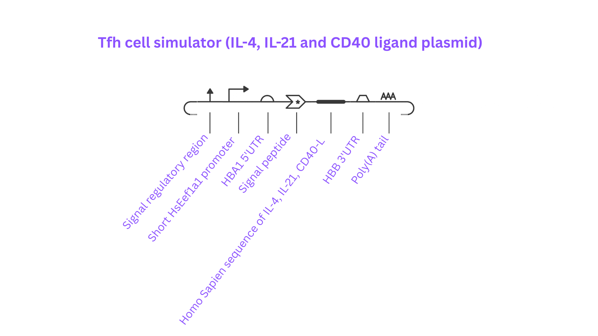
Construct: Tfh cell simulator (IL-4, IL-21 and CD40-ligand Combination plasmid)

Part 1: Define your expression target

What molecule are you expressing?

  • Cytokine Interleukin-4 (IL-4)
  • Interleukin-21 (IL-21)
  • CD40-Ligand

What is the sequence?

What host or chassis?

  • Mammalian Cell-free system

Where does the product need to go?

  • Combined plasma will go in a cytoplasm in a cell-free system that needs to be secreted from the cell-free system

Part 2: Design your genetic parts

Vector context- Backbone appropriate to host

  • Part name: Signal regulatory region
  • Why? This part is a Regulatory DNA element that will respond to an external signal thus is a Signal-responsive regulator

Transcription start - Promoter (+ enhancer / intron if used)

  • Part name: Short HsEef1a1 promoter
    • From Kernel part Repository: Asimov Public
  • Why? Promotes DNA transcription in homo sapiens; Host: Mammalian cell-free system

Translation start - 5’ UTR with Kozak

  • Part name: HBA1 5’UTR
    • From Kernel part Repository: Mariana Diaz Luna_William&Mary
  • Why? Provides correct initiation of the translation.

Targeting - Signal peptide (secretion), tag

  • Part name: Signal peptide (sig-peptide)
    • From Homosapiens IL-4 gene from GenBank
  • Why? Tags the proteins in the Golgi apparatus to signal that proteins will be placed in vesicles for secretion.

Coding sequence - CDS (+start +stop)

  • Part name: 13677 bp
    • From Homo Sapien Gene sequence of IL-4 (8693 bp) from GenBank
    • From Homo Sapien mRNA of IL-21 (3137 bp)from GenBank
    • From Homo Sapien Gene sequence of CD40-Ligand mRNA (1852 bp) from GenBank:
  • Why? Provides the mRNA sequences of IL-4, IL-21 and CD40-Ligand each with appropriate start and stop codon for translation.

Transcription stop Terminator + Poly(A) signal

  • Part name: HBB 3’UTR
    • From Kernel part Repository: Mariana Diaz Luna_William&Mary
  • Part name: Poly(A) tail
    • From Kernel part Repository: Mariana Diaz Luna_William&Mary
  • Why for both transcription stop parts? Assures the termination of the translation.

Part 3: Assemble your construct

Figure 1 above

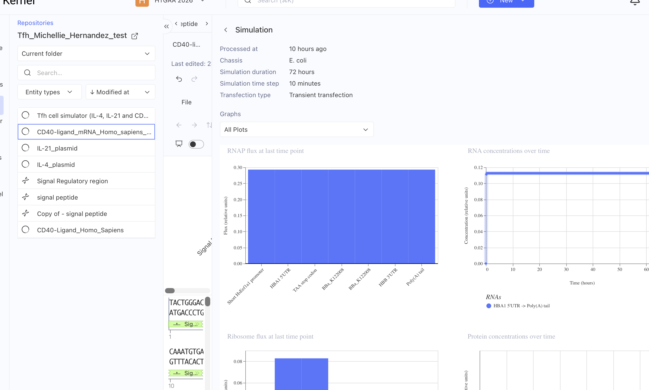
This construct is a mammalian cell-free system plasmid providing the substrates for the LF-OOAC organ-on-a-chip maintenance such as cytokines (IL-4 and IL-21) and costimulating CD40-Ligand that are normally produced in Follicular T helper cells (Tfh cells) as part of the HOPE factory workflow.

Part 4: Optimize and quality-check

  • Optimization of Codons: portion of IL-4 without stop codons in sequence Michellie Hernandez

ATC,GTT,AGC,TTC,TCC,ACT,AAT,TGC,CTC,ACA,TTG,TCA,CTG,CAA,ATC,GAC,ACC,TAT,TGG,GTC,TCA,CCT,CCC,AAC,TGC,TTC,CCC,CTC,TGT,TCT,TCC,TGC,CAT,GTG,CCG,GCA,ACT,TTG,TCC,ACG,GAC,ACA,AGT,GCG,ATA,TCA,CCT,TAC,AGG,AGA,TCA,TCA,AAA,CTT,ACA,GCC,TCA,CAG,AGC,AGA,AGG,GTA,CCT,ATC,TGG,CAC,CAT,CTC,TCC,AGA,TGT,TCT,GGT,GAT,GCT,CTC,AGT,ATT,TCT,AGG,CAT,GAA,AAC,GTT,AAC,AGC,TGC,AGA,AGT,TGG,AAC,TGG,TGG,TTG,GTG,GCA,GTC,CAG,GGC,ACA,CAG,CGA,GGC,TTC,TCC,CCT,GCC,ACT,CTT,TTT,TCT,GAG,GGT,TTG,GAA,GTT,TCC,TCA,GTT,GGA,GGG,AGT,GAG,AGC,TGC,TCA,TCA,AGG,ACT,TCT,CTG,TCC,GGT,TGG,AGG,TTA,ACT,CTG,TCT,CTT,GCT,CTC,TCA,TTT,CTG,CCT,GGA,CCA,AGA,CTC,TGT,GCA,CCG,AGT,CCG,CAG,ACA,TCT,TTG,CTG,CCT,CCA,AGG,GAA,GCC,GTC,CCA,CGG,TCT,GTT,TTA,GCA,AAT,GGG,GAG,ATC,CAT,CCC,CAA,ATG,TCT,GAA,CAA,GAA,ACT,TGT,CTA,ATG,GAA,AAC,GAG,CGG,GCC,CAA,ATT,AAC,TCT,AAG,GTG,TTA,GAT,GTT,TTC,AAA,GAA,CGA,GAA,GTC,TCT,TTA,CTC,TTA,AGC,ATG,TTT,TGG,TCT,TTC,TGG,TTT,CAC,TTG,ATT,AAG,ACA,TGT,AAT,AGA,AAG,CTT,ACA,TGC,TGT,AGT,CCT,GAC,TCA,GAT,CCT,GGT,CAA,AGA,AAA,GCC,CTC,TTG,GGT,TTT,ACT,CTT,TGG,CAT,AGT,GCC,TGG,AAC,GTA,GGA,GGC,ACT,CAA,ATG,CCT,TGT,ATG,AGA,GAA,TTT,TTC,TGG,CCC,ATA,CAT,TTC,AAA,ACC,AAA,TAC,TCT,CAC,AGA,AAC,AGA,TAT,GAT,GAC,AGG,TTG,AGG,GAG,CTT,TCA,TTT,TGT,CTA,AGA,GAC,TTC,CTA,TGG,CAA,CAG,AAA,AGG,TAT,CGC,CAG,AGC,CCC,TCC,TCT,TCC,ACA,GCC,TGG,CCA,CCT,AAC,AGC,CCT,CTG,GGT,TCC,GGG,GCT,GGC,CGT,CCA,GAG,CTC,CTC,AGC,TTG,CTC,TGG,CCG,GCC,GAA,CTC,CCC,TCC,AGC,TCG,GTC,TGG,AAC,CAT,CCT,GCT,GGG,CAG,CGT,CCA,GCA,CAT,CCC,TGC,TTC,GGG,CTG,CCT,GGG,CAC,CTC,GCC,TCT,CTG,CCT,CCT,GTG,CTG,CCT,CAC,CCC,CAC,CCC,TCT,ATC,TGT,AGT,GGG,AGA,GAG,ATA,GAT,TTG,ACA,GCT,GAT,AGT,GCA,TTT,TCT,CTG,ACA,AAC,ACA,CTA,CAG,CCG,TAT,CAA,TTT,TGT,GCA,TTT,CAG,TTC,CTG,TTT,TCA,TGG,AAA,CAC,ACG,GCT,GAG,AAT,GAA,AGC,CCA,AAG,CCT,CAA,TTT,CAC,AGT,GGT,CTC,CTA,ACT,ACC,TGC,TTT,CCA,TGC,AAA,CTA,GGG,AGA,TAT,GGC,CAG,GAG,AGC,CCT,GTG,TGT,TGG,GCA,GGG,TCA,CAC,TCC,AGC,ACC,CAG,ACC,ATA,GAA,CAG,GGC,CCA,TCC,TGC,TTC,ATG,AGG,GAA,ACT,GCT,CTT,CGG,GCC,TTT,AGC,TGG,ACT,ATC,TCA,TTT,CAT,TTA,TCC,CGG,GAG,TCC,GAT,ACA,GGA,GAT,TCT,GAA,GGG,CAA,ATT,ACA,CAC,TTT,TTT,TTT,TTT,TTG,AGA,GGT,CTT,GTT,CTG,TCA,CCC,AGG,CTG,GAG,TGC,AGT,GGT,GCG,ATT,TCA,GCT,CAT,AGC,AGC,CTC,CAC,CTC,CCA,GGC,TCA,AGC,TAT,CTT,CCT,ACC,TCA,GCC,TCC,CAA,GTA,GCC,GGG,ACG,ACA,GGT,GTG,CAC,CAC,CAC,GCC,TGG,CTA,ATT,TTT,GTA,TTT,TTT,TTG,AGA,TGG,AGT,CTT,GCC,ATT,TTG,CCC,AGG,CTT,GTC,TCG,AAC,TTC,TGG,GCT,CAA,GCA,ATC,CGT,CCA,CCT,CGG,CCT,CTC,AAA,GTG,CTG,GGA,TTA,GCC,ACT,GCA,CCT,GGG,CAA,CAG,TTT,ATG,TGT,GTG,TGT,GTG,TGT,GTG,TGT,GTG,TAT,ATA,TGT,GTG,TGT,ATA,TAT,ATG,TGT,GTA,TGT,ATA,TAT,GTG,TGT,GTG,TGT,GTG,TGT,GTG,TGT,GTA,AAT,CTC,CAA,GTC,CAT,CCA,ACC,GAG,AGG,CTC,CTA,CTA,GAA,GCC,AAG,AGT,CCA,CCG,GGT,GCA,CGG,GTC,TCT,GGA,GGC,CTG,TGG,CAC,TGC,GAA,GGC,TCT,AAC,AAA,GCC,AAG,GGA,AGG,GCC,ACC,TCA,CTA,GAA,GCC,AGG,CCT,GGA,GGA,AGG,GTG,AGG,GCT,GAG,GGC,CTG,GAG,GTA,AGA,CTG,CCT,GTG,GTT,TTA,GAC,CCA,GCT,CTG,CCA,CTG,ACT,AGC,TGT,GTG,GCT,GGC,CTT,CAG,CAC,ATC,TTC,ACA,CCT,CTC,TGC,ACC,TCA,GTT,TCC,ACA,TGT,GAA,GAT,ATG,AAA,GTG,ATT,CTG,AAG,GTG,ATT,GCA,AGG,TTG,ATT,GGA,ATC,CAG,CTC,TTG,AGT,TGC,AAA,GTG,TTA,TTG,GAT,GAT,ATA,ACC,ACG,ATT,AAA,AGC,AAG,AAC,AGG,TGC,AGA,GAA,GCG,TTC,GAA,GGA,GGG,GAC,CGG,GTT,GGA,AAG,GAT,CAA,ACC,TCC,AGG,ATG,CCG,AGT,CTG,GGG,CAA,TCC,ATC,TGG,GCT,GTT,TCT,GGA,AGA,CCC,CGG,GTT,GCA,GGC,CAG,GAC,ACT,GCT,GCC,CTC,CCG,TCC,TTA,ACT,CCC,CTC,TTC,ACT,CAG,TCC,TCA,CTC,ACC,TCC,CTC,TCA,CAC,ACA,CAA,CAT,CTC,CTA,GAA,TCC,CCA,CTG,CCT,GCT,TCA,CTC,TTA,CCC,GTC,TCA,TTT,GCC,TCC,CCT,GAA,CTT,CAT,CCT,CCT,GGA,GTT,CAC,GAT,CTC,ACT,CTT,CAC,TCT,TTT,CTT,CCC,CTC,GAA,GAT,TCA,GCA,CTG,CTT,ACT,TAC,ATG,TTA,AGA,TAT,TTC,AGA,ACA,GTG,AAA,TGT,TGC,TAT,TTT,CAA,AAA,CCT,ACA,AAG,GTG,GTA,TGC,AGA,GGA,AAA,GGT,ACT,TCT,TTG,TGT,TCC,CAA,AGA,AAA,CAT,CTT,TCC,AAA,ATC,CAG,CCT,ATT,GAT,TTT,ATT,TCT,TCG,GGG,GAA,CAA,GAA,TTT,TAT,CTC,GTT,GGG,CAT,TCT,ACT,CTT,GGC,AGT,TGC,TGG,AAG,AAG,GCA,CTG,GTC,GTC,CTG,GGC,TTC,ACA,GGT,AAC,ACC,TGT,CAG,GGT,GTC,TAT,GAA,GTC,AAG,GCT,GTC,GGA,ACA,GCA,AAG,TGG,GAA,GAA,GCA,GCT,GGC,TGG,CTG,ATG,AAG,GGT,TTC,TTG,GGT,GGA,CAA,GTA,GTT,GGA,GCT,ATT,TCC,TAT,TTA,CCA,AAG,AGA,GCT,AAA,GTT,CAT,AAT,TCT,ACA,GAG,AGT,TCC,ATA,ATG,AAC,CTC,AAA,TAC,CTC,TGT,TTT,TTG,AAG,GAG,TTT,CTC,ATA,TAC,AGC,ACT,AGC,CTA,TCC,TGG,GCA,GGA,TGG,GAG,ATG,ATG,CAG,TGC,CAA,TCG,GGC,TGG,ATT,TAT,ATG,GTC,CTC,AGT,GAG,GCT,GGT,CAA,GAA,CCG,AGT,AAC,TCT,CAC,AGA,GTC,ACT,GCC,ACA,GAA,GAA,ATC,TCC,CAA,GTG,GCT,GTT,TCC,CAT,TCC,CGG,GAG,GGA,CAG,GCC,TCC,TTC,GTC,ACT,CCC,GCA,GTT,CTG,AAC,TGT,GAG,GTC,AGC,CAG,GCT,GTC,CAA,GTG,CAC,TCC,CTG,AGC,CAC,TGG,CAG,ACA,CAC,TCA,GCA,GCC,AGA,GCT,AGA,CAG,GCA,GGT,GGT,AGG,AGT,CCA,GGG,CCA,CGG,CAG,GGA,TGG,AGT,GTC,GCC,CCC,CTC,GCT,GCG,ATA,CCA,GAG,CAA,CTA,AAA,CGT,GGC,CTT,GCA,CTA,AAG,CTG,CCC,CTA,GGA,TGC,ATT,CTT,TTA,AAG,TTT,TCC,ATT,TGC,AGA,CTC,TTT,TCA,ATT,CTT,ATT,TTA,TCC,TTG,TTT,CCT,TTA,GAA,AGT,CCT,TTC,AAA,AAT,ATC,TTT,AGA,GGG,TTT,TTC,CAT,ACT,ATG,TGG,CCA,TAT,ACG,GGT,CAA,AAT,GTT,TTT,CCA,GGC,TCC,AAG,CCA,GCG,TTT,CAG,AAA,ATC,TCA,CCA,AGG,TTG,TGG,AGA,AGC,AAA,GGC,CTT,TTT,GGT,TTC,CTT,GAA,TCT,CAC,TGT,TCC,CTC,TGC,AGC,AGC,ATG,CAT,GTC,TGC,CCA,CCT,CCA,GAC,ACA,CAG,GCA,CCA,TCT,GCC
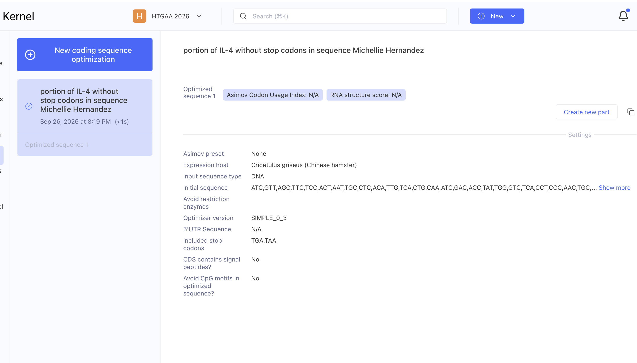
image image
  • Search your sequence AAC,ACA,ACT,GAG,AAG,GAA,ACC,TTC,TGC,AGG,GCT,GCG,ACT,GTG,CTC,CGG,CAG,TTC,TAC,AGC,CAC,CAT,GAG,AAG,GAC,ACT,CGC,TGC,CTG,GGT,GCG,ACT,GCA,CAG,CAG,TTC,CAC,AGG,CAC,AAG,CAG,CTG,ATC,CGA,TTC,CTG,AAA,CGG,CTC,GAC,AGG,AAC,CTC,TGG,GGC,CTG,GCG,GGC,TTG
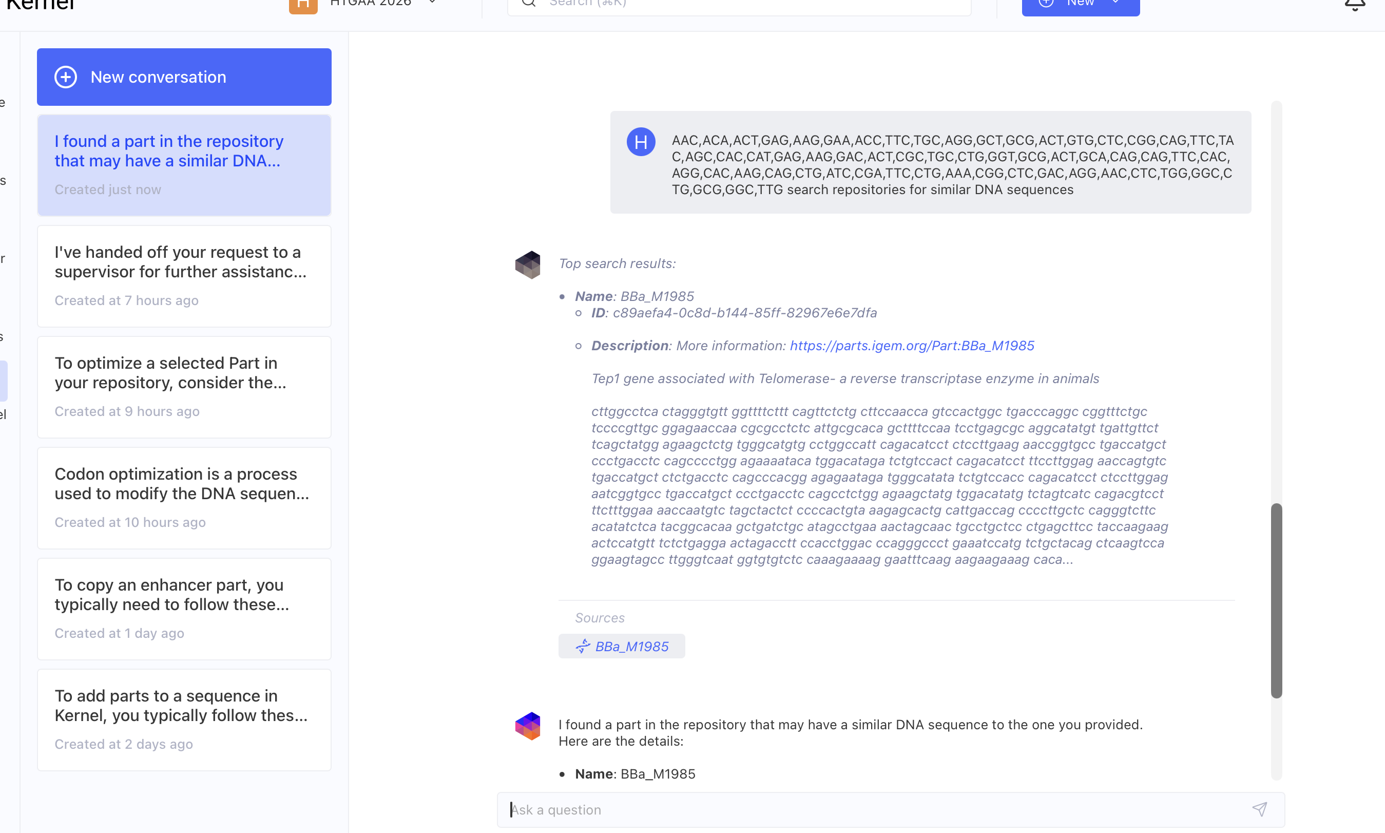
image image
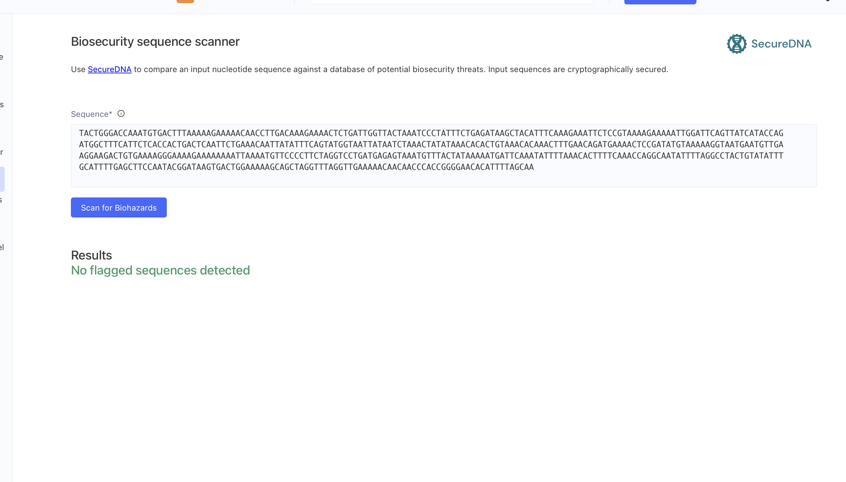
  • Run the safety screen
image image

Part 5: From design to build

How would you build it? I would suggest building it from a pool of oligos synthesized on a chip due to the length and complexity of reducing the amount of error during DNA synthesis.

What does synthesis constrain? Synthesis has error rates thus the amount of errors in the synthesis would have to be reduced drastically and multiple mRNA sequences and its corresponding signalling tag might require the introduction of a plasmid per each substrate instead of a combination plasmid with all substrates in one plasmid.

Prototype vs. scale. The microfluidic automation in the HOPE factory workflow will measure the substrate concentration in the LF-OOAC and send the external signal that are detected by the vector context providing the signaling needed to enhance the substrate production. The plasmid itself will not need to be replicated, as only transcription and translation will occur upon stimulation from the automation.

Part 6: What don’t you know yet?

2–3 biggest unknowns or risks in your construct:

  • Vector context (Signal Regulatory region) is from Homo sapiens BRCA1 intron 2 regulatory region thus from a totally different gene from the genes of IL-4, IL-21 or CD40-Ligand. Thus it is unknown if it will function as detailed.
  • The signal peptide is from IL-4 gene not found in IL-21 nor CD40-Ligand. Thus it is unknown if it will function as detailed.
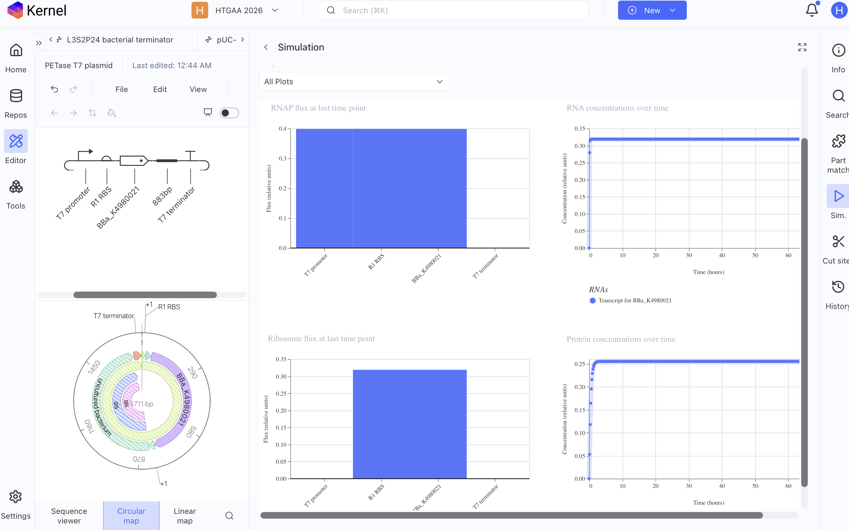
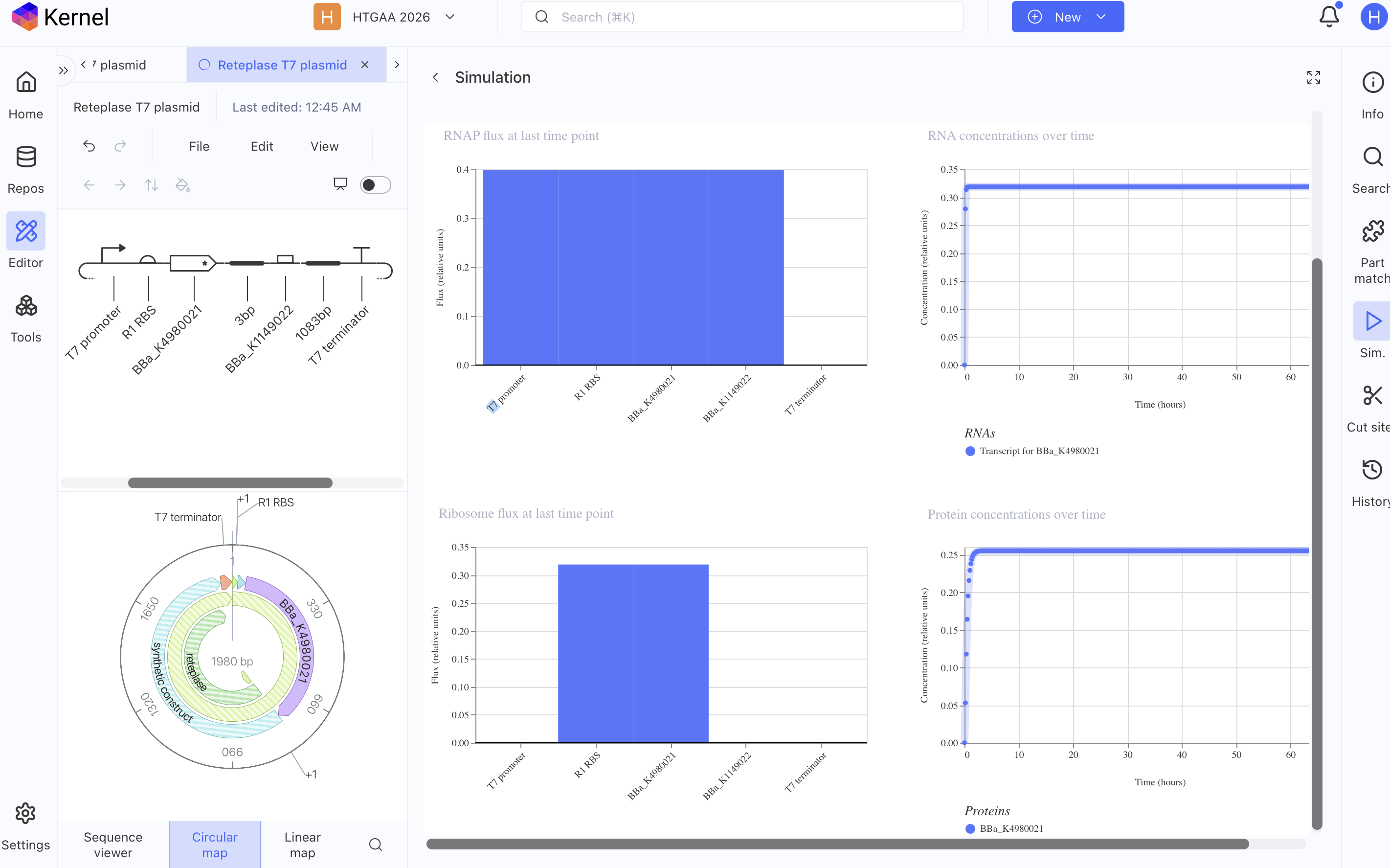
  • Although the simulator shows successful transcription of mRNA, I do not know if the Tfh cell simulator plasmid will successfully produce IL-4, IL-21 and CD40-Ligand when prompted to do so externally via automation.
  • Another unknown are the post translational modification issues that may arise during translation, since this is a common issue even in chassis of mammalian cells thus one must suspect the same issue in mammalian cell-free systems as well.
image image

AI Prompts

Kernel AI

  • How to extract mRNA sequence from the gene from GenBank

  • From repository Tfh_Michellie_Hernandez how can I extract mRNA

  • how to add parts to a sequence

  • 5’URL in public repository

  • Parts of the IL-4 gene in humans

  • which parts in a plasmid sequence are needed to stimulate the transcription and translation of mRNA for IL-4

  • How to optimize the codons

  • AAC,ACA,ACT,GAG,AAG,GAA,ACC,TTC,TGC,AGG,GCT,GCG,ACT,GTG,CTC,CGG,CAG,TTC,TAC,AGC,CAC,CAT,GAG,AAG,GAC,ACT,CGC,TGC,CTG,GGT,GCG,ACT,GCA,CAG,CAG,TTC,CAC,AGG,CAC,AAG,CAG,CTG,ATC,CGA,TTC,CTG,AAA,CGG,CTC,GAC,AGG,AAC,CTC,TGG,GGC,CTG,GCG,GGC,TTG search repositories for similar DNA sequences
OpenAI ChatGPT 5

  • Do Natural bacterial plasmids all start with 5’UTR and end with 3’UTR and a Poly A tail

  • What is the plasmid architecture in cell-free systems

  • How can I stimulate a mammalian cell-free system DNA architecture?

  • What is a Vector context backbone in the construct of a plasmid

  • What vector context can be used in cell-free system plasmid that needs to be enhanced by exterior signaling

  • What Signal peptide (secretion), tag are available in cell-free systems

Week 3 HW: Cell-Free Protocols

Author: Dr. Michellie Hernandez, MD Assisted by ChatGPT for tutorial and broaden understanding. Content ideation and creation was done without AI and solely done by Dr. Michellie Hernandez

Release date: October 5, 2026

A. COMPARE CELL-FREE REACTION COMPOSITIONS

Why does the concentration of potassium glutamate and magnesium glutamate differ between compositions? Potassium Glutamate PanOx system: 200 mM PanOx SP system: 175 mM Cytomim system: 130 mM

Magnesium Glutamate PanOx system: 0 PanOx SP system: 20 mM Cytomim system: 8 mM

Which is more sensitive? Maximum protein synthesis yield decreases at higher or lower concentrations from those listed above on each of the systems.

“Glutamate is the principal anion produced as a result of osmotic stress in E. coli, and it has been shown that protein – nucleic acid interactions are more favorable in glutamate than in acetate salt solutions (Record et al., 1998b, Jewett et. al 2004)”

Protein synthesis yield varies by concentrations of potassium and magnesium per system.

Explain how a PEP+NTP reaction formulation is different from a Ribose+NMP reaction formulation. PEP+NTP reaction formulation utilizes PEP as the energy reagent with NTP reagents. Ribose+NMP reaction formulation utilizes ribose as the energy source reagent and utilizes NMP reagents. Per Smith et. al (2026), GMP can replace GTP and AMP can replace ATP without a loss in protein titer. However CMP cannot replace CTP nor UMP replace UTP without a change in titer. Thus partial replacement of NTP by NMP can yield a more cost effective reaction formulation. Hence I propose a reaction formulation of a combination of NTP and NMP; specifically containing GMP, AMP, CTP and UTP reagents.

“In concurrence with Olsen et al.,42 GPT-5 concluded that use of NTPs is not a cost-effective route to high protein titers and that NMPs yield a better specific cost. The model also noted that NMPs cannot be fully replaced by the corresponding nucleosides or bases, but partial replacement does reduce costs while preserving titers. Again, this finding agrees with Olsen et al., who note that GMP may be replaced by guanine and ribose without loss of titer, a result we confirmed (Tables 1 and 2, reaction compositions 177786377 and 179366574). GPT-5 also found that AMP may be partially replaced by adenosine (Tables 1 and 2, reaction composition 1793122_35). In Step 5, reactions with adenosine had a median titer of 0.279 (95% confidence interval 0.273–0.285) and those without had a median titer of 0.138 (95% confidence interval 0.136–0.139). However, attempts to replace CMP and UMP with their pyrimidine nucleoside counterparts failed. (Smith et. al 2026).”

Choose two cell-free reaction compositions from the literature, compare and contrast the choice of reagents and the concentration of reagents.

Cytomim system is a cell free system that allows pyruvate as an energy source and closely resembles the E. coli natural cytoplasm environment (Jewett et. al 2004). The cytomim does so by synthesizing chloramphenicol acetyltransferase (CAT) and removing PEG, pH buffers and polyethylene glycol from the PANOx system. Acetate accumulates in the Cytomim system. Optimal pH for CAT production is 6.8. Cytomim has a 5-fold yield increase of CAT production in comparison to the PANOx system, but has the same yield as the PANOx-SP system. Unnatural reagents that are useful: oxalic acid. Genetically engineered E. coli removing the pps gene will reduce the need for oxalic acid (Jewett et. al 2004). I propose that the E. coli lysate be obtained from genetically engineered E. coli that has removed the pps gene from the E. coli genome. I propose we then reevaluate the yield of the Cytomim system in this genetically modified E. coli lysate. Concentration of reagents: 1.2 mM adenosine triphosphate (ATP); 0.85 mM each of GTP, UTP, and CTP; 34 Ag/mL folinic acid; 170.6 Ag/mL of E. coli tRNA mix- ture; 13.3 Ag/mL plasmid; 100 Ag/mL T7 RNA polymerase; 5 AM L-[U-14C]-leucine; 2 mM each of 20 unlabeled amino acids; 0.33 mM nicotinamide adenine dinucleotide (NAD); 0.26 mM coenzyme-A (CoA); and 0.24 volume of S30 extract.

PANOx energy regeneration system (Kim et. al 2001) utilizes phosphoenolpyruvate (PEP) as the energy source, but the accumulation of phosphate and acidic environment hinders protein synthesis.

“In the PANOx energy regeneration system, 57 mM HEPES – KOH (pH 7.5), 2% (w/v) polyethylene-glycol 8000 (PEG), 200 mM potassium glutamate, 80 mM ammonium acetate, 16 mM magnesium acetate, 33 mM PEP, and 2.7 mM sodium oxalate were added. In the PANOx-SP system, 175 mM potassium glutamate, 10 mM ammonium glutamate, 20 mM magnesium glutamate, 33 mM PEP, 1.5 mM spermidine, 1 mM putrescine, and 2.7 mM sodium oxalate were added. Solutes added to the Cytomim system included 130 mM potassium glutamate, 10 mM ammonium glutamate, 8 mM magnesium glutamate, 33 mM sodium pyruvate, 1.5 mM spermidine, 1 mM putrescine, and 4 mM sodium oxalate. There was approximately an additional 3.3 mM magnesium, 14.4 mM potassium, 2.4 mM Tris, and 23.5 mM acetate in each reaction originating from the cell extract (Jewett et. al 2004).”

B. DESIGN ONE REACTION COMPOSITION FOR EACH OF THE THREE PROTEINS

Concentration Rank of sfGFP Composition:

  1. Potassium Glutamate 279.84 mM
  2. HEPES-KOH pH 7.5 57.50 mM
  3. Ammonium Glutamate 10.00 mM
  4. Magnesium Glutamate 8.22 mM
  5. Potassium phosphate 1.6:1 monobasic:dibasic (molar ratio) 5.00 mM
  6. 17 Amino Acid Mix 2.81 mM
  7. Glucose 2.78 mM
  8. Spermidine 1.56 mM
  9. Putrescine Dihydrochloride 1.25 mM
  10. NAD 375.00 uM
  11. Coenzyme A 250.00 uM
  12. AMP 125.00 uM
  13. CMP 125.00 uM
  14. GMP 125.00 uM
  15. UMP 125.00 uM
  16. Adenosine 62.50 uM
  17. Cytidine 62.50 uM
  18. Guanosine 31.25 uM
  19. Uridine 31.25 uM
  20. Folinic Acid 0.025 g/L
  21. Water 6.575 uL|

Reagent supplement JSON of sfGFP: { “sample_id”: “michellie_hernandez_2026-10-04_0aqv1h”, “sample_type”: “experimental”, “reagent_list”: { “adenosine”: 50, “amino_acid_mix_17”: 725, “ammonium_glutamate”: 200, “amp”: 25, “base_buffer”: 2000, “cmp”: 25, “coa”: 100, “cytidine”: 50, “folinic_acid”: 50, “glucose”: 50, “gmp”: 25, “guanosine”: 25, “hepes_koh”: 550, “kpo_monobasic_mix”: 200, “lysate”: 5000, “magnesium_glutamate”: 225, “nad”: 75, “nuclease_free_water”: 6575, “potassium_glutamate”: 1825, “putrescine”: 50, “spermidine”: 125, “template_sfgfp”: 2000, “ump”: 25, “uridine”: 25 }, “metadata”: { “ai_model”: “”, “protein_target”: “sfGFP”, “acknowledgements”: “”, “comments”: "" } } |

Composition PETase

Concentration Rank of PETase composition

  1. HEPES-KOH pH 7.5 57.50 mM
  2. Magnesium Glutamate 10.10 mM
  3. Ammonium Glutamate 10.00 mM
  4. Glucose 5.55 mM
  5. Potassium Oxalate 4.38 mM
  6. Oxidized Glutathione (GSSG) 4.04 mM
  7. 17 Amino Acid Mix 2.00 mM
  8. Spermidine 1.56 mM
  9. ATP 1.25 mM
  10. Putrescine Dihydrochloride 1.25 mM
  11. PEP mono (0.1M stock) 1.00 mM
  12. CTP 875.00 uM
  13. GTP 875.00 uM
  14. UTP 875.00 uM
  15. NAD 375.00 uM
  16. Coenzyme A 250.00 uM
  17. DsbC (E. coli, 25.5 kDa) 1.13 uM
  18. Folinic Acid 0.025 g/L
  19. Water 7.200 uL

Reagent supplement JSON of PETase composition { “sample_id”: “michellie_hernandez_2026-10-05_scv2ia”, “sample_type”: “experimental”, “reagent_list”: { “amino_acid_mix_17”: 400, “ammonium_glutamate”: 200, “atp”: 250, “base_buffer”: 2000, “coa”: 100, “ctp”: 175, “dsbc_ecoli”: 225, “folinic_acid”: 50, “glucose”: 100, “gtp”: 175, “hepes_koh”: 550, “lysate”: 5000, “magnesium_glutamate”: 300, “nad”: 75, “nuclease_free_water”: 7200, “oxidized_glutathione”: 475, “pep_mono”: 200, “potassium_oxalate”: 175, “putrescine”: 50, “spermidine”: 125, “template_petase”: 2000, “utp”: 175 }, “metadata”: { “ai_model”: “”, “protein_target”: “PETase”, “acknowledgements”: “”, “comments”: "" } }

Composition Reteplase:

Concentration Rank of Reteplase Composition

  1. HEPES-KOH pH 7.5 57.50 mM
  2. Ammonium Glutamate 10.00 mM
  3. Magnesium Glutamate 8.22 mM
  4. Glucose 5.55 mM
  5. Oxidized Glutathione (GSSG) 4.04 mM
  6. Potassium Oxalate 3.75 mM
  7. 17 Amino Acid Mix 2.00 mM
  8. Spermidine 1.56 mM
  9. ATP 1.25 mM
  10. Putrescine Dihydrochloride 1.25 mM
  11. CTP 875.00 uM
  12. GTP 875.00 uM
  13. UTP 875.00 uM
  14. NAD 375.00 uM
  15. Coenzyme A 250.00 uM
  16. DsbC (E. coli, 25.5 kDa) 5.00 uM
  17. Folinic Acid 0.025 g/L
  18. Water 6.725 uL

Reagent supplement JSON of Reteplase Composition { “sample_id”: “michellie_hernandez_2026-10-04_lm3vpf”, “sample_type”: “experimental”, “reagent_list”: { “amino_acid_mix_17”: 400, “ammonium_glutamate”: 200, “atp”: 250, “base_buffer”: 2000, “coa”: 100, “ctp”: 175, “dsbc_ecoli”: 1000, “folinic_acid”: 50, “glucose”: 100, “gtp”: 175, “hepes_koh”: 550, “lysate”: 5000, “magnesium_glutamate”: 225, “nad”: 75, “nuclease_free_water”: 6725, “oxidized_glutathione”: 475, “potassium_oxalate”: 150, “putrescine”: 50, “spermidine”: 125, “template_reteplase”: 2000, “utp”: 175 }, “metadata”: { “ai_model”: “”, “protein_target”: “Reteplase”, “acknowledgements”: “”, “comments”: "" } }

▸Concentration Rank of Reteplase Composition ⧉

  1. Potassium Glutamate 200.00 mM
  2. HEPES-KOH pH 7.5 57.50 mM
  3. Magnesium Glutamate 20.10 mM
  4. Ammonium Glutamate 10.00 mM
  5. Cysteine 5.00 mM
  6. Oxidized Glutathione (GSSG) 4.46 mM
  7. 17 Amino Acid Mix 4.00 mM
  8. Potassium Oxalate 3.75 mM
  9. Spermidine 1.56 mM
  10. AMP 1.50 mM
  11. GMP 1.50 mM
  12. Putrescine Dihydrochloride 1.25 mM
  13. Tyrosine pH 12 1.00 mM
  14. CTP 875.00 uM
  15. UTP 875.00 uM
  16. Folinic Acid 52.81 uM
  17. DsbC (E. coli, 25.5 kDa) 1.00 uM
  18. Ribose 3.125 g/L
  19. Glucose 1.000 g/L
  20. CMP 0.0 nM
  21. UMP 0.0 nM
  22. Guanine 0.0 nM
  23. Potassium phosphate dibasic 0.0 nM
  24. Potassium phosphate monobasic 0.0 nM
  25. Nicotinamide 0.0 nM
  26. Water 5.175 uL

Reagent supplement JSON of Reteplase Composition

{ “sample_id”: “michellie_hernandez_2026-10-06_gt4yxb”, “sample_type”: “experimental”, “reagent_list”: { “amino_acid_mix_17”: 1200, “ammonium_glutamate”: 200, “amp”: 300, “base_buffer”: 2000, “ctp”: 175, “cysteine”: 400, “dsbc_ecoli”: 200, “folinic_acid”: 50, “glucose”: 100, “gmp”: 300, “hepes_koh”: 550, “lysate”: 5000, “magnesium_glutamate”: 700, “nuclease_free_water”: 5175, “oxidized_glutathione”: 525, “potassium_oxalate”: 150, “putrescine”: 50, “ribose”: 625, “spermidine”: 125, “template_reteplase”: 2000, “utp”: 175 }, “metadata”: { “ai_model”: “”, “protein_target”: “Reteplase”, “acknowledgements”: “”, “comments”: "" } }

Comment

– Substituted ATP for AMP and GTP for GMP, but maintained CTP and UTP due to results in Smith et. al 2026. Thus last composition combines ribose NMP with partial NTP to include in the composition: AMP, GMP, CTP and UTP. Composition was adjusted to reflect in sfGFP, Petase and Reteplase in the coopetition platform.

Suggestion of new reagents to add to the cell-free reagent inventory

Template GSSG Redox category Reasoning: To experiment determine whether or not the hypothesis of adding a template of enzymatic reagents may reduce costs. We can test with another enzymatic reagent that is not already added to the lysate, as DsbC is already in the repletase lysate. We can add to the GSSG template a tag and regulatory enzymes to the plasmid so that translation the GSSG only occurs when needed, instead of producing GSSG constantly. The regulatory gene ideally will respond to an external additive that is added to the well or in response to measurements of GSSG within the well.

C. ASIMOV KERNAL (OPTIONAL)

Build T7 compatible plasmids for cell-free expression using the amino acid coding sequences below:
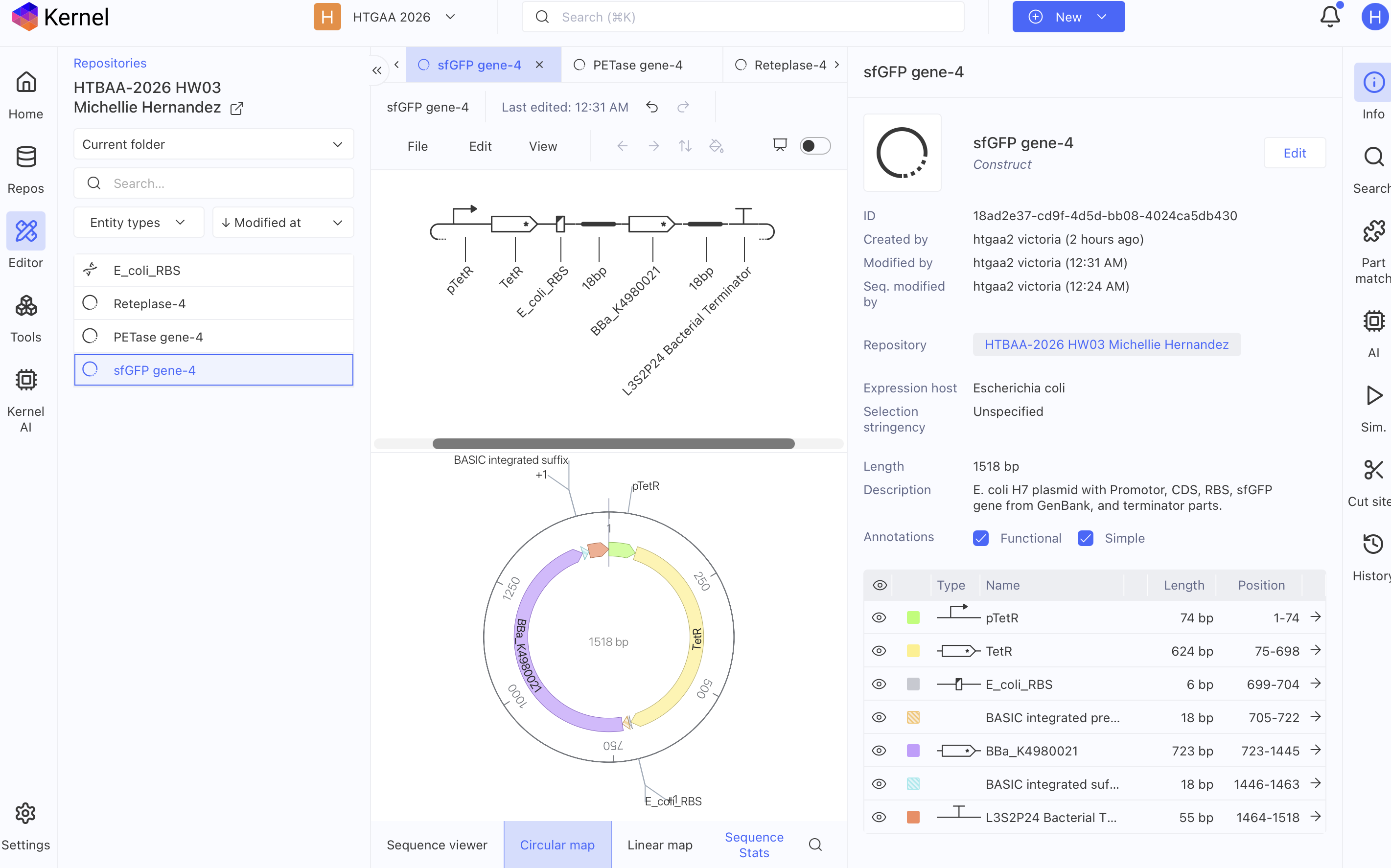
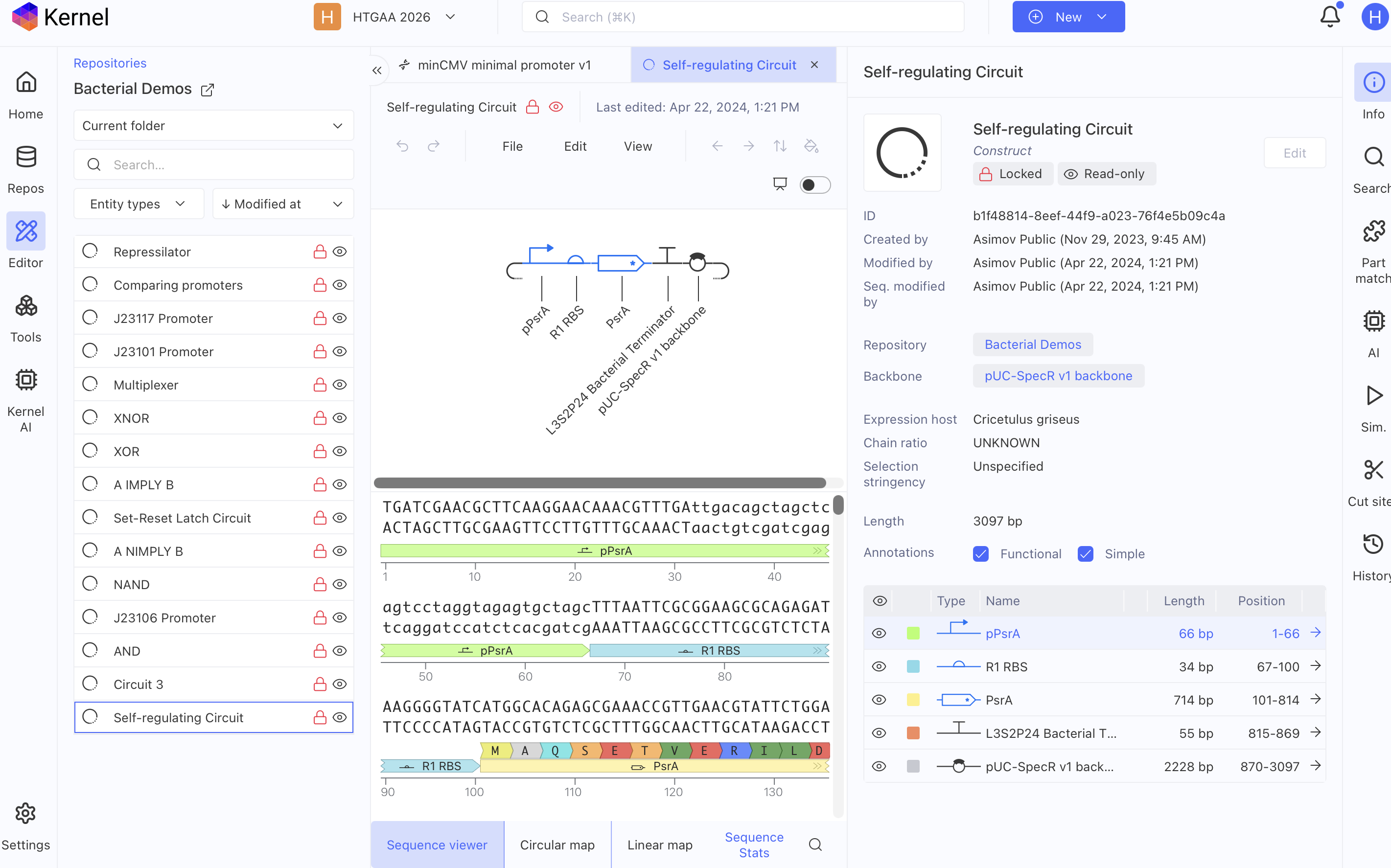
image imageimage imageimage imageimage imageimage image
  1. Use of AI

You are encouraged to use AI and online tools for this assignment to assist your reagent composition development process. As you will present your work during recitation, you must be able to explain and justify the reasoning behind your design.

If you use AI, ensure that you cite the AI client and your prompts used.

Rosalind AI in HGTAA

– Rosalind, How does glutamate affect the protein synthesis yield?

– Roselind, how does glutamate affect the protein synthesis yield?

– Whether leftover metabolites accumulate in cell-free system from the use of NMP that can inhibit protein synthesis

– Whether AMP use makes the formulation cheaper.

– Whether ribose in the Ribose+NMP formulation serves as the energy source, and which paper covers the formulation.

– Whether the cost-effective formulation must be a combination of GMP, AMP, CTP, and UTP?

Chat GPT

– Review oxidation reduction reaction inside the Endoplasmic reticulum and Golgi apparatus

– What makes the interior of the Golgi apparatus more oxidizing than the cytosol? What chemicals or molecules allow a more oxidizing environment?

– What must a E. coli H7 plasmid contain so that the E. coli can translate a mammalian protein?

– Does a cds do the function of ATG and stop codon?

– Which organism produces petase naturally?

– Which organism produces sfGFP naturally?

– Which organism produces reteplase naturally?

  1. REFERENCES AND RESOURCES
  • Smith, A. A., et al. (2026). Using a GPT-5-driven autonomous lab to optimize the cost and titer of cell-free protein synthesis (Ginkgo Bioworks × OpenAI). bioRxiv preprint. DOI: 10.64898/2026.02.05.703998. https://www.biorxiv.org/content/10.64898/2026.02.05.703998v1

  • Olsen, M. L., Copeland, C. E., Sundberg, C. A., Aw, R., Shaver, Z. M., Rao, G., Swartz, J. R., Karim, A. S., &

  • Jewett, M. C. (2026). Design-driven optimization of low-cost reagent formulations for reproducible and high-yielding cell-free gene expression. Nature Communications. DOI: 10.1038/s41467-026-69605-8.

  • Jewett, M. C., & Swartz, J. R. (2004). Mimicking the Escherichia coli cytoplasmic environment activates long-lived and efficient cell-free protein synthesis. Biotechnology and Bioengineering, 86(1), 19–26. DOI: 10.1002/bit.20026.

  • Silverman, A. D., Karim, A. S., & Jewett, M. C. (2020). Cell-free gene expression: an expanded repertoire of applications. Nature Reviews Genetics, 21, 151–170. DOI: 10.1038/s41576-019-0186-3.

  • Rasor, B. J., Vögeli, B., Landwehr, G. M., Bogart, J. W., Karim, A. S., & Jewett, M. C. (2021). Toward sustainable, cell-free biomanufacturing. Current Opinion in Biotechnology, 69, 136–144. DOI: 10.1016/j.copbio.2020.12.012.

Week 4 LAB: Cell-Free Manufacturing

Author: Michellie Hernandez

CONTRIBUTING HYPOTHESIS

– I hypothesize that mimicking the microenvironment composition where the desired protein to biomanufacture occurs naturally and adjust the components of its composition to the host lysate that is used to enhance proper folding of the desired protein within the host lysate. In the case of the coopetition, enzymatic ratios of naturally occuring microenvironment composition where the desired protein is produced and folded should be adjusted to functionally equivalent enzymes in the E. Coli. The ratios and proportions of the composition should be similar within the E. coli lysate. I hypothesize that this might enhance proper folding of the protein. Thus for cell free protein synthesis (CFPS) of reteplase the microenvironment to mimic will be the interior of mammalian Endoplasmic Reticulum, while CFPS of Petase the microenvironment to mimic will be the cytosol of Ideonella sakainsis where Petase occurs naturally. In CFSP of sfGFP the natural micro environment composition to mimic will be the cytosol of the Aequorea Victoria jellyfish.

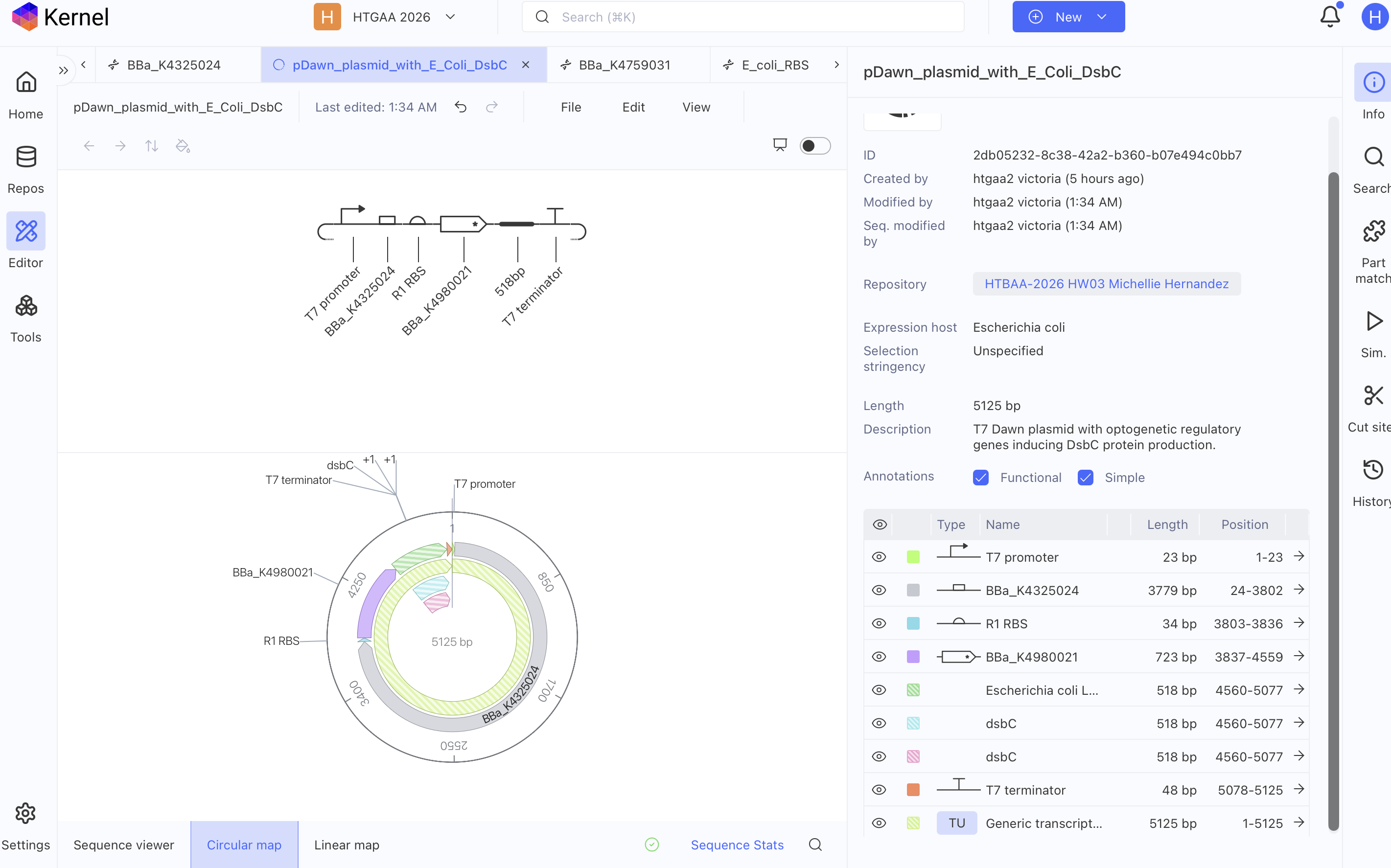
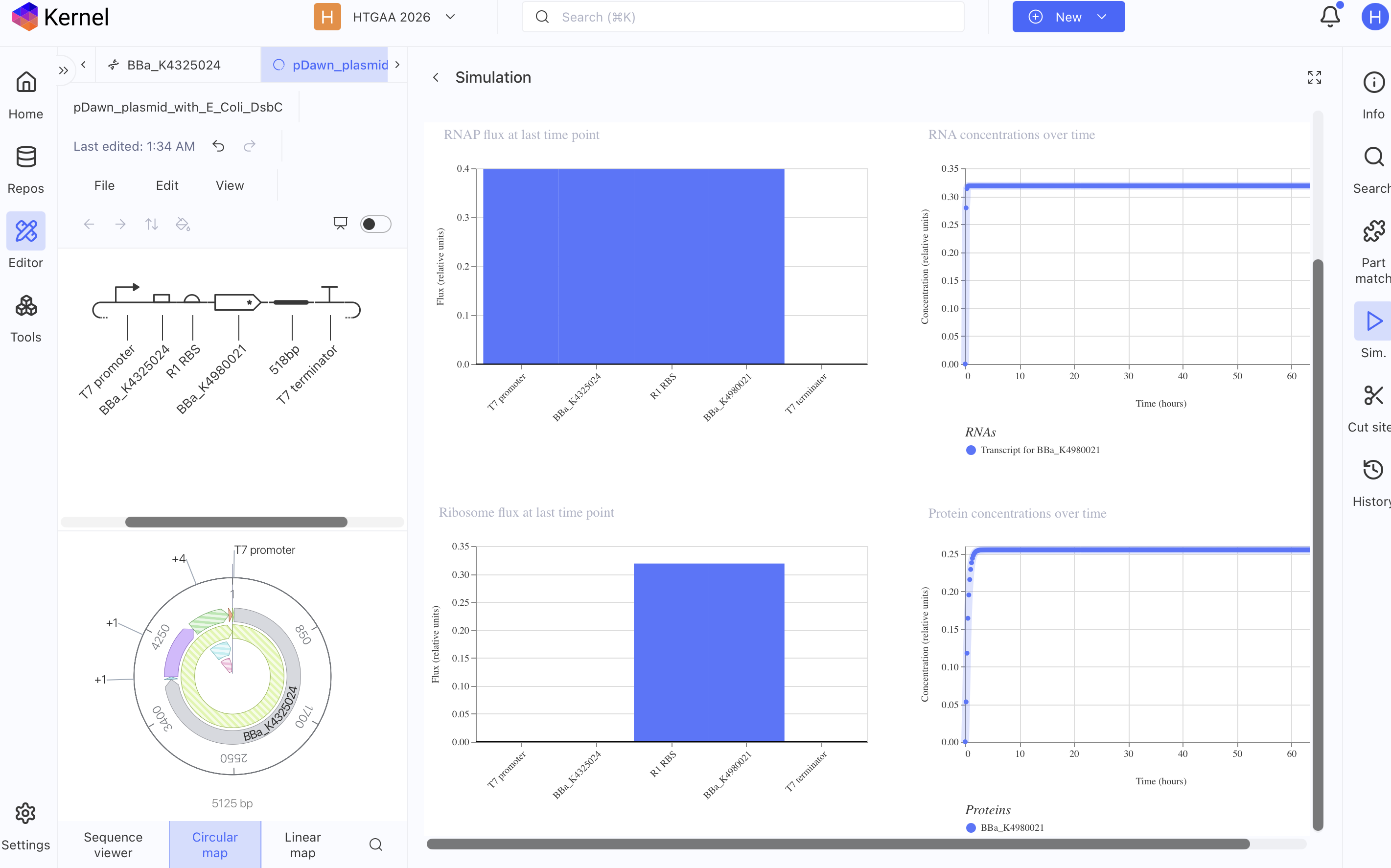
– I hypothesize that the cost of biomanufacturing will reduce upon adding a plasmid with regulatory genes and templates of protein reagents that can be induced with blue light whenever the reagent concentrations are low within the cell free system. I suggest the use of pDusk and pDawn plasmids to use optogenetics for light-regulated gene expression of enzymatic substrates like GSSG and DsbC per resource: Ohlendorf et. al 2014 “From dusk till dawn: one-plasmid system for light-regulated gene expression. Alternatively the regulatory genes of Aequorea Victoria jellyfish that induces GFP naturally in response to blue light can also be used if pDusk or pDawn plasmids are not available.

pDawn T7 Plasmid with E. coli DsbC gene

image imageimage image

COMPOSITIONS SUBMITTED

Composition Rank for sfGFP

  1. Sodium Pyruvate 40.89 mM
  2. Ammonium Glutamate 10.00 mM
  3. Magnesium Glutamate 8.85 mM
  4. Potassium Oxalate 4.38 mM
  5. 17 Amino Acid Mix 4.00 mM
  6. Tyrosine pH 12 2.00 mM
  7. Cysteine 2.00 mM
  8. Spermidine 1.56 mM
  9. CTP 1.50 mM
  10. UTP 1.50 mM
  11. AMP 1.50 mM
  12. GMP 1.50 mM
  13. Putrescine Dihydrochloride 1.25 mM
  14. Folinic Acid 0.037 g/L
  15. Water 6.325 uL

REAGENT SUPPLEMENT JSON FOR sfGFP

{ “sample_id”: “michellie_hernandez_2026-10-11_xwi7a8”, “sample_type”: “experimental”, “reagent_list”: { “amino_acid_mix_17”: 1200, “ammonium_glutamate”: 200, “amp”: 300, “base_buffer”: 2000, “ctp”: 300, “cysteine”: 100, “folinic_acid”: 75, “gmp”: 300, “lysate”: 5000, “magnesium_glutamate”: 250, “nuclease_free_water”: 6325, “potassium_oxalate”: 175, “putrescine”: 50, “sodium_pyruvate”: 900, “spermidine”: 125, “template_sfgfp”: 2000, “tyrosine”: 400, “utp”: 300 }, “metadata”: { “ai_model”: “”, “protein_target”: “sfGFP”, “acknowledgements”: “”, “comments”: “sfGFP: Changed pyruvate as energy substrate per cytomim system. Equalized NTP and NMP combination. Substituted ATP for AMP and GTP for GMP, but maintained CTP and UTP due to results in Smith et. al 2026. Role: medical doctor new to Cell free systems.” } }

COMPOSITION RANK FOR PETASE

  1. Sodium Pyruvate 34.08 mM
  2. Ammonium Glutamate 10.00 mM
  3. Magnesium Glutamate 8.85 mM
  4. Ribose 5.00 mM
  5. 17 Amino Acid Mix 4.00 mM
  6. Potassium Oxalate 3.75 mM
  7. Oxidized Glutathione (GSSG) 1.70 mM
  8. Spermidine 1.56 mM
  9. CTP 1.50 mM
  10. UTP 1.50 mM
  11. AMP 1.50 mM
  12. GMP 1.50 mM
  13. Putrescine Dihydrochloride 1.25 mM
  14. DsbC (E. coli, 25.5 kDa) 375.0 nM
  15. Folinic Acid 0.037 g/L
  16. Water 6.575 uL

REAGEANT COMPOSITION JSON FOR PETASE

{ “sample_id”: “michellie_hernandez_2026-10-11_xwi7a8”, “sample_type”: “experimental”, “reagent_list”: { “amino_acid_mix_17”: 1200, “ammonium_glutamate”: 200, “amp”: 300, “base_buffer”: 2000, “ctp”: 300, “dsbc_ecoli”: 75, “folinic_acid”: 75, “gmp”: 300, “lysate”: 5000, “magnesium_glutamate”: 250, “nuclease_free_water”: 6575, “oxidized_glutathione”: 200, “potassium_oxalate”: 150, “putrescine”: 50, “ribose”: 150, “sodium_pyruvate”: 750, “spermidine”: 125, “template_sfgfp”: 2000, “utp”: 300 }, “metadata”: { “ai_model”: “”, “protein_target”: “sfGFP”, “acknowledgements”: “”, “comments”: “Petase: Combined Jewett and Ribose+NMP system, but with CTP and UTP. As Smith et. al 2026 noted that CMP cannot replace CTP nor UMP cannot replace UTP without affecting the titers. Pyruvate and ribose as energy substrates. Role: medical doctor background new to cell free systems.” } }

COMPOSITION RANK FOR RETEPLASE

  1. Sodium Pyruvate 32.94 mM
  2. Ammonium Glutamate 10.00 mM
  3. Magnesium Glutamate 8.85 mM
  4. Cysteine 5.00 mM
  5. Oxidized Glutathione (GSSG) 4.46 mM
  6. Potassium Oxalate 4.38 mM
  7. 17 Amino Acid Mix 4.00 mM
  8. Spermidine 1.56 mM
  9. CTP 1.50 mM
  10. UTP 1.50 mM
  11. AMP 1.50 mM
  12. GMP 1.50 mM
  13. Putrescine Dihydrochloride 1.25 mM
  14. Reduced Glutathione (GSH) 1.00 mM
  15. Nicotinamide 1.00 mM
  16. DsbC (E. coli, 25.5 kDa) 1.00 uM
  17. Folinic Acid 0.025 g/L
  18. Water 5.500 uL

REAGENT SUPPLEMENT JSON FOR RETEPLASE

{ “sample_id”: “michellie_hernandez_2026-10-11_9p2lna”, “sample_type”: “experimental”, “reagent_list”: { “amino_acid_mix_17”: 1200, “ammonium_glutamate”: 200, “amp”: 300, “base_buffer”: 2000, “ctp”: 300, “cysteine”: 400, “dsbc_ecoli”: 200, “folinic_acid”: 50, “gmp”: 300, “lysate”: 5000, “magnesium_glutamate”: 250, “nicotinamide”: 200, “nuclease_free_water”: 5500, “oxidized_glutathione”: 525, “potassium_oxalate”: 175, “putrescine”: 50, “reduced_glutathione”: 200, “sodium_pyruvate”: 725, “spermidine”: 125, “template_reteplase”: 2000, “utp”: 300 }, “metadata”: { “ai_model”: “”, “protein_target”: “Reteplase”, “acknowledgements”: “”, “comments”: “Reteplase: Adjusted some reagents to be more similar to Jewett cytomim system. Added Nicotinamide to help oxidative stress from H2O2 during oxidative state for reteplase. Equalized concentration of the Nucleotides NTP and NMP Changed energy source to pyruvate to mimic Cytomim system.Role: Medical doctor background new to cell free systems.” } }

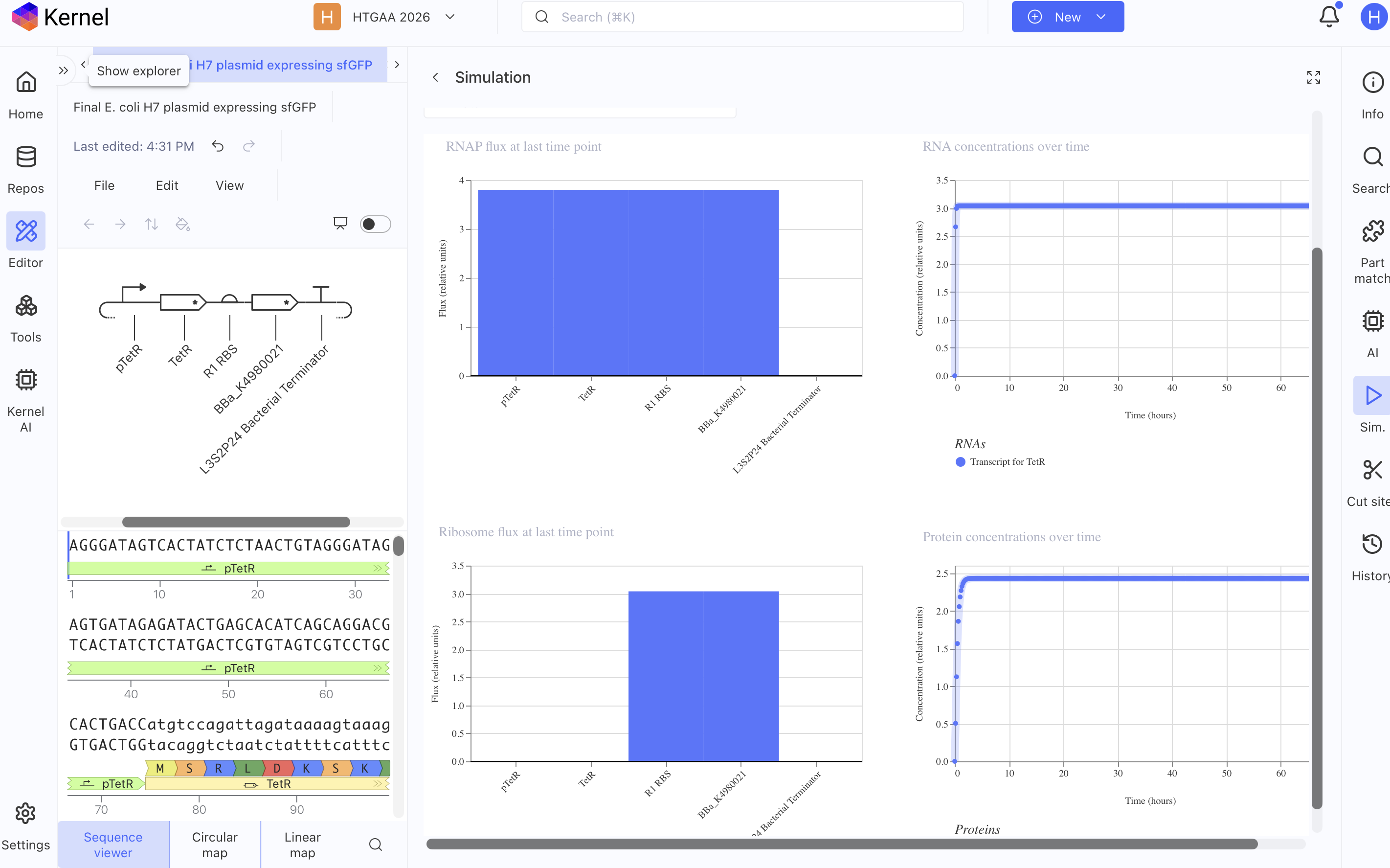
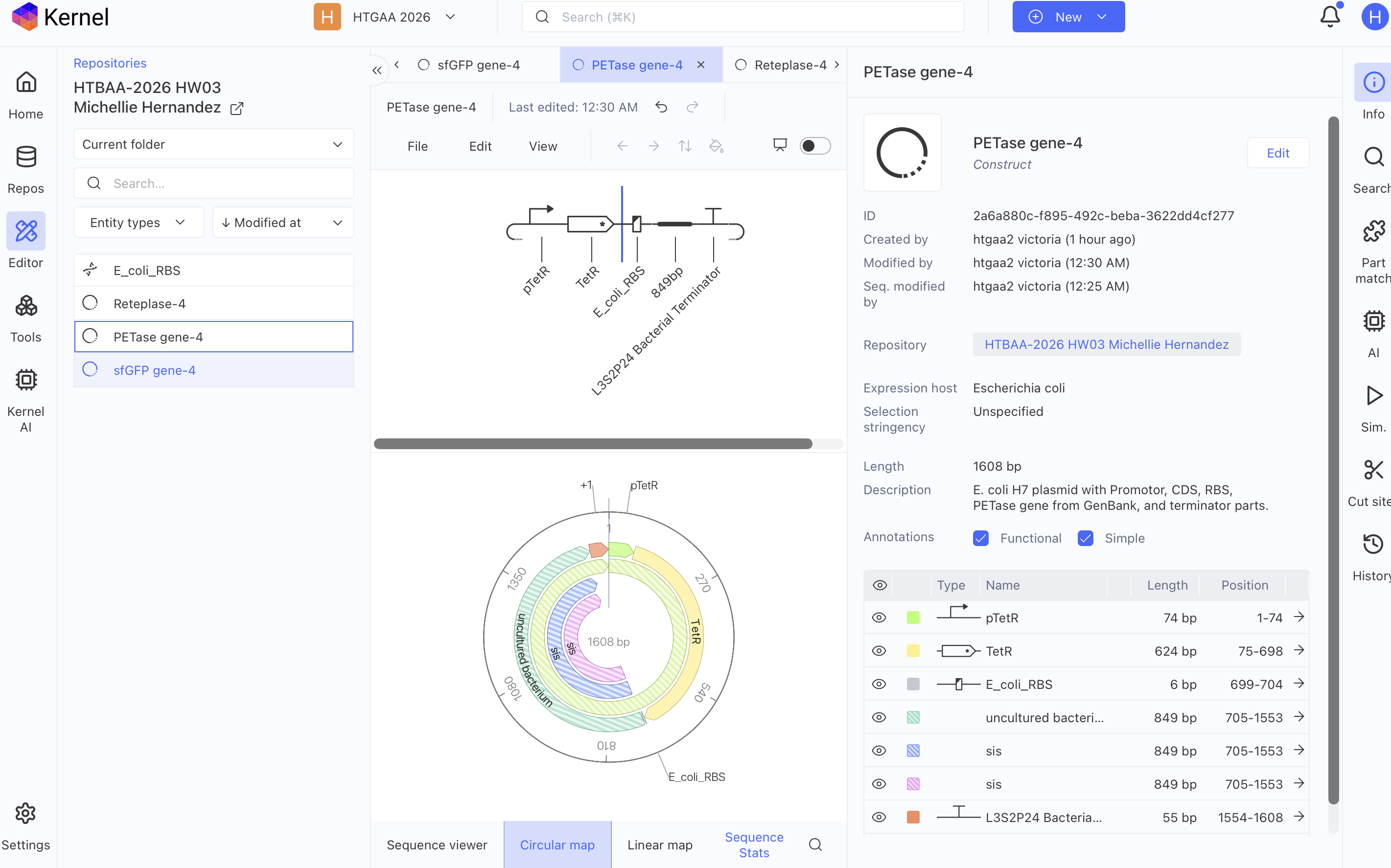
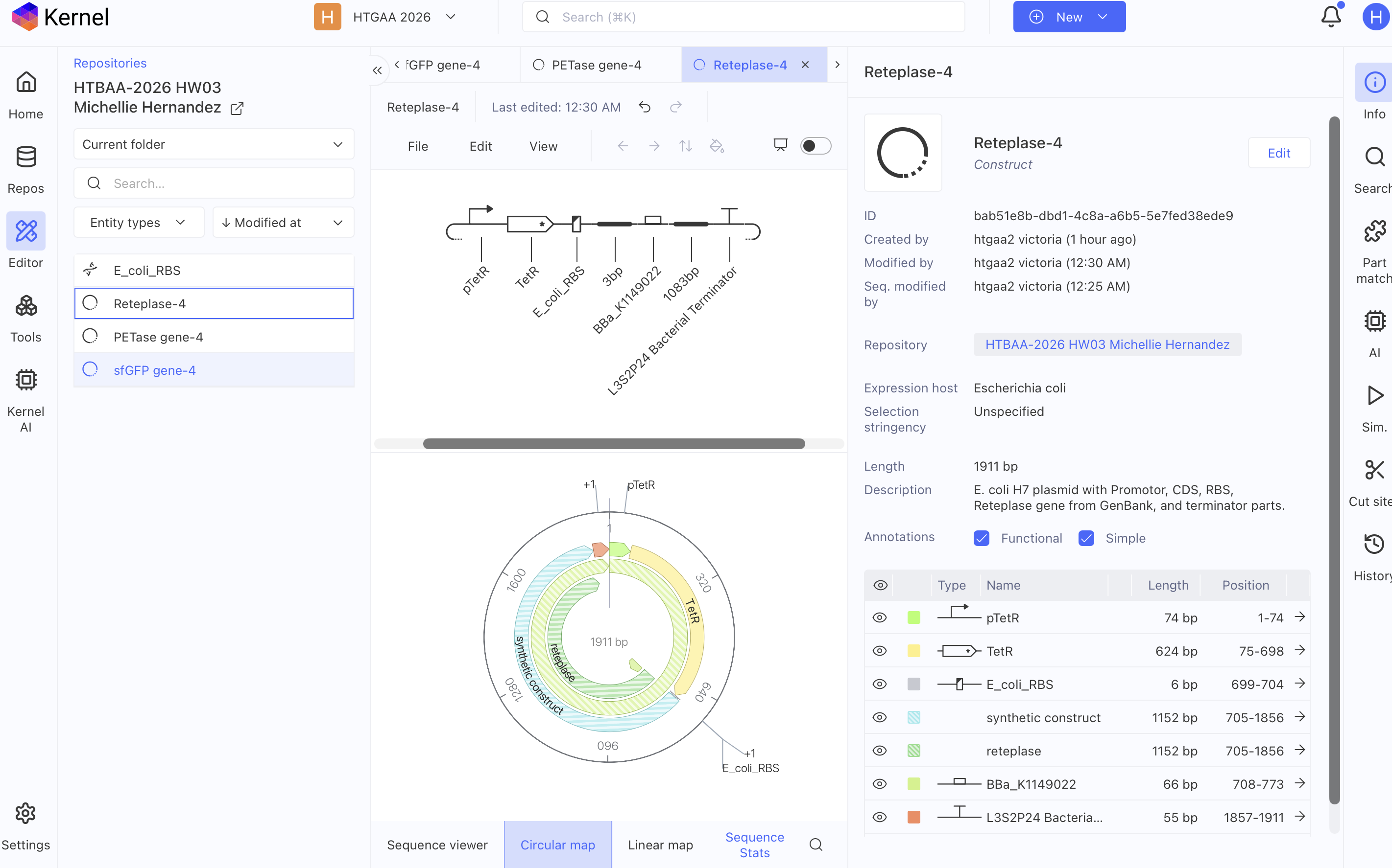
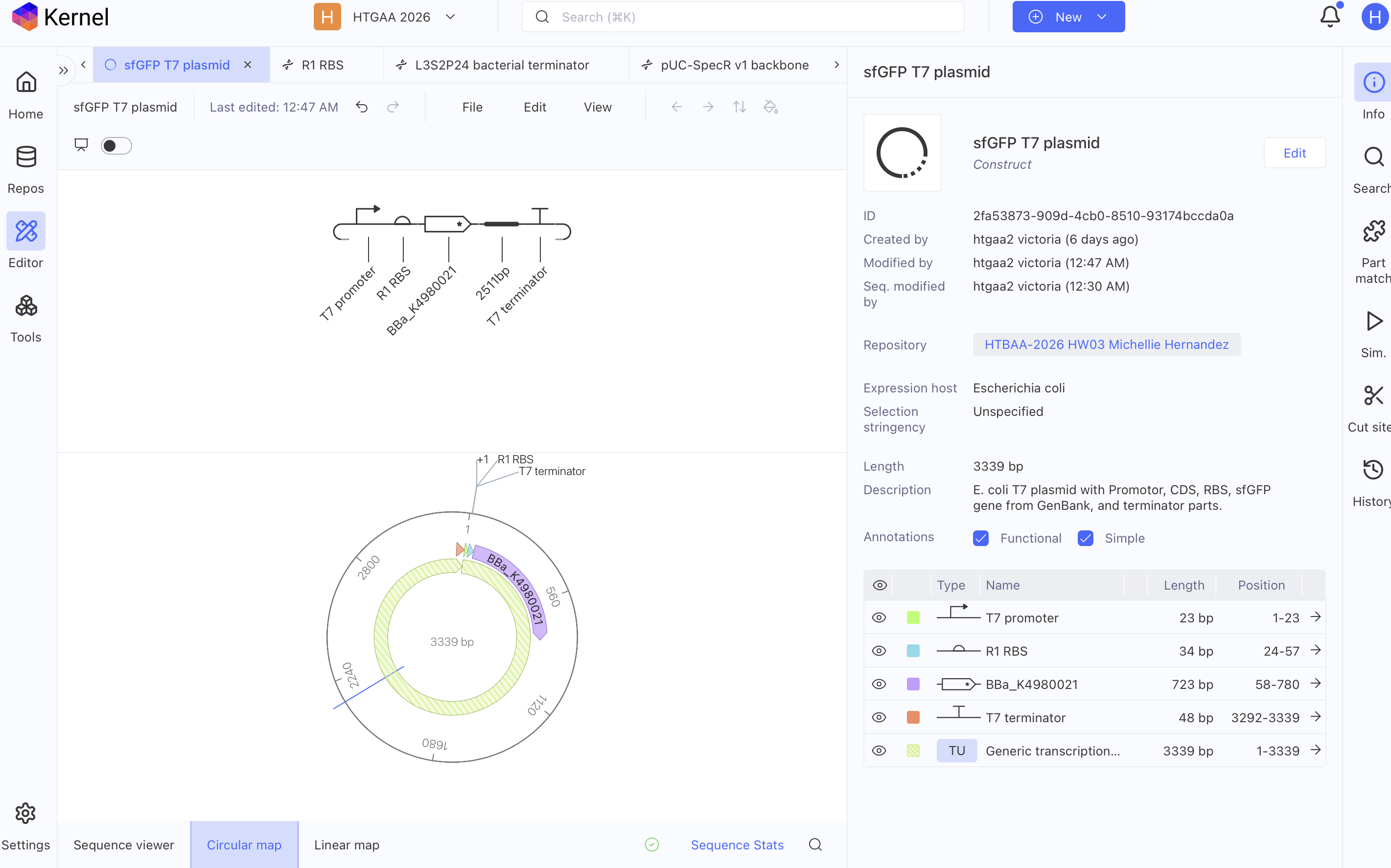
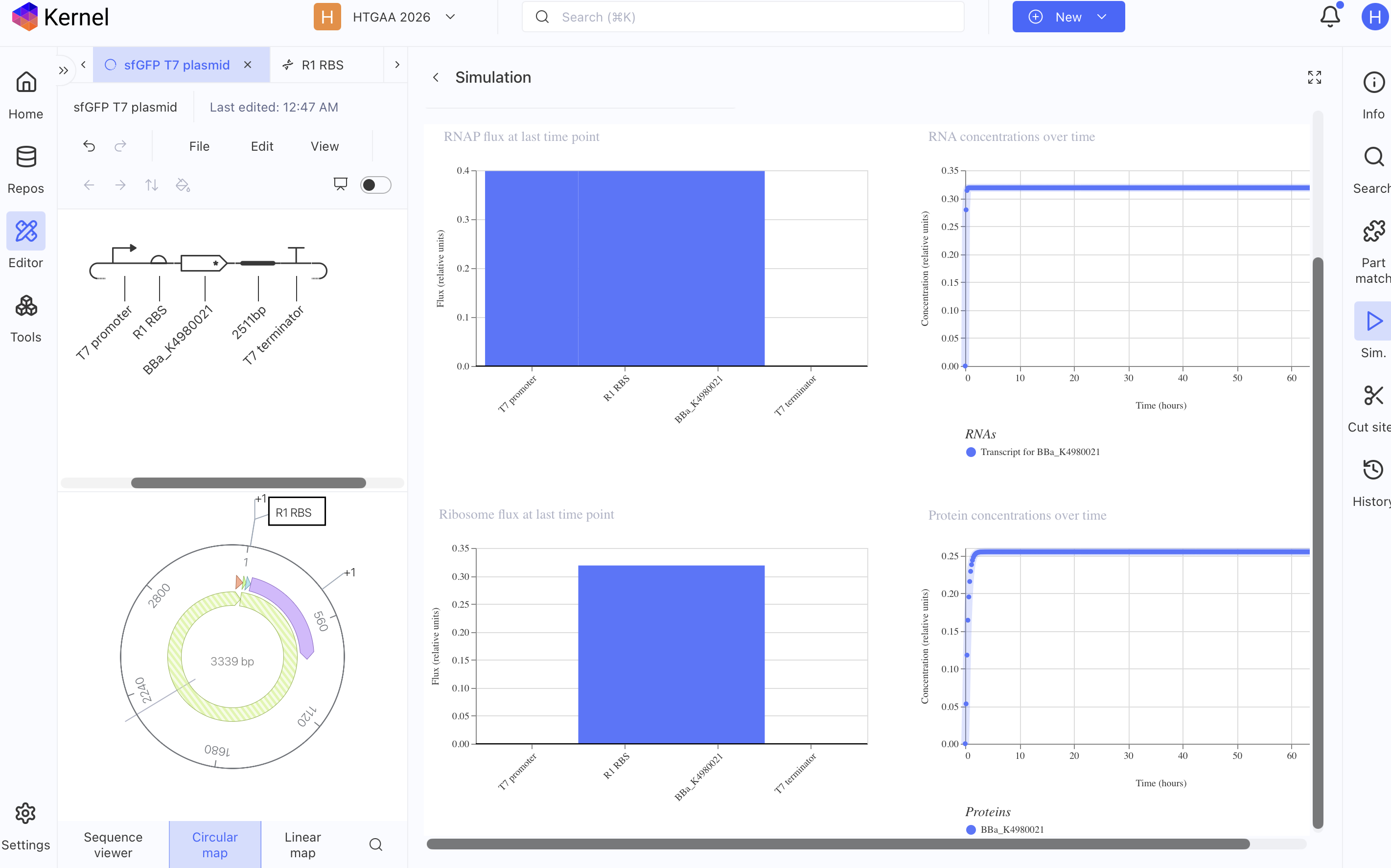
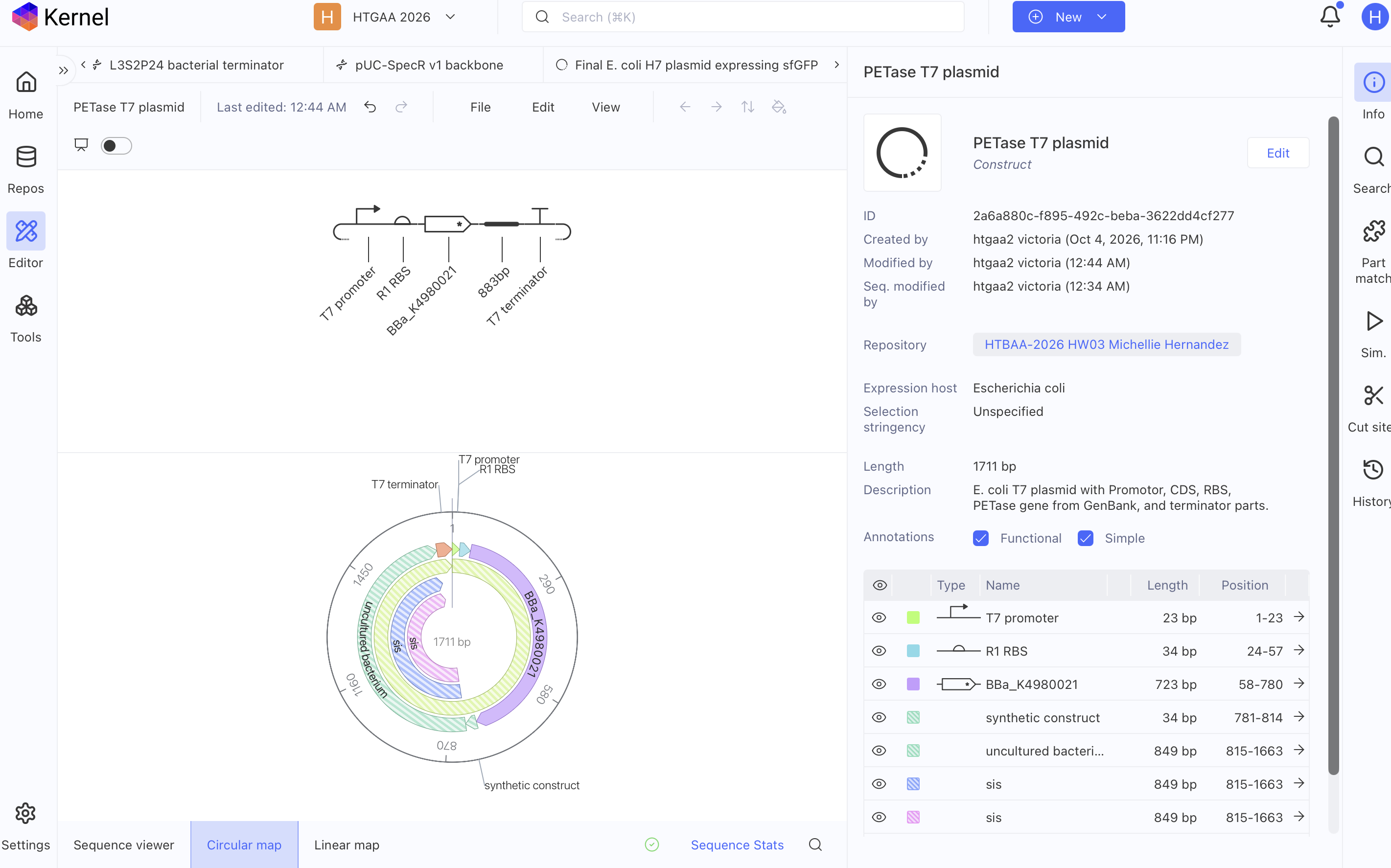
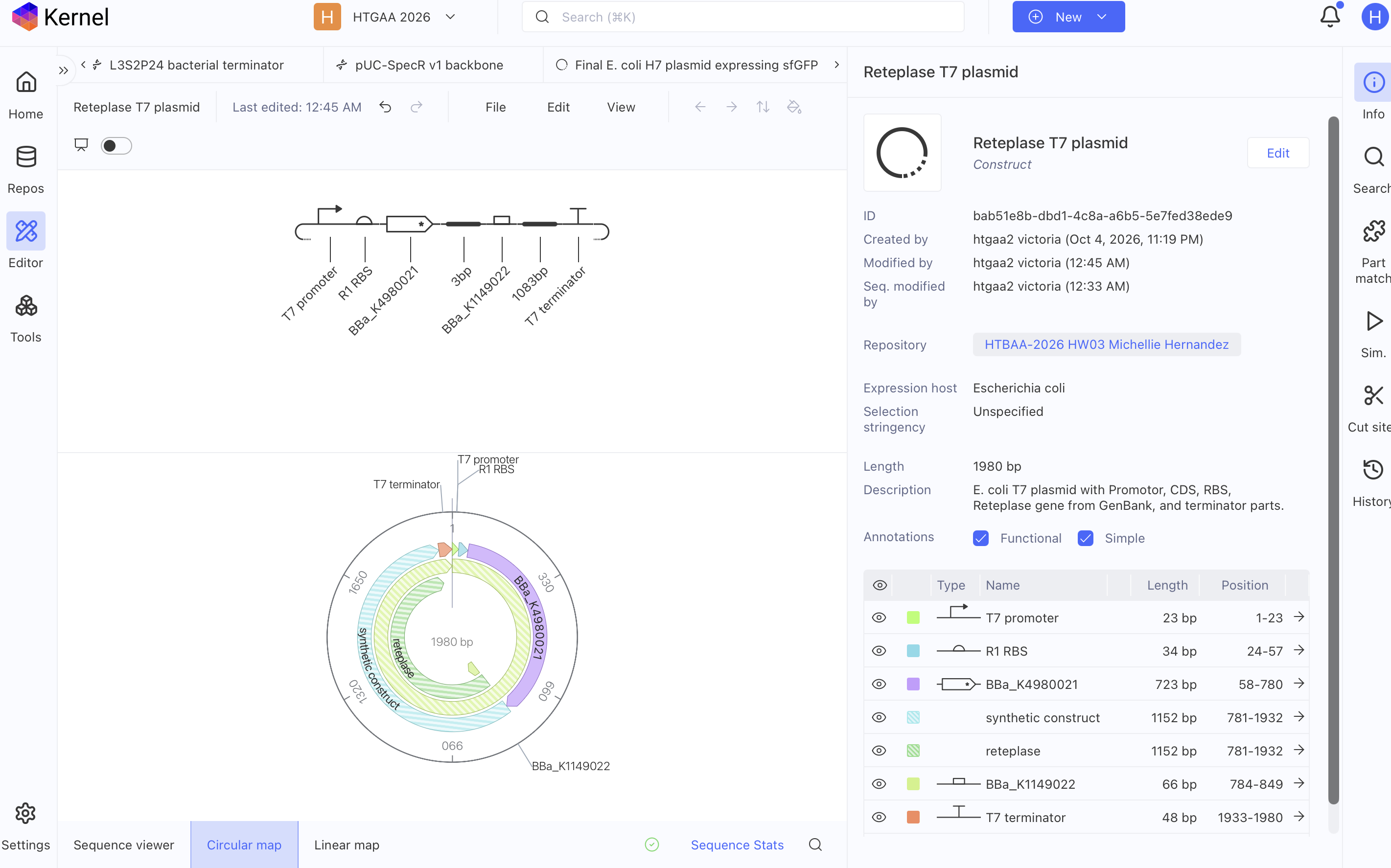
FUNCTIONING T7 PLASMIDS PER KERNEL SIMULATOR

sfGFP T7 Plasmid

image imageimage image

PETase T7 Plasmid

image imageimage image

Reteplase T7 Plasmid

image imageimage image

AI PROMPTS

CHAT GPT

  1. Where does petase protein occur naturally?
  2. What is the composition of the cytosol of Ideonella sakaiensis?
  3. I hypothesize that mimicking the microenvironment composition where the protein occurs naturally in an E. Coli lysate cell free system one might enhance proper folding of that desired protein one is trying to produce, what sources can validate my hypothesis?
  4. What papers state the native jellyfish cytosol’s exact composition?
  5. Does GFP folding occur in the ER or the cytosol of A. Victoria jellyfish?
  6. Where is sfGFP produced naturally and how is it regulated genetically
  7. Where can I find A. Victoria regulatory dna?

MIT Rosalind AI

  1. Papers on the microenvironment of the interior of an Endoplasmic Reticulum.
  2. Summarize in a paragraph Endoplasmic reticulum oxidoreductin (ERO) provides resilience against reductive stress and hypoxic conditions by mediating luminal redox dynamics
  3. Why enzymatic enzyme or chaperon helped manage oxidative stress in the paper Endoplasmic reticulum oxidoreductin (ERO) provides resilience against reductive stress and hypoxic conditions by mediating luminal redox dynamics
  4. In E. coli what functions as an ERO as described in 1. Endoplasmic reticulum oxidoreductin (ERO) provides resilience against reductive stress and hypoxic conditions by mediating luminal redox dynamics — https://www.biorxiv.org/content/10.1101/2021.12.13.472397v1
  5. Paper on E. coli periplasmic disulfide oxidation system (DsbA–DsbC–DsbB/DsbD pathway) regulation
  6. How is a oxidative state stabilized in the E. coli periplasmic disulfide oxidation system (DsbA–DsbC–DsbB/DsbD pathway)
  7. Going back to the Endoplasmic reticulum oxidoreductin (ERO) provides resilience against reductive stress and hypoxic conditions by mediating luminal redox dynamics — https://www.biorxiv.org/content/10.1101/2021.12.13.472397v1 paper what was the ratio of disulfide isomerase and ERO to stabilize the oxidative state
  8. is there a paper that states what was the ratio of disulfide isomerase and ERO to stabilize the oxidative state in an ER
  9. How is H2O2 stabilized in the redox regulatory system in the ER?
  10. In E. coli what functions as glutathione to eliminate H2O2 overproduction in an oxidative state
  11. What papers state the Aequorea victoria jellyfish cytosol’s exact composition

RESOURCES

– Ohlendorf, R., Vidavski, R. R., Eldar, A., Moffat, K., & Möglich, A. (2012). From dusk till dawn: One-plasmid systems for light-regulated gene expression. Journal of Molecular Biology, 416(4), 534–542. https://doi.org/10.1016/j.jmb.2012.01.001

– Ugalde, J. M., Aller, I., Kudrjasova, L., Schmidt, R. R., Schlößer, M., Homagk, M., Fuchs, P., Lichtenauer, S., Schwarzländer, M., Müller-Schüssele, S. J., & Meyer, A. J. (2021). Endoplasmic reticulum oxidoreductin (ERO) provides resilience against reductive stress and hypoxic conditions by mediating luminal redox dynamics. https://doi.org/10.1101/2021.12.13.472397

Final Project

cover image cover image