Week 4 LAB: Cell-Free Manufacturing
Author: Michellie Hernandez
CONTRIBUTING HYPOTHESIS
– I hypothesize that mimicking the microenvironment composition where the desired protein to biomanufacture occurs naturally and adjust the components of its composition to the host lysate that is used to enhance proper folding of the desired protein within the host lysate. In the case of the coopetition, enzymatic ratios of naturally occuring microenvironment composition where the desired protein is produced and folded should be adjusted to functionally equivalent enzymes in the E. Coli. The ratios and proportions of the composition should be similar within the E. coli lysate. I hypothesize that this might enhance proper folding of the protein. Thus for cell free protein synthesis (CFPS) of reteplase the microenvironment to mimic will be the interior of mammalian Endoplasmic Reticulum, while CFPS of Petase the microenvironment to mimic will be the cytosol of Ideonella sakainsis where Petase occurs naturally. In CFSP of sfGFP the natural micro environment composition to mimic will be the cytosol of the Aequorea Victoria jellyfish.
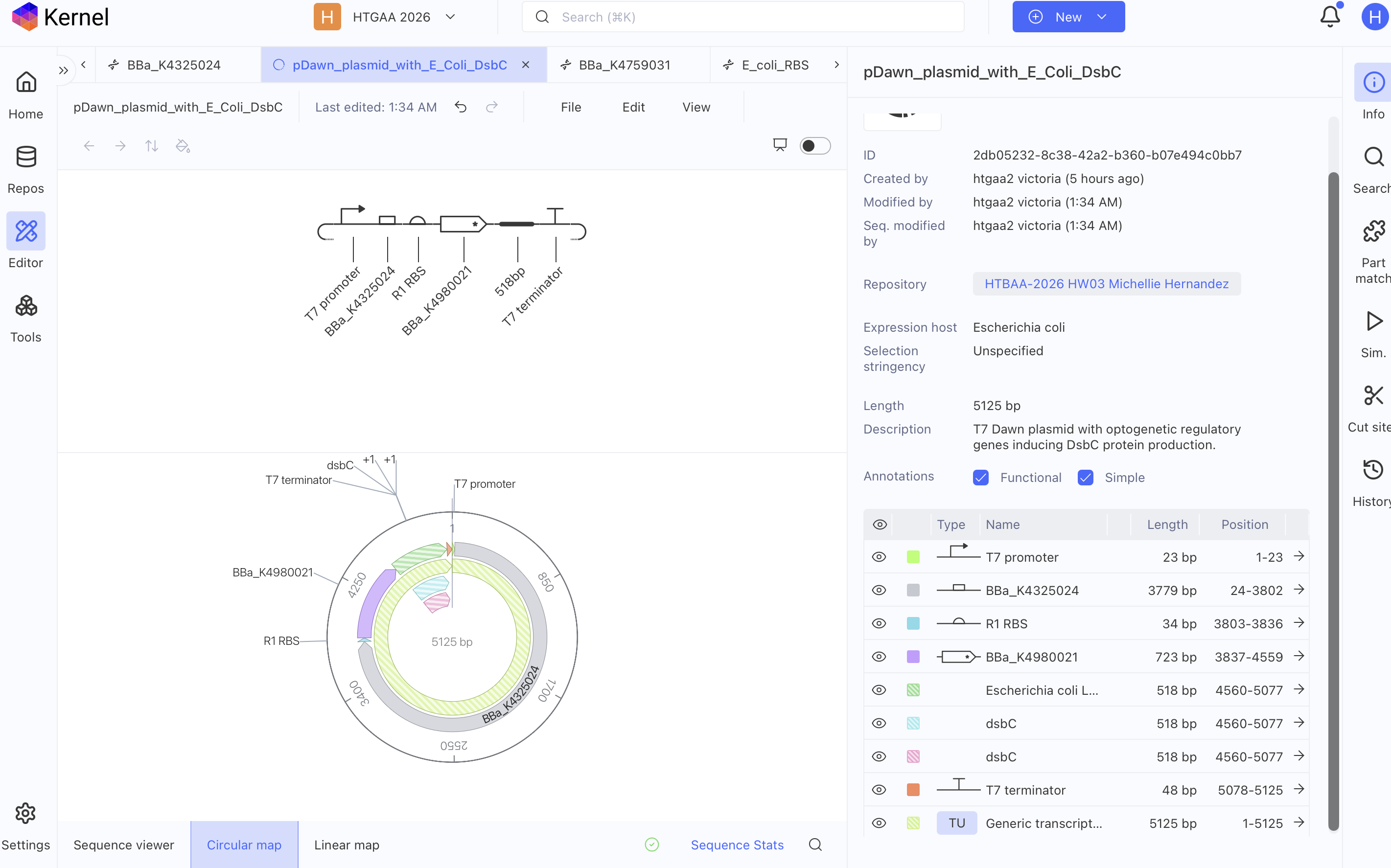
– I hypothesize that the cost of biomanufacturing will reduce upon adding a plasmid with regulatory genes and templates of protein reagents that can be induced with blue light whenever the reagent concentrations are low within the cell free system. I suggest the use of pDusk and pDawn plasmids to use optogenetics for light-regulated gene expression of enzymatic substrates like GSSG and DsbC per resource: Ohlendorf et. al 2014 “From dusk till dawn: one-plasmid system for light-regulated gene expression. Alternatively the regulatory genes of Aequorea Victoria jellyfish that induces GFP naturally in response to blue light can also be used if pDusk or pDawn plasmids are not available.
pDawn T7 Plasmid with E. coli DsbC gene

COMPOSITIONS SUBMITTED
Composition Rank for sfGFP
- Sodium Pyruvate 40.89 mM
- Ammonium Glutamate 10.00 mM
- Magnesium Glutamate 8.85 mM
- Potassium Oxalate 4.38 mM
- 17 Amino Acid Mix 4.00 mM
- Tyrosine pH 12 2.00 mM
- Cysteine 2.00 mM
- Spermidine 1.56 mM
- CTP 1.50 mM
- UTP 1.50 mM
- AMP 1.50 mM
- GMP 1.50 mM
- Putrescine Dihydrochloride 1.25 mM
- Folinic Acid 0.037 g/L
- Water 6.325 uL
REAGENT SUPPLEMENT JSON FOR sfGFP
{ “sample_id”: “michellie_hernandez_2026-10-11_xwi7a8”, “sample_type”: “experimental”, “reagent_list”: { “amino_acid_mix_17”: 1200, “ammonium_glutamate”: 200, “amp”: 300, “base_buffer”: 2000, “ctp”: 300, “cysteine”: 100, “folinic_acid”: 75, “gmp”: 300, “lysate”: 5000, “magnesium_glutamate”: 250, “nuclease_free_water”: 6325, “potassium_oxalate”: 175, “putrescine”: 50, “sodium_pyruvate”: 900, “spermidine”: 125, “template_sfgfp”: 2000, “tyrosine”: 400, “utp”: 300 }, “metadata”: { “ai_model”: “”, “protein_target”: “sfGFP”, “acknowledgements”: “”, “comments”: “sfGFP: Changed pyruvate as energy substrate per cytomim system. Equalized NTP and NMP combination. Substituted ATP for AMP and GTP for GMP, but maintained CTP and UTP due to results in Smith et. al 2026. Role: medical doctor new to Cell free systems.” } }
COMPOSITION RANK FOR PETASE
- Sodium Pyruvate 34.08 mM
- Ammonium Glutamate 10.00 mM
- Magnesium Glutamate 8.85 mM
- Ribose 5.00 mM
- 17 Amino Acid Mix 4.00 mM
- Potassium Oxalate 3.75 mM
- Oxidized Glutathione (GSSG) 1.70 mM
- Spermidine 1.56 mM
- CTP 1.50 mM
- UTP 1.50 mM
- AMP 1.50 mM
- GMP 1.50 mM
- Putrescine Dihydrochloride 1.25 mM
- DsbC (E. coli, 25.5 kDa) 375.0 nM
- Folinic Acid 0.037 g/L
- Water 6.575 uL
REAGEANT COMPOSITION JSON FOR PETASE
{ “sample_id”: “michellie_hernandez_2026-10-11_xwi7a8”, “sample_type”: “experimental”, “reagent_list”: { “amino_acid_mix_17”: 1200, “ammonium_glutamate”: 200, “amp”: 300, “base_buffer”: 2000, “ctp”: 300, “dsbc_ecoli”: 75, “folinic_acid”: 75, “gmp”: 300, “lysate”: 5000, “magnesium_glutamate”: 250, “nuclease_free_water”: 6575, “oxidized_glutathione”: 200, “potassium_oxalate”: 150, “putrescine”: 50, “ribose”: 150, “sodium_pyruvate”: 750, “spermidine”: 125, “template_sfgfp”: 2000, “utp”: 300 }, “metadata”: { “ai_model”: “”, “protein_target”: “sfGFP”, “acknowledgements”: “”, “comments”: “Petase: Combined Jewett and Ribose+NMP system, but with CTP and UTP. As Smith et. al 2026 noted that CMP cannot replace CTP nor UMP cannot replace UTP without affecting the titers. Pyruvate and ribose as energy substrates. Role: medical doctor background new to cell free systems.” } }
COMPOSITION RANK FOR RETEPLASE
- Sodium Pyruvate 32.94 mM
- Ammonium Glutamate 10.00 mM
- Magnesium Glutamate 8.85 mM
- Cysteine 5.00 mM
- Oxidized Glutathione (GSSG) 4.46 mM
- Potassium Oxalate 4.38 mM
- 17 Amino Acid Mix 4.00 mM
- Spermidine 1.56 mM
- CTP 1.50 mM
- UTP 1.50 mM
- AMP 1.50 mM
- GMP 1.50 mM
- Putrescine Dihydrochloride 1.25 mM
- Reduced Glutathione (GSH) 1.00 mM
- Nicotinamide 1.00 mM
- DsbC (E. coli, 25.5 kDa) 1.00 uM
- Folinic Acid 0.025 g/L
- Water 5.500 uL
REAGENT SUPPLEMENT JSON FOR RETEPLASE
{ “sample_id”: “michellie_hernandez_2026-10-11_9p2lna”, “sample_type”: “experimental”, “reagent_list”: { “amino_acid_mix_17”: 1200, “ammonium_glutamate”: 200, “amp”: 300, “base_buffer”: 2000, “ctp”: 300, “cysteine”: 400, “dsbc_ecoli”: 200, “folinic_acid”: 50, “gmp”: 300, “lysate”: 5000, “magnesium_glutamate”: 250, “nicotinamide”: 200, “nuclease_free_water”: 5500, “oxidized_glutathione”: 525, “potassium_oxalate”: 175, “putrescine”: 50, “reduced_glutathione”: 200, “sodium_pyruvate”: 725, “spermidine”: 125, “template_reteplase”: 2000, “utp”: 300 }, “metadata”: { “ai_model”: “”, “protein_target”: “Reteplase”, “acknowledgements”: “”, “comments”: “Reteplase: Adjusted some reagents to be more similar to Jewett cytomim system. Added Nicotinamide to help oxidative stress from H2O2 during oxidative state for reteplase. Equalized concentration of the Nucleotides NTP and NMP Changed energy source to pyruvate to mimic Cytomim system.Role: Medical doctor background new to cell free systems.” } }
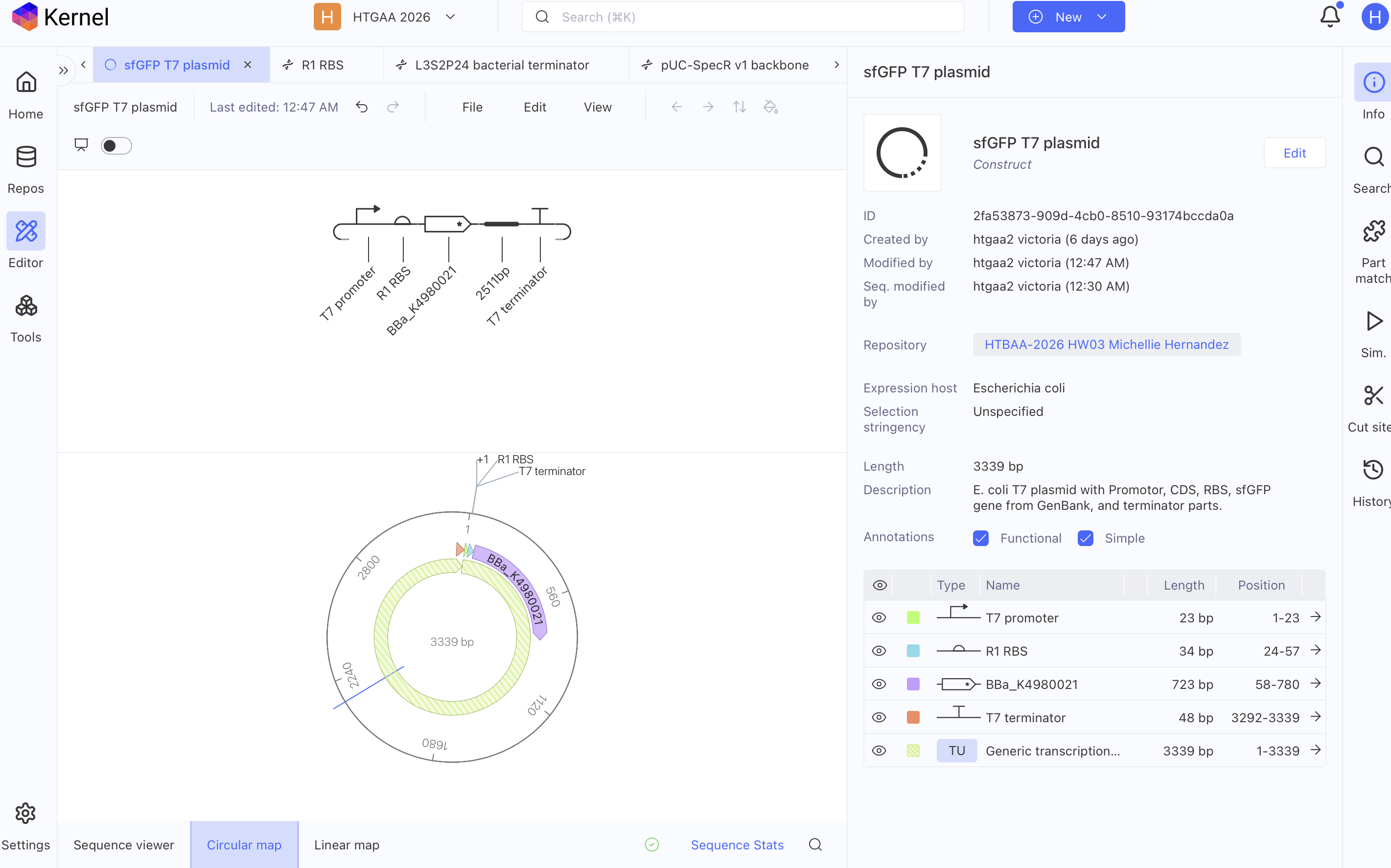
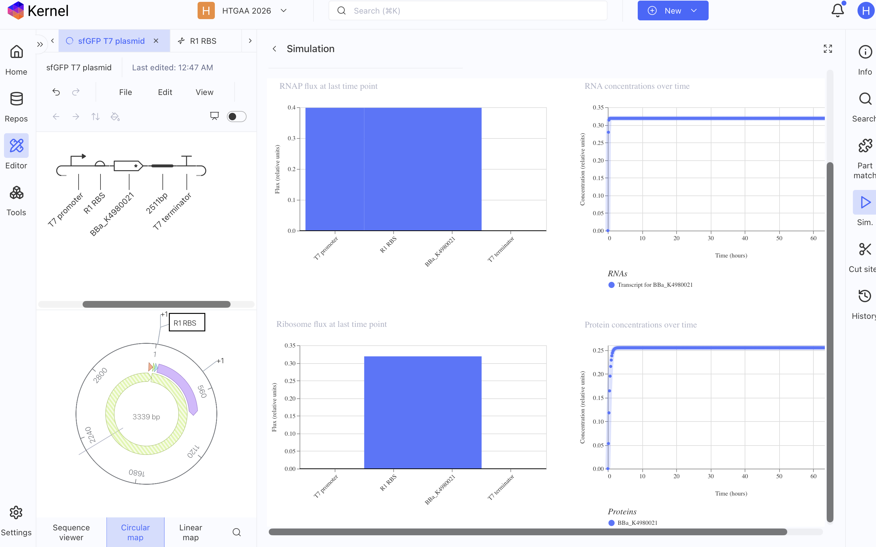
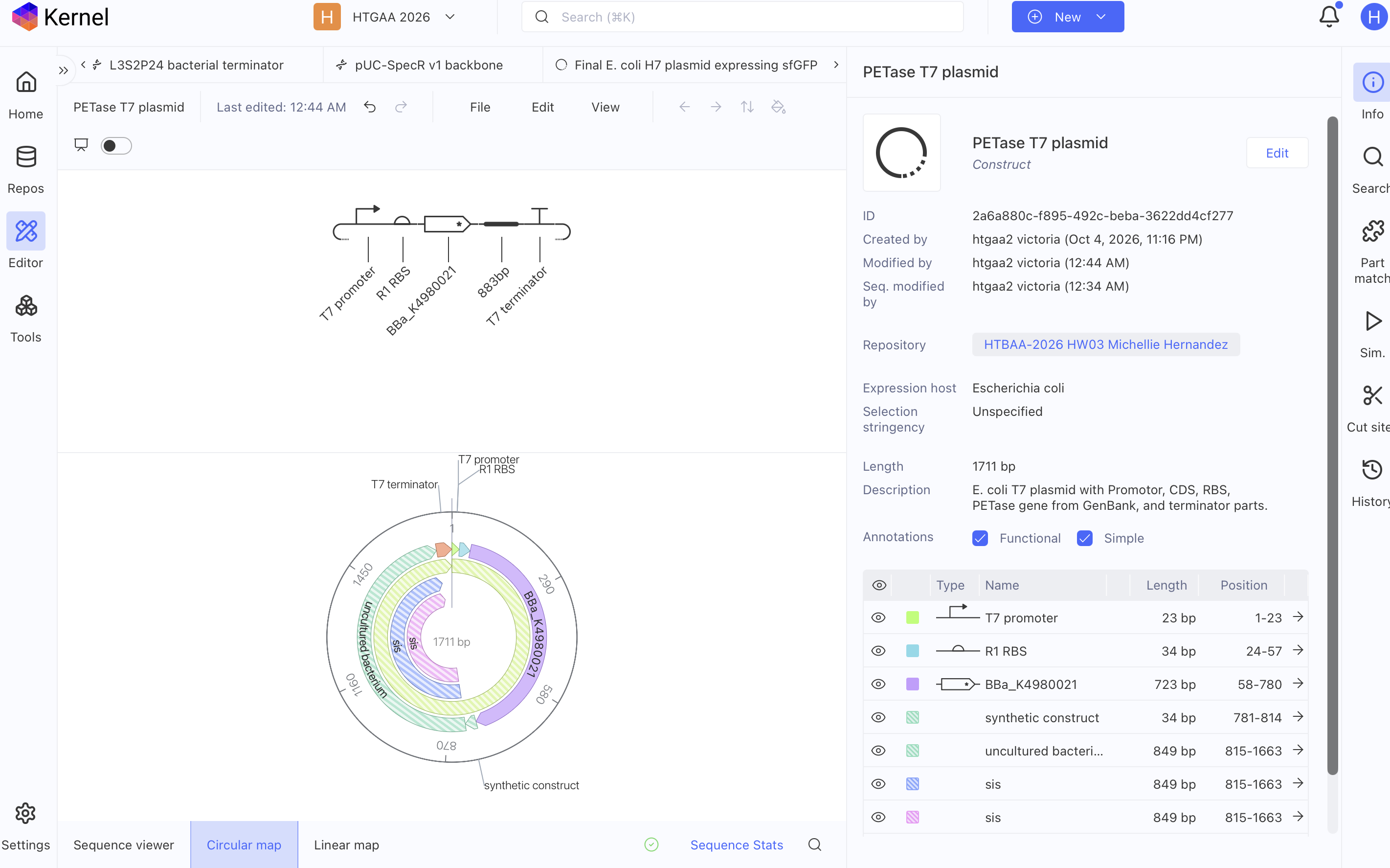
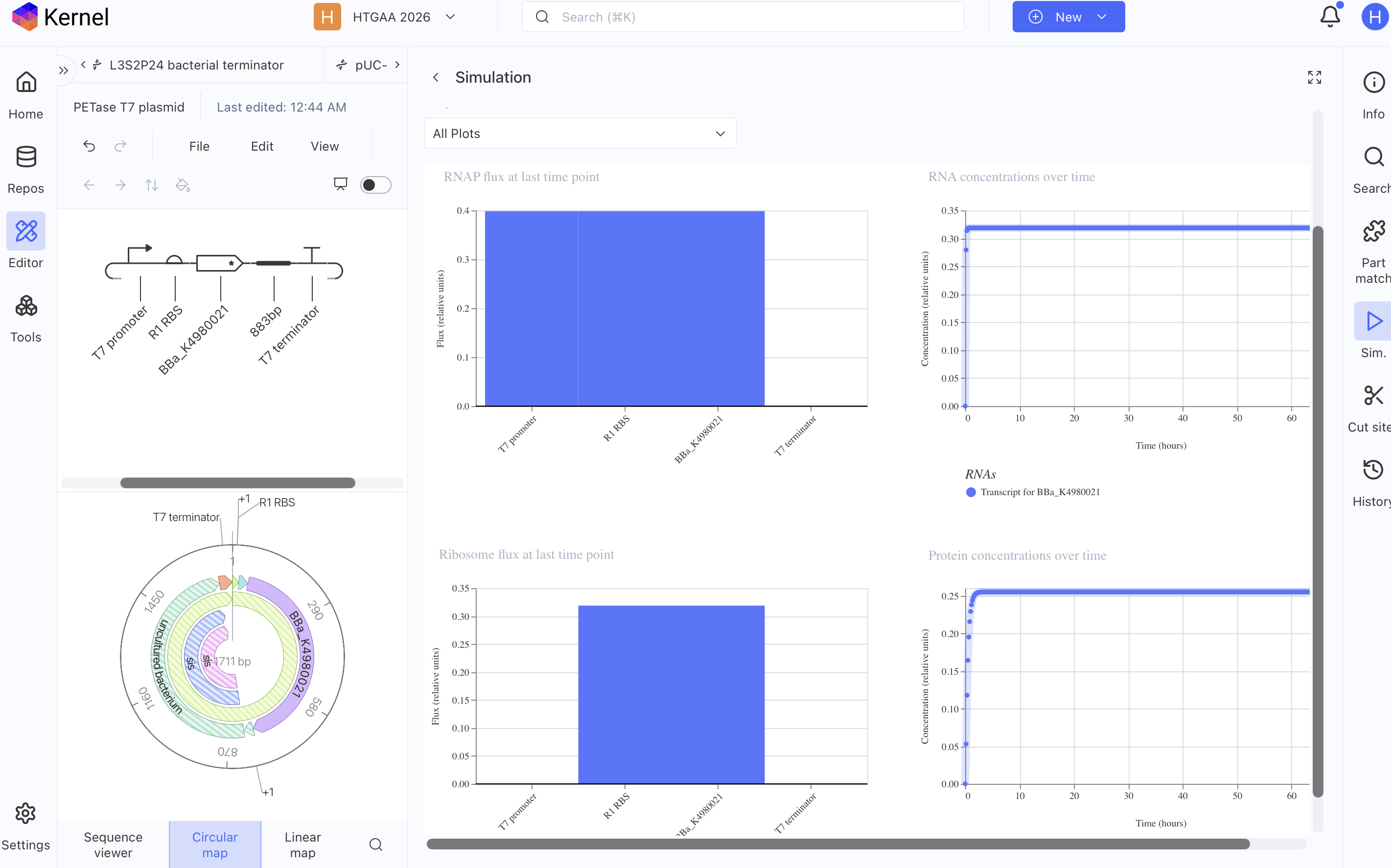
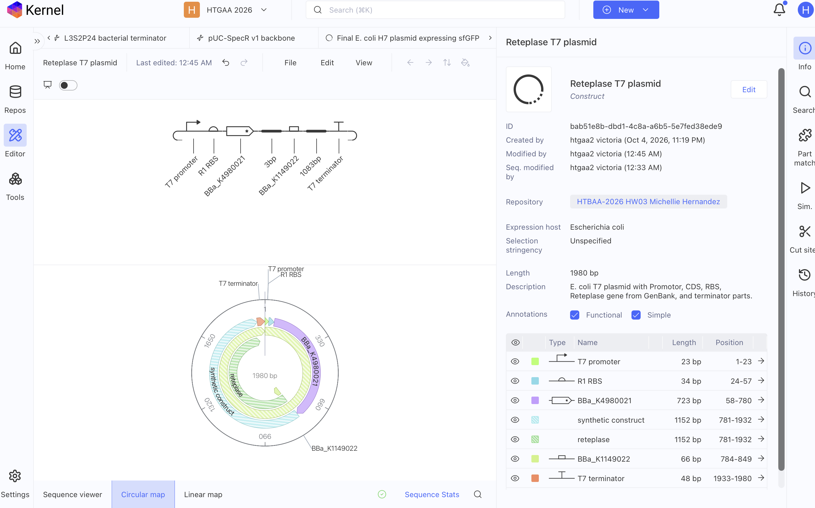
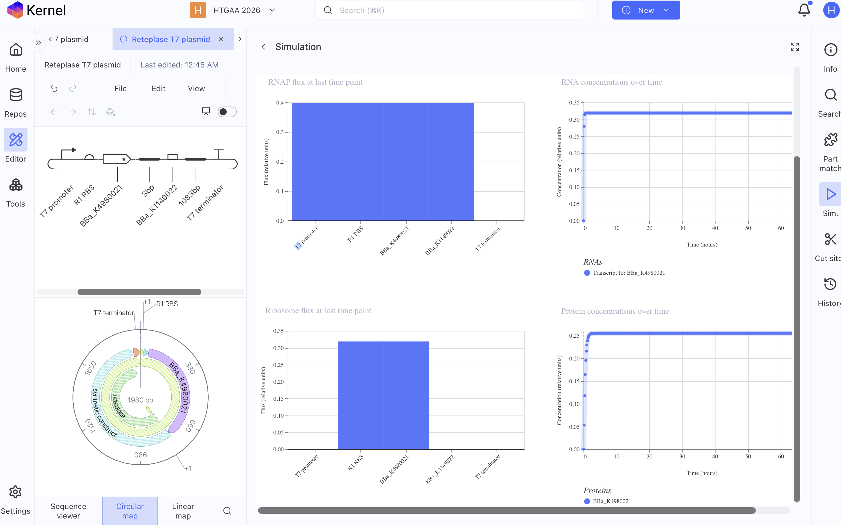
FUNCTIONING T7 PLASMIDS PER KERNEL SIMULATOR
sfGFP T7 Plasmid
PETase T7 Plasmid
Reteplase T7 Plasmid
AI PROMPTS
CHAT GPT
- Where does petase protein occur naturally?
- What is the composition of the cytosol of Ideonella sakaiensis?
- I hypothesize that mimicking the microenvironment composition where the protein occurs naturally in an E. Coli lysate cell free system one might enhance proper folding of that desired protein one is trying to produce, what sources can validate my hypothesis?
- What papers state the native jellyfish cytosol’s exact composition?
- Does GFP folding occur in the ER or the cytosol of A. Victoria jellyfish?
- Where is sfGFP produced naturally and how is it regulated genetically
- Where can I find A. Victoria regulatory dna?
MIT Rosalind AI
- Papers on the microenvironment of the interior of an Endoplasmic Reticulum.
- Summarize in a paragraph Endoplasmic reticulum oxidoreductin (ERO) provides resilience against reductive stress and hypoxic conditions by mediating luminal redox dynamics
- Why enzymatic enzyme or chaperon helped manage oxidative stress in the paper Endoplasmic reticulum oxidoreductin (ERO) provides resilience against reductive stress and hypoxic conditions by mediating luminal redox dynamics
- In E. coli what functions as an ERO as described in 1. Endoplasmic reticulum oxidoreductin (ERO) provides resilience against reductive stress and hypoxic conditions by mediating luminal redox dynamics — https://www.biorxiv.org/content/10.1101/2021.12.13.472397v1
- Paper on E. coli periplasmic disulfide oxidation system (DsbA–DsbC–DsbB/DsbD pathway) regulation
- How is a oxidative state stabilized in the E. coli periplasmic disulfide oxidation system (DsbA–DsbC–DsbB/DsbD pathway)
- Going back to the Endoplasmic reticulum oxidoreductin (ERO) provides resilience against reductive stress and hypoxic conditions by mediating luminal redox dynamics — https://www.biorxiv.org/content/10.1101/2021.12.13.472397v1 paper what was the ratio of disulfide isomerase and ERO to stabilize the oxidative state
- is there a paper that states what was the ratio of disulfide isomerase and ERO to stabilize the oxidative state in an ER
- How is H2O2 stabilized in the redox regulatory system in the ER?
- In E. coli what functions as glutathione to eliminate H2O2 overproduction in an oxidative state
- What papers state the Aequorea victoria jellyfish cytosol’s exact composition
RESOURCES
– Ohlendorf, R., Vidavski, R. R., Eldar, A., Moffat, K., & Möglich, A. (2012). From dusk till dawn: One-plasmid systems for light-regulated gene expression. Journal of Molecular Biology, 416(4), 534–542. https://doi.org/10.1016/j.jmb.2012.01.001
– Ugalde, J. M., Aller, I., Kudrjasova, L., Schmidt, R. R., Schlößer, M., Homagk, M., Fuchs, P., Lichtenauer, S., Schwarzländer, M., Müller-Schüssele, S. J., & Meyer, A. J. (2021). Endoplasmic reticulum oxidoreductin (ERO) provides resilience against reductive stress and hypoxic conditions by mediating luminal redox dynamics. https://doi.org/10.1101/2021.12.13.472397