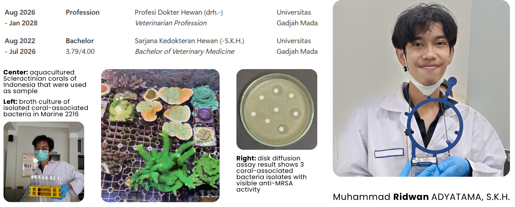
Profession (veterinary medicine) student with interest in genome mining and bioprospecting of marine microbiome microorganism for biomanufacturing of novel drugs. He previously isolated coral-associated bacteria with anti-MRSA secondary metabolite for bioprospecting of novel antimicrobial. As a bachelor of veterinary medicine (S.K.H.) graduate, he is also passionate about One Health and wildlife conservation
See the navigation bar on the left to look through my work for How To
Biomanufacture Almost Anything (Fall 2026)!
Subsections of Muhammad Ridwan Adyatama — HTBAA Fall 2026
Week 1 HW: Final Project Ideas
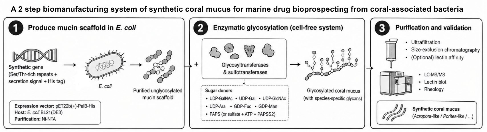
A 2 Step Biomanufacturing System of Synthetic Coral Mucus for Marine Drug Bioprospecting from Coral-Associated Bacteria
Figure 1. Visual abstract of the project
(1) What you would like to biomanufacture and why?
I want to make a synthetic coral mucus platform that can reproduce the important physical and chemical properties of coral mucus without having to continuously collect mucus from living corals. My main reason for making this is related to coral-microbe interactions in the context of coral-associated microorganism bioprospecting. The presence of coral mucus affects secondary metabolite production of its associated microbial community. This challenges bioprospecting research of coral-associated microorganism, as it is often non-viable or doesn’t produce wanted metabolite in conventional culture condition. However, natural mucus is biologically complex and can vary depending on coral species, environmental conditions, and physiological state. This makes it difficult to use natural mucus as a standardized material for experiments.
The synthetic mucus would therefore not just be a replacement for natural mucus. It would become a standardized and modular experimental material that could be changed according to coral species. For example, I could make an Acropora-like mucus with relatively high arabinose, moderate mannose and GlcNAc, and sulfated glycans, and then make another Porites-like mucus with relatively higher galactose. I could then compare how different microbial communities respond to different mucus glycoforms.
I think the most interesting part of this project is that the product can be made using two stages of biomanufacturing:
First, I would use E. coli as a production chassis to manufacture a recombinant mucin-like protein scaffold.
Second, I would take the purified scaffold and glycosylate it using a cell-free enzymatic system containing different glycosyltransferases and sulfotransferases.
The reason I prefer this two-step approach is that I do not expect E. coli to naturally reproduce the complex glycosylation of coral mucus. Instead of trying to genetically engineer the entire glycosylation pathway into E. coli, I can separate protein production from glycan production and make the second stage modular.
In the long term, I want this to become something like a coral mucus “Lego” system, where the same mucin scaffold can be produced repeatedly, while the glycosylation module can be changed to create different coral mucus compositions. This means that theoretically I would not need to develop a completely new manufacturing process for every coral species. I would mainly need to change the glycosyltransferases, sulfotransferases, and sugar donors used in the second stage.
The first proof-of-concept would therefore focus on two mucus types:
Acropora-like synthetic mucus, using a glycan program enriched toward arabinose, mannose, GlcNAc and sulfated glycans.
Porites-like synthetic mucus, using a glycan program with higher galactose, moderate GlcNAc and mannose, and sulfation on galactose.
If this works, the same platform could theoretically be expanded to Favia/Favites-like mucus and potentially other coral species or even different coral physiological states.
Who is it for? Fundamentally, it is for researchers like me who is working on marine natural product discovery, but is can also be used for researchers studying coral microbiomes, coral disease, and coral conservation. In my particular case, I would use it to improve the discovery of coral-associated microbial organisms and potentially identify bacteria with useful secondary metabolites.
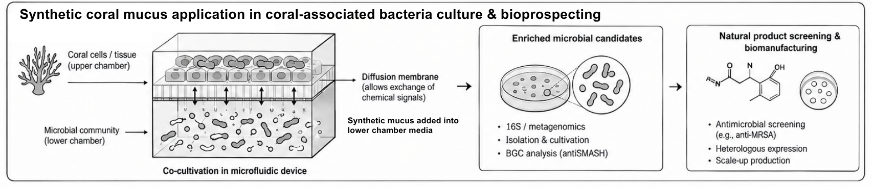
Figure 2. Biomanufactured synthetic coral mucus application in marine-drug bioprospecting
The specific application that I am interested in is to use this synthetic mucus in a host-guided microbial cultivation system, where coral cells and a mixed microbial community are physically separated by a diffusion membrane, but can still exchange diffusible chemical molecules. The idea is that instead of simply growing coral-associated bacteria on conventional laboratory media, I could expose them to a more realistic chemical environment generated by a synthetic coral mucus and eventually by the coral host itself. This could potentially help select for microbial organisms that are associated with the host but are difficult to recover using conventional cultivation.
I would use coral cells or coral tissue on one side of a diffusion membrane and a mixed microbial culture on the other side. The synthetic mucus could be incorporated into this system to provide a defined chemical environment and to test whether particular microbial populations become enriched.
coral cells/tissue ⇄ diffusion membrane ⇄ microbial community
(2) What living system would you use?
The actual production system would use two different systems because the two manufacturing stages have different requirements.
For the first stage, I would use E. coli as the chassis to produce the mucin scaffold. The reason for using E. coli is mainly that it is already a very well-established recombinant protein production system. The synthetic mucin-like sequence could be designed to contain Ser/Thr-rich repeats to provide sites that can later be glycosylated. I would also include a secretion signal such as PelB and a purification tag such as 6×His so that the protein could potentially be recovered from the periplasm and purified using affinity chromatography.
The important thing is that at this stage I would not try to make the final coral mucus. I would only make the protein scaffold. The target would be a recombinant mucin-like protein perhaps in the 50–200 kDa range rather than trying to reproduce the extremely large and heterogeneous size of native coral mucins.
The second stage would be a cell-free enzymatic system. The purified mucin scaffold would be mixed with a selected panel of glycosyltransferases and sulfotransferases, together with the corresponding sugar donors. For example, ppGalNAc-T could initiate mucin-type glycosylation on Ser/Thr, followed by enzymes that add galactose, GlcNAc, fucose, mannose, arabinose, or sulfate groups depending on the desired coral glycan profile.
I think this cell-free stage is especially important because it gives the production system modularity. I could use the same E. coli process to make the mucin scaffold and then produce multiple different mucus types simply by changing the enzymes and donor substrates in the cell-free reaction.
So the overall system would look something like:
Stage 1:
E. coli → recombinant mucin scaffold → purification
Automation would be integral to both the manufacturing process and the research process.
For the manufacturing side, I imagine the whole system as a modular production line. The first module would be automated E. coli cultivation and mucin production. The culture conditions, induction, harvesting, and purification could be standardized so that the same mucin scaffold could be produced repeatedly. After purification, the material would move to a second automated module for cell-free glycosylation.
The second module would basically function like a programmable glycan assembly line. Instead of producing only one type of synthetic mucus, a liquid-handling robot could automatically add different glycosyltransferases, sulfotransferases, and nucleotide-sugar donors according to a predefined recipe.
For example:
Mucin scaffold → Acropora glycan program or Porites glycan program or Favia/Favites glycan program
This could eventually allow me to produce a large library of synthetic mucus compositions without changing the first manufacturing step.
Automation would also be useful for optimization. There are many variables in the cell-free system, including enzyme concentration, donor concentration, enzyme ratios, reaction time, pH, temperature, and order of enzyme addition. Instead of testing these manually one at a time, an automated liquid-handling system could generate many different reaction conditions simultaneously.
I would then combine this with automated quality control. For example, LC-MS could be used to check whether the expected glycan modifications were actually added, while other measurements could be used to check molecular size, aggregation, and viscosity. The results could then be fed back into the next round of reactions.
I also think automation becomes even more interesting when the synthetic mucus is connected to the microbial-selection experiment. A microfluidic device could contain many microbial chambers, each separated from coral cells or tissue by a diffusion membrane. Different chambers could receive different synthetic mucus glycoforms, and automated imaging or sampling could identify which bacterial populations become enriched.
This would allow me to connect biomanufacturing directly to discovery. Instead of producing synthetic mucus only as a final product, the same automated platform could use the product to screen microbial communities.
However, I still do not know exactly which parts of this process would benefit most from automation. In particular, it is not yet clear whether the biggest bottleneck would be mucin production, purification, glycosylation, or downstream quality control.
(4) How much would you need to make, and how long would it take?
How much would I need to make?
Because this synthetic mucus is initially intended as a research and microbial cultivation reagent rather than a therapeutic product, the required amount is much smaller than something like a pharmaceutical protein.
For an initial proof of concept, I would like to be able to run around 10 membrane-separated microbial cultivation experiments for each mucus type.
Assuming each experiment requires around 1 mL of synthetic mucus at 1 mg/mL, each experiment would require approximately 1 mg of synthetic mucus.
This means:
To perform 10 experiments using one mucus type, I would need approximately 1 gram of synthetic mucus.
To make both an Acropora-like and a Porites-like mucus, I would need approximately 2 grams of final product.
If I later expand the platform to four different coral glycoforms, I would need approximately 4 grams of final product for 10 experiments per glycoform.
The biggest production requirement would actually be the first stage, because according to my current expected E. coli production range, the mucin scaffold yield could be around 5–50 mg/L of culture.
Using the 2 gram target as an example:
20 mg : 50 mg/L = 0.4 L
at the high end of the expected production rate, while:
20 mg : 5 mg/L = 4 L
at the lower end.
Therefore, theoretically I would need around 0.40–4 L of E. coli culture to produce enough mucin scaffold for 20 experiments, assuming the final mucus requirement is approximately equivalent to the scaffold requirement. In reality, I would need more because there would be losses during purification and the glycosylation step, and the glycans themselves add additional mass.
How long would it take?
For the first stage, expression and purification would probably take around one week per production batch. The glycosylation-enzyme preparation would initially take longer, around 2–4 weeks, because the enzymes would also have to be expressed, purified, and tested.
Once the enzymes and the mucin production pipeline were already established, the actual glycosylation reaction itself could potentially be completed within 1–2 days, followed by purification and quality control.
The first complete prototype would therefore probably take around 4–6 weeks, mainly because the initial enzyme preparation and optimization would be the bottlenecks. After the system is established, producing another batch of a previously characterized mucus type could potentially be reduced to around 1–2 weeks, especially if several production steps are performed in parallel.
The interesting part is that the same 2 grams of material could be used repeatedly across many experiments. Therefore, unlike manufacturing a therapeutic protein where the ultimate goal is kilograms of product, the important metric here is how many reproducible host-microbe experiments can be supported by each batch of synthetic mucus.
(5) What don’t you know yet?
There are a few crucial things that are still unknown for this project to actually come into fruition.
The exact composition of coral mucin and its glycans is still not completely clear to me. I can design an Acropora-like or Porites-like glycan profile based on available composition data, but this does not necessarily mean that the final synthetic mucus will reproduce the exact molecular structures of native coral mucus.
I do not yet know whether the proposed glycosyltransferases can actually build all of the desired coral glycan structures on the recombinant mucin scaffold. The arabinose component in particular is a major uncertainty because the appropriate arabinosyltransferase may be difficult to identify.
I do not know whether the synthetic mucus will reproduce the biological function of native coral mucus. Matching the monosaccharide composition does not necessarily mean matching the three-dimensional structure, charge, rheological properties, or biological activity of natural mucus.
I do not yet know how effective the membrane-separated co-cultivation system will be at selecting coral-associated microbes. A bacterium enriched by the system might be responding to generic nutrients or stress-related compounds rather than being a genuine coral symbiont.
I do not know how important dynamic chemical exchange is compared with simply using coral-conditioned medium. One of the reasons I want to use the membrane system is that it allows continuous chemical interaction between the host and microbes, but I still need to experimentally demonstrate that this gives a different or more useful selection than static conditioned medium.
The actual production yield of the mucin scaffold is uncertain. The 5–50 mg/L estimate is useful for planning, but real yields will depend heavily on the scaffold sequence, expression system, solubility, secretion efficiency, and purification losses.
Automation is still a major unknown. I know that automation could make the glycan assembly and microbial screening much more scalable, but I do not yet know which parts should actually be automated first or whether a fully automated system would be economically justified.
Real-world inefficiencies are not included in my calculations. The current numbers assume relatively ideal production and do not fully account for failed expression batches, incomplete glycosylation, enzyme degradation, purification losses, or the amount of material needed for quality control.
Generative AI declaration
Generative AI was used to create the two figures of the page. All figures have been reviewed and revised manually to correction.
References
Here are some papers that significantly shaped my idea and used as basis of this page
Couceiro, J. F., Costa, R., & Keller-Costa, T. (2025). Beyond restoration: Coral microbiome biotechnology. In R. S. Peixoto & C. R. Voolstra (Eds.), Coral reef microbiome (Vol. 20, pp. 110–121). Springer. https://doi.org/10.1007/978-3-031-76692-3_15
Jatkar, A. A., Brown, B. E., Bythell, J. C., Guppy, R., Morris, N. J., & Pearson, J. P. (2010). Coral mucus: The properties of its constituent mucins. Biomacromolecules, 11(4), 883–888. https://doi.org/10.1021/bm9012106
Kightlinger, W., Warfel, K. F., DeLisa, M. P., & Jewett, M. C. (2020). Synthetic glycobiology: Parts, systems, and applications. ACS Synthetic Biology, 9(7), 1534–1562. https://doi.org/10.1021/acssynbio.0c00210
Lee, S. T. M., Davy, S. K., Tang, S.-L., & Kench, P. S. (2016). Mucus sugar content shapes the bacterial community structure in thermally stressed Acropora muricata. Frontiers in Microbiology, 7, Article 371. https://doi.org/10.3389/fmicb.2016.00371
Thobor, B. M., Tilstra, A., Mueller, B., et al. (2024). Mucus carbohydrate composition correlates with scleractinian coral phylogeny. Scientific Reports, 14, Article 14019. https://doi.org/10.1038/s41598-024-64828-5
van de Water, J. A. J. M., Tignat-Perrier, R., Allemand, D., & Ferrier-Pagès, C. (2022). Coral holobionts and biotechnology: From Blue Economy to coral reef conservation. Current Opinion in Biotechnology, 74, 110–121. https://doi.org/10.1016/j.copbio.2021.10.013
Wiederkehr, F., Paoli, L., Richter, D., Racunica, D., Ruscheweyh, H.-J., Sperfeld, M., O’Brien, J., Miravet-Verde, S., Streiff, A. B., Ransome, J., Chepkirui, C., Priest, T., Sintsova, A., Salazar, G., Bistolas, K. S. I., Sawyer, T., Labadie, K., Mayer, K.-I., Perdereau, A., Reddy, M. M., … Sunagawa, S. (2026). Coral microbiomes as reservoirs of unknown genomic and biosynthetic diversity. Nature, 652(8110), 686–693. https://doi.org/10.1038/s41586-026-10159-6
1. What molecule are you expressing? A protein, an RNA, or an enzyme (or small set of enzymes) for a pathway. If your HW1 product is a material, food, or chemical, identify the protein(s) that produce it and design for those.
A recombinant protein. An unglycosylated coral mucin-like scaffold designed for subsequent in vitro enzymatic glycosylation. This is the protein backbone that will be added with coral species-specific glycans in the next step of biomanufacturing using cell-free system.
2. What is the sequence? Give the protein or RNA sequence, or the specific gene, you will encode, and say where it comes from (a database accession such as UniProt or NCBI, a known part, or de novo). Cite it.
de-novo sequence based on Thobor et al., 2024 research (DOI: 10.1038/s41598-024-64828-5)
Note that the sequence before does not include pelB signal sequence and codons
2. What host or chassis? The living system from HW1, for example E. coli, S. cerevisiae, CHO or another mammalian line, or a cell-free system. Your host drives almost every part choice that follows.
Escherichia coli (K-12 derivative, e.g., BL21(DE3) or SHuffle for periplasmic expression). It offers gast growth, low cost, scalable fermentation, well-characterized periplasmic secretion via PelB signal, and is compatible with downstream osmotic shock purification
3. Where does the product need to go? Cytoplasm, secreted, membrane, or a compartment? This decides whether you need a signal peptide or targeting sequence.
It’s gonna be localized in perisplasmic space. Periplasmic export via PelB signal peptide enables osmotic shock purification, reducing contamination from cytoplasmic proteins. It also avoids inclusion body formation in cytoplasm (common for repetitive, aggregation-prone proteins). Disulfide bond formation possible in SHuffle strains if needed. Thus, the construct must include an N-terminal PelB signal and be cloned into a vector that supports periplasmic export (e.g., pET22b(+)).
Part 2: Design your genetic parts
Element
Choice
Source
Reasoning
Promoter (+ enhancer / intron if used)
T7 promoter
Benjamin Arias Almeida
Strong, IPTG-inducible T7 promoter for high-level expression in BL21 DE3
etc.
Lac operator
Xiaoyi Feng
Lac operator to control the expression of synthetic coral mucin.
Ser/Thr-rich tandem repeats (≈45% Ser+Thr) to mimic coral mucin acceptor sites plus proline for rigidity, glycine for flexibility
Terminator
T7 terminator
BioClub Tokyo
Strong rho-independent terminator to prevent readthrough transcription into vector backbone.
Part 3: Assemble your construct
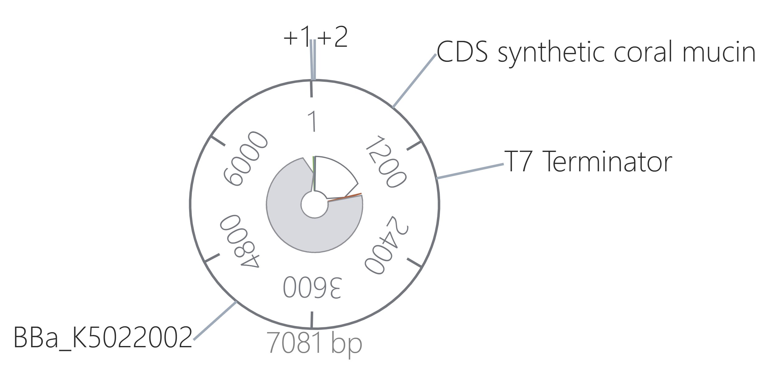
All parts assembled within the pET22b(+) backbone context.
This construct expresses a periplasmic, His-tagged coral mucin scaffold under T7 control. The PelB signal directs secretion to the periplasm, where the protein can be purified by osmotic shock and Ni-NTA chromatography. The T7 terminator ensures clean transcription termination.
Part 4: Optimize and quality-check
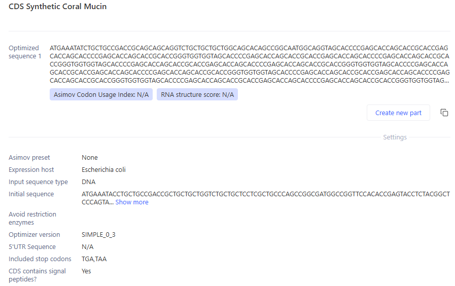
Codon-optimize your CDS for your chosen host. Compare before and after: what changed, and why does codon usage matter for expression in this organism? Note any constraints you applied (e.g. avoiding certain restriction sites, GC content).
E. coli has biased tRNA pools; rare codons (e.g., AGG/AGA for Arg, CUA for Leu) cause ribosome stalling, truncated proteins, and low yields. Codon optimization increases translation efficiency and protein solubility, critical for a repetitive, aggregation-prone scaffold.
The optimization successfully replaced rare codons with E. coli-preferred synonyms. For Tyr, it changed TAC to TAT, for Pro, it changed CCA and CCT to CCG, for Ser it changed the originals to AGC and TCC. All of the changed codons are now optimized as the new ones are preferred in E. coli
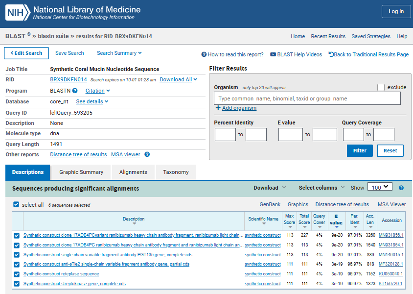
Search your sequence (BLAST / sequence search) against known sequences. Is your CDS what you think it is? Is it identical to a natural gene, or novel? What is the closest match?
There is no exact matches as expected from a de novo synthetic gene. The CDS is novel, not identical to any natural gene. This confirms we are engineering a synthetic mimic, not cloning a native coral gene.
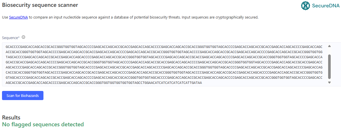
Run the safety screen on your construct. Summarize what it checks for and what it reports. If it flags something, explain whether it is expected for your design and how you would respond.
None as construct contains only standard lab parts. There is no changes needed as the construct passes safety screening for BSL-1 lab use. No toxin, allergen, or pathogenicity concerns.
Part 5: From design to build
How would you build it? Would you order it as synthetic DNA, assemble it from smaller fragments (e.g. Gibson assembly), or build it from a pool of oligos synthesized on a chip? Justify your choice given your construct’s length and complexity. What does synthesis constrain? Synthesis has error rates and struggles with certain sequences. Does your design contain anything hard to synthesize? How would error correction or fragment-based assembly change your approach? Prototype vs. scale. Mirroring HW1: the way you’d build one construct to test your idea may differ from how you’d produce DNA at the scale your product needs. Briefly describe both if they differ.
I would order it as synthetic DNA (from gene synthesis) from a vendor (e.g., Twist Bioscience, GenScript, IDT). Repetitive CDS (20× tandem repeats) is challenging for PCR-based assembly due to recombination and slippage, but commercial gene synthesis providers routinely handle repetitive sequences up to 5–10 kb using chip-based oligo pools and error correction
Constraints might be at repetitive sequences, GC content, homopolymers, and secondary structure.
Error rates of chemical synthesis is about ~1 error per 300–1,000 bp. For the CDS, I expect 1–5 errors per clone. I would sequence 6–10 colonies to find error-free clone, or request clonal isolation + NGS validation from vendor
For prototype scale I would order synthetic CDS as gBlocks or chip-synthesized oligo pool (~$200–500), clone into pET22b via Gibson assembly, transform into E. coli, sequence 6 clones, and express in 50 mL cultures, purify via osmotic shock + Ni-NTA.
For scaled (industrial production, >100 L fermentation), I would do de novo gene synthesis of full plasmid with error-corrected, clonal isolation, remove AmpR (replace with auxotrophic marker or antibiotic-free selection) for regulatory compliance, use high-copy origin (e.g., pUC-derived) for maximal yield, and codon-optimize further based on empirical expression data (e.g., adjust rare codons in repeat regions)
Part 6: What don't you know yet?
Expression level & solubility. Repetitive Ser/Thr proteins often aggregate or express poorly in E. coli, even with codon optimization.
Periplasmic export efficiency. PelB signal may not efficiently secrete a 45 kDa repetitive protein. it could accumulate in cytoplasm or form inclusion bodies.
Glycosylation site accessibility. In vitro GTs may not access Ser/Thr residues if scaffold folds into aggregates or beta-sheets.
Week 3 HW: Cell-Free Reaction Design
ASSIGNMENT: CFPS REACTION DESIGN FOR SFGFP, PETASE, RETEPLASE
Goal: using the collective intelligence of the HTBAA community and accessible AI systems, design novel CFPS
reaction compositions for each of the three target proteins: sfGFP, PETase, and Reteplase to reduce the cost
per gram of functional protein. Each design should be compatible with the experimental framework described
above and should reflect the distinct expression and folding requirements of that protein.
A. COMPARE CELL-FREE REACTION COMPOSITIONS
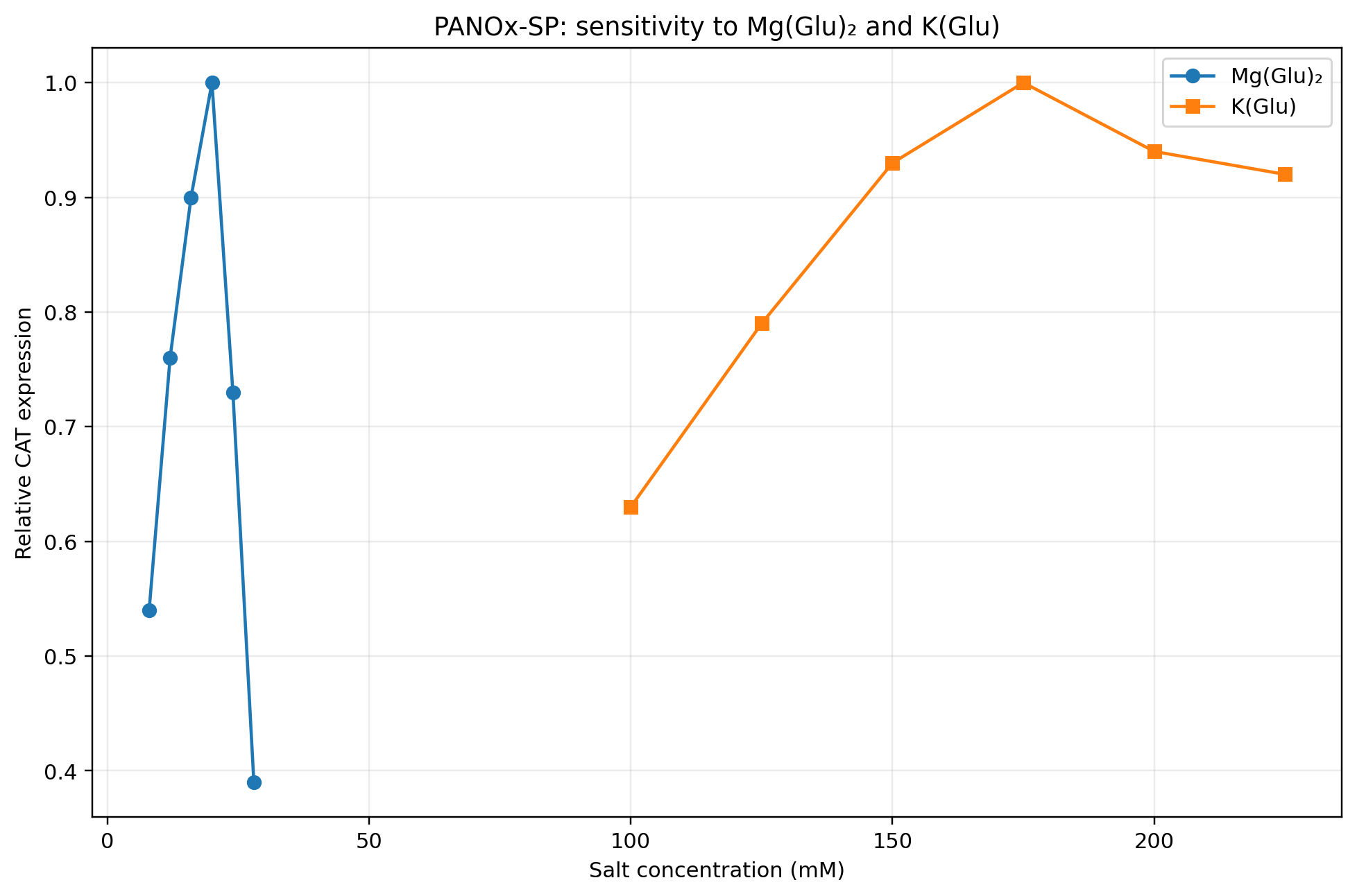
Why does the concentration of potassium glutamate and magnesium glutamate differ between compositions? Which is more sensitive?
The concentrations differ because each formulation establishes a different overall ionic and metabolic environment, and K⁺ and Mg²⁺ are therefore co-optimized rather than fixed at one universal concentration. Magnesium glutamate is more sensitive as its concentration has a much narrower optimum, while potassium glutamate tolerates a broader range around its optimum.
In Jewett & Swartz (2004), the original PANOx system used 200 mM potassium glutamate but no magnesium glutamate, instead using 19.3 mM magnesium acetate. PANOx-SP changed this to 175 mM potassium glutamate and 20 mM magnesium glutamate, while Cytomim used 130 mM potassium glutamate and 8 mM magnesium glutamate. Cytomim also replaced PEP with pyruvate and removed HEPES and PEG. In other words, the salt concentrations were optimized as part of a larger change in the reaction environment, rather than independently fixed values.
Figure 1. Magnesium and potassium concentration in relation to relative expression from Jewett & Swartz (2004).
Magnesium glutamate is clearly more sensitive than potassium glutamate in the optimization data provided.
Explain how a PEP+NTP reaction formulation is different from a Ribose+NMP reaction formulation.
The main difference between PEP+NTP and Ribose+NMP is that these formulations approach nucleotide supply and energy generation in different ways.
In a PANOx-SP PEP+NTP formulation from Jewett & Swartz (2004), PEP is supplied at approximately 30–33 mM, while ATP, CTP, GTP and UTP are also supplied directly. PEP provides energy for ATP generation, but phosphate accumulates as PEP is consumed.
In a RFopt Ribose+NMP formulation from Olsen et al. (2026), RFopt contains 50 mM ribose and AMP, CMP, GMP and UMP, but no externally supplied NTP set or PEP. Optimized formulation could generate NTPs from ribose and nitrogenous bases and that it could maintain ATP production without starting with exogenous ATP.
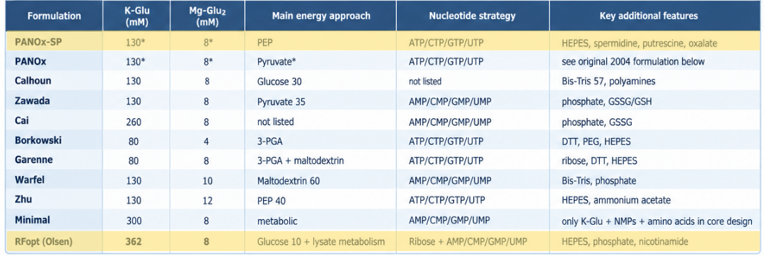
Choose two cell-free reaction compositions from the literature, compare and contrast the choice of reagents and the concentration of reagents.
I chose PANOx-SP from Jewett and Swartz and RFopt from Olsen et al. as two contrasting cell-free reaction formulations. PANOx-SP represents a conventional phosphorylated-energy-substrate approach, whereas RFopt replaces the conventional PEP+NTP strategy with a lower-cost system based on ribose and nucleotide monophosphates. PANOx-SP contains approximately 30–33 mM PEP and directly supplied ATP, CTP, GTP, and UTP, while RFopt contains 50 mM ribose and AMP, CMP, GMP, and UMP but no exogenous PEP or NTP set. RFopt also uses substantially higher potassium glutamate and amino-acid concentrations than PANOx-SP in the Olsen formulation comparison.
Figure 2. Comparison between PANOx-SP (Jewett & Swartz, 2004) and RFopt formulations (Olsen et al., 2026)
The difference is especially striking in cost and yield. Under the standardized testing performed by Olsen et al., PANOx-SP produced 1.11 ± 0.13 g/L sfGFP at a reagent cost of $4,083/g protein, whereas RFopt produced 2.39 ± 0.28 g/L at only $60/g protein. Therefore, RFopt simultaneously increased yield and reduced reagent cost by approximately two orders of magnitude. The tradeoff is that RFopt depends more heavily on the lysate’s endogenous metabolism, whereas PANOx-SP directly supplies PEP and NTPs.
B. DESIGN ONE REACTION COMPOSITION FOR EACH OF THE THREE PROTEINS
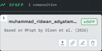
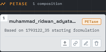
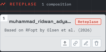
Sign in to coopetition.htgaa.org and publish your first unique 20 µL CFPS reaction composition for each of the following (don’t worry, you can resubmit later):
As a HTBAA research team, we will have the opportunity to suggest new reagents to add to the cell-free reagent inventory. Contribute (or upvote) at least one reagent to the “Community Reagents” section of the website and provide a short rationale. Include in your homework documentation how you decided on that specific reagent and provide links to any relevant research.
Figure 3. Community reagents contribution of mine is upvote for polyphosphate and FkpA
I upvoted polyphosphate because the Smith et al. study identified NTP/ATP regeneration as an important design consideration and specifically listed polyphosphate among potential additional reagents. Unlike simply increasing the concentration of an existing nucleotide substrate, polyphosphate could provide another route for phosphate transfer during energy regeneration. The current formulation work demonstrates that changing the energy and nucleotide-generation strategy can substantially reduce CFPS cost, so polyphosphate would be a useful reagent to test in the same optimization framework.
I also upvoted FkpA since the Reteplase formulation already includes DsbC and FkpA reduce aggregation and works with DsbC to improve antibody folding and assembly in a cell-free system
Visit the cfps-coopetition forum page to ensure you will be able to take part in the discussion. Bonus:
send a note in the channel about a CFPS optimization idea that you have! If you would like clarification
on aspects of the project, please message in this dedicated channel.
Done!
C. ASIMOV KERNEL
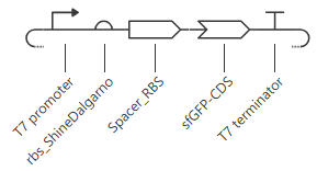
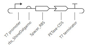
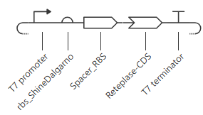
Build T7 compatible plasmids for cell-free expression using the amino acid coding sequences below:
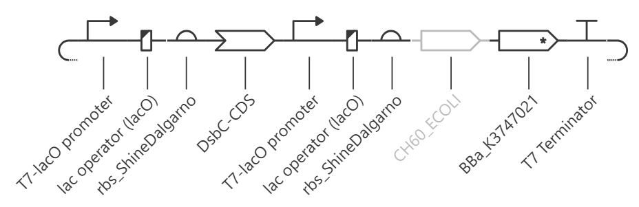
Bonus question: design a T7 helper plasmid that you could add to the cell-free reaction to improve the
yield of functional reteplase (general chaperones, disulfide-bond machinery, etc).
I tried to design a pACYCDuet-1 based helper plasmid with two separate T7 cassettes (MCS1 and MCS2), each with its own promoter and RBS. A clean design is:
MCS1: DsbC (cytosolic)
MCS2: GroEL and GroES as a single operon-like insert under one promoter.
Together with its co-chaperonin GroES, chaperonin GroEL plays an essential role in assisting protein folding (PubMed:2897629, PubMed:2573517, PubMed:10532860, PubMed:1676490, PubMed:8104102, PubMed:9285593, PubMed:16751100, PubMed:18418386, PubMed:18987317, PubMed:20603018, PubMed:24816391). The GroEL-GroES system forms a nano-cage that allows encapsulation of the non-native substrate proteins and provides a physical environment optimized to promote and accelerate protein folding, probably by preventing aggregation and by entropically destabilizing folding intermediates (PubMed:16751100, PubMed:18418386, PubMed:18987317, PubMed:20603018, PubMed:24816391). Rapid binding of ATP, followed by slower binding of the non-native substrate protein and GroES to the cis open ring of GroEL initiates productive folding of the non-native protein inside a highly stable GroEL-ATP-GroES complex (PubMed:9285593, PubMed:9285585, PubMed:19915138, PubMed:22445172). Binding of ATP and GroES induces conformational changes that result in the release of the substrate protein into a nano-cage compartment, within the GroEL central cavity, for folding in isolation (PubMed:8861908, PubMed:9285585, PubMed:16684774, PubMed:22445172).
Reference
Jewett, M.C. & Swartz, J.R. (2004). Mimicking the Escherichia coli cytoplasmic environment activates long-lived and efficient cell-free protein synthesis. Biotechnol. Bioeng., 86: 19-26. https://doi.org/10.1002/bit.20026
Olsen, M.L., Copeland, C.E., Sundberg, C.A. et al. (2026). Design-driven optimization of low-cost reagent formulations for reproducible and high-yielding cell-free gene expression. Nat. Commun., 17: 3478. https://doi.org/10.1038/s41467-026-69605-8
Smith, A. A., Wong, E. L., Donovan, R. C., Chapman, B. A., Harry, R., Tirandazi, P., … & Reuel, N. F. (2026). Using a GPT-5-driven autonomous lab to optimize the cost and titer of cell-free protein synthesis. bioRxiv. https://doi.org/10.64898/2026.02.05.703998