Qingyun Liu — HTBAA Fall 2026
About me
Contact info
Homework, Labs, and Projects
See the nav bar on the left to look through my work for How To Biomanufacture Almost Anything (Fall 2026)!
See the nav bar on the left to look through my work for How To Biomanufacture Almost Anything (Fall 2026)!
HTBAA Homework 1
I want to make a soft patch for people who lift and move heavy objects at work. It would be worn on the upper arm and would combine two kinds of measurements in one circular design. A small reusable electronic core in the center would use light to measure heart rate and pulse shape. A replaceable ring around it would collect sweat and measure sweat rate, sodium level, and skin temperature. The Bio-skin would be designed to last for three work shifts, while the core would be removed and charged after each 8–12 hour shift. The patch would help a worker notice patterns related to heat, hydration, and physical effort, and provide timely reminders when signs of increasing strain appear.
The part touching the skin would be called the Bio-skin. It would be made partly from a soft sheet of cellulose grown by bacteria. The Bio-skin could carry printed artwork, so it could look more like a temporary tattoo or personal accessory than a medical device. My goal is to combine a useful workplace wearable with self-expression.
The whole device would be modular. Later versions could use other removable cores, such as a movement sensor or a sensor that measures muscle activity.
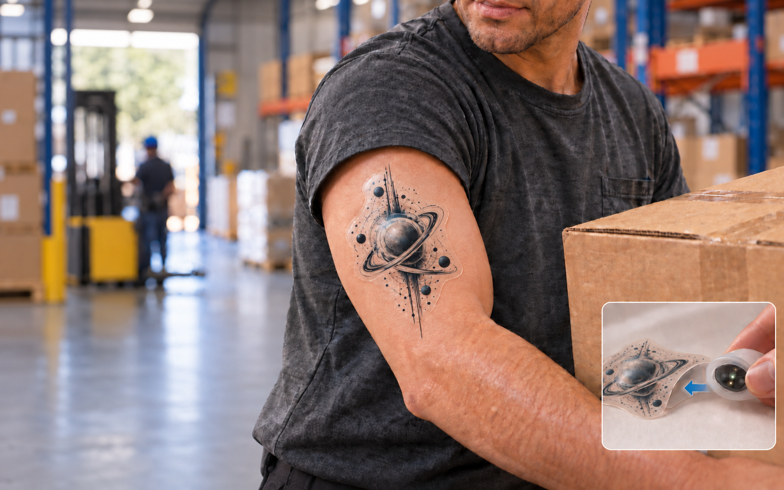
Warehouse, construction, factory, and delivery workers may repeat the same lifting tasks for many hours. Their bodies also respond differently to heat and heavy work. Many wearable devices look technical or medical. I am interested in whether a modular wearable can reduce unnecessary replacement, feel more personal, and still provide useful information.
The design is inspired by Swatch. The electronic core would stay the same, but the outer Bio-skin could change in color, pattern, and artwork. In the future, artists could create different collections. The worker would be the person wearing the patch, while an employer or safety team might buy the system. This means privacy and consent would be important. A worker should know what is being measured and who can see the data. The system should support personal awareness and workplace safety, not hidden productivity monitoring or unsupported medical diagnosis. It also have potential to be a fashion item.
I want to biomanufacture the replaceable skin-facing part of a modular wearable. The Bio-skin would be about 70 millimeters in diameter and worn on the outside of the upper arm. It would contain:
The fluid channels are very small paths that guide fresh sweat from the skin to the sensors.
| Part | Use cycle | Purpose | |
|---|---|---|---|
| Central electronic core | Reusable | PPG light sensor, small computer, Bluetooth, and rechargeable battery. It is removed and charged after each work shift. | |
| Bio-skin | Material and art layer | Three shifts | Soft bacterial-nanocellulose base, artist-designed graphic, central optical opening, opaque fluid-resistant barrier, and a pocket for the 25–30 millimeter core. |
| Bio-skin | Sweat-sensing ring | Three shifts | Direct sweat inlets, ring-shaped small channels, printed sensors for sweat rate and sodium, and contact for skin-temperature measurement. |
| Bio-skin | Adhesive edge | Three shifts | A patterned medical-grade adhesive border that holds the complete Bio-skin against the skin. |
The three Bio-skin rows describe parts of one replaceable patch, not three separate products. The base, sweat-sensing ring, and adhesive edge would be assembled together, so the user could peel off and replace the complete Bio-skin at once. They can still use different materials: bacterial nanocellulose for the soft base and an existing medical-grade adhesive for the skin-contact border.
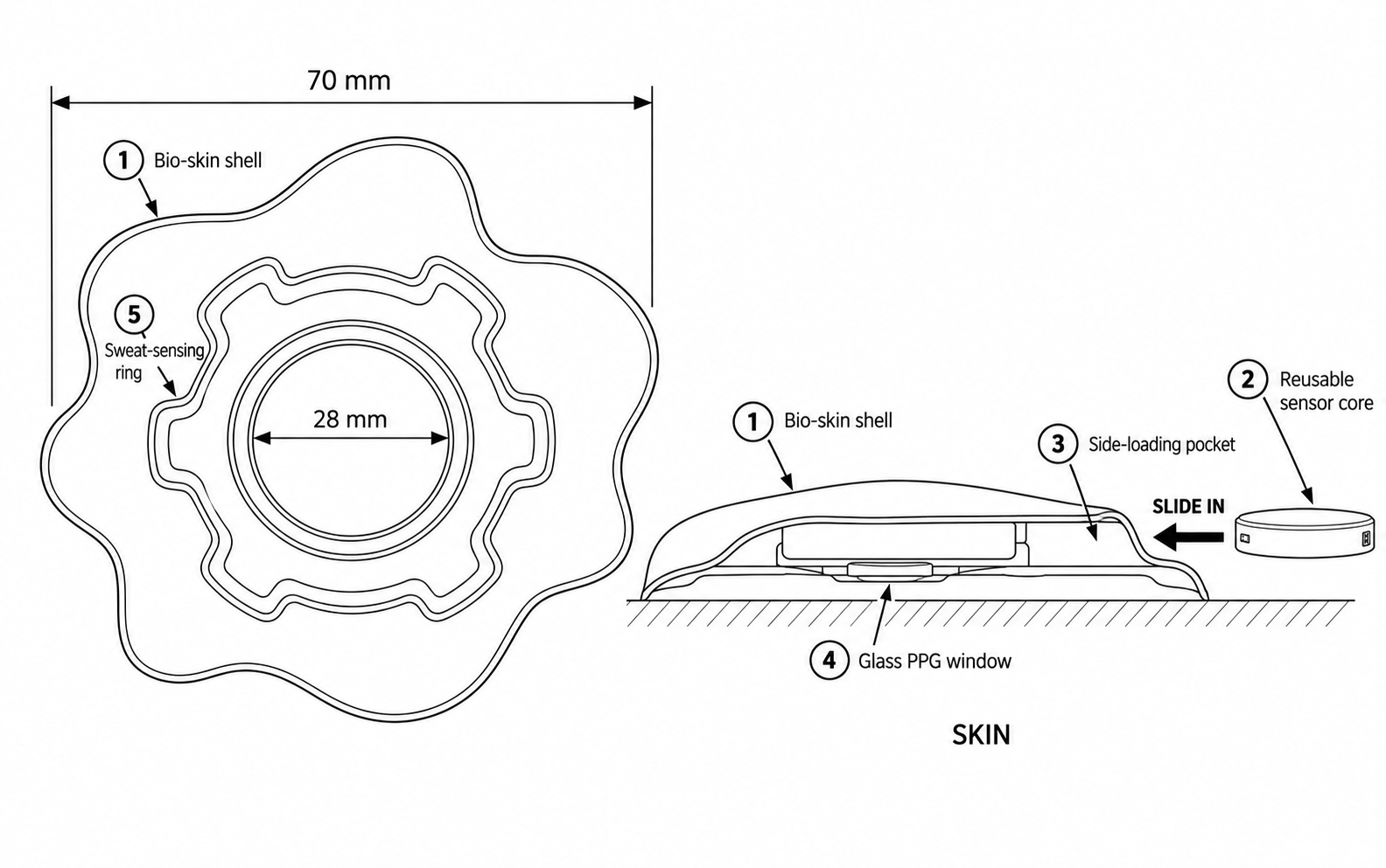

The patch has a circle within a circle design. The reusable sensor core is in the center. A fluid resistant ring around it keeps sweat away from the light path. The sweat sensing area forms the outer ring, and the adhesive sit around the outer edge. On the surface, it is the artwork.
PPG uses a small light and a light detector to observe changes in blood volume under the skin. So, the electronic core would sit above the Bio-skin, but its optical face would reach the skin through a direct opening or a tested transparent window. Studies suggest that the upper arm can be a useful place for PPG, although rapid full-body movement can still create errors [1].
The sweat sensors must also reach fresh sweat. A solid cellulose or gel layer placed between the skin and sensor could dilute sweat, hold sodium, mix old and new sweat, or slow the reading. The sweat inlets would therefore touch the skin directly and guide sweat through thin water-attracting channels. Research devices have used controlled channels and a replaceable sweat layer connected to reusable electronics [2,3].
For three-shift use, sweat cannot remain trapped in the sensing ring. A small channel could move sweat past the sensors toward a drainage or evaporation area so that newer sweat continues to arrive. Other sweat patches have used capillary force and evaporation for continuous flow [4]. This idea still needs to be tested with my material.
The core would work for one 8–12 hour shift and charge overnight. To save battery power, it could measure PPG during selected time windows, read the sweat sensors at intervals, and send data in groups. Lifting creates movement errors in PPG, so the core should also report when the signal is unreliable. A small movement sensor could help remove those periods from the results.
The first version would still use a conventional soft circuit board, silicon chips, LEDs, a light detector, Bluetooth, and a rechargeable battery. Its adhesive and conductive sensor inks would also be conventional materials. The biomanufactured material would provide the skin-contact surface, sweat collection and transport, a base for the printed sensors, and the artwork. It would not replace all of the electronics.
In this project, the living producer would be bacteria from the genus Komagataeibacter. The culture would need water, sugar, other nutrients, oxygen, bacteria, and shallow trays or shaped molds. The bacteria make very small cellulose fibers. Near the air and liquid surface, the fibers join together and form a sheet called bacterial nanocellulose. Researchers have made sheets of 0.2 square meters or more and have also guided the material to grow with surface patterns [6,7].
The basic process would be:
The bacteria would act as the factory, but the final patch would not contain living bacteria. This feels more realistic for a first version because a living patch would need food, water, containment, and safety controls while it is worn. The amount and quality of cellulose will depend on the food source, oxygen, tray shape, and growth time. Bacterial nanocellulose has already been explored as a thin base that carries body fluids toward flexible electrolyte sensors [8].
For early experiments, I would use a small liquid-handling robot. It could prepare many small cultures while changing one condition at a time, such as the amount of sugar, pH, or growth time. A camera could record how quickly each sheet grows and whether it has holes, shrinkage, or uneven areas. I would then test thickness, strength, bending, water uptake, sweat movement, sodium recovery, and whether printed sensors stay attached. Automation would make the experiments more consistent and let me compare many recipes [11].
A production system would use different machines from the early experiments. The main steps could be:
The electronic core would still use normal electronics manufacturing and automated testing for the PPG, battery, wireless connection, sensor contacts, and protection from sweat. Early automation would help me discover a good design. Production automation would help make the same design consistently.
I will define a meaningful scale as enough patches for 1,000 workers for one year. I assume that each worker has 250 work shifts each year, one Bio-skin lasts three shifts, 10 percent extra finished patches are kept as reserve, and 85 percent of the pieces made pass quality control.
A 70 millimeter patch has a radius of 3.5 centimeters. Its area is approximately 3.14 × 3.5 × 3.5, or 38.5 square centimeters.
If one shallow tray provides 0.1 square meter of growth surface, one weekly group would need about 80 trays. After the line is operating, 52 overlapping weekly production groups would make about 108,000 gross pieces in one year. The estimate of 50 grams of dry cellulose per square meter is only an early assumption. These calculations do not yet include all cutting waste, failed cultures, cleaning time, or the space between trays. They are mainly useful for understanding the rough size of the production system.
One production batch might take:
The total time for one batch would be about eight to twelve days. Different batches could overlap, so a small factory could still finish one group each week.
I estimate about twelve weeks for a basic bench demonstration:
This would be a bench prototype, not a clinically tested product. So, testing the patch on people would require a separate safety plan, informed consent, and the appropriate institutional approval.
After the material and sensor design are fixed, I estimate another six to twelve months to build and test a repeatable small production system. Its long-term target would be about 92,000 Bio-skins that pass quality checks each year.
I do not yet know how to grow bacterial nanocellulose with consistent thickness, softness, and strength, or how to shape it into the sweat channels, central opening, and side-loading pocket that my design needs. I need to learn how to culture Komagataeibacter and grow bacterial nanocellulose with consistent thickness, softness, and shape. How culture conditions affect the material and whether these structures should be grown directly, molded, cut, or added later? I would measure thickness, flexibility, shrinkage, strength, and liquid transport across multiple samples.
I do not yet know how to assemble the Bio-skin, sweat channels, printed sensors, adhesive, artwork, and electronic-core pocket into one working prototype. The layers must stay together without blocking sweat, disturbing the PPG light path, or allowing sweat to enter the electronics. I would first test each part separately and then measure leakage, sensor response, optical alignment, and pocket durability in an integrated prototype.
I do not know whether the patch can provide useful signals while a worker is lifting and sweating, or whether it can remain comfortable and attached for three work shifts. Sweat may become trapped or mixed with older sweat, bacterial nanocellulose may change the sodium reading, and movement may disturb the PPG signal. I would test the patch with artificial sweat, repeated arm movement, repeated core insertion, and extended wear before considering a human study.
I used OpenAI GPT5.6 Sol as a discussion and writing tool while developing this proposal. I began with the problem of workers becoming tired or hurt while lifting heavy objects and an idea for a Band-Aid-like product with a reusable core. I asked whether sweat sensing and PPG could be placed in the same patch. Through the discussion, I chose a circle-within-a-circle structure: a PPG sensor in the center and a sweat-sensing ring around it.
I also asked where the electronic core should sit and whether sweat could pass through a lower biological material layer. The discussion helped me understand that both sensing areas need a clear path to the skin. The PPG needs an optical opening, while the sweat ring needs direct collection openings and small channels. We then narrowed the first version to sweat rate, sodium, skin temperature, heart rate, and pulse waveform. We decided not to claim that sweat lactate alone can measure muscle fatigue, and not to promise blood pressure or medical-grade blood oxygen measurement.
We compared a living patch, a cell-free patch, and a non-living material grown by bacteria. I chose to use bacteria to manufacture the cellulose sheet, then wash the cells out before the patch is worn. We also discussed how often to replace the Bio-skin. I first considered replacing it after every work shift, but that seemed expensive. I changed the goal to one Bio-skin for three shifts, while removing and charging the electronic core each night.
Examples of prompt topics I used were:
HTBAA Homework 2
My biomanufactured material is bacterial nanocellulose (BNC). BNC is not a protein; it is an extracellular polysaccharide composed of β-1,4-linked glucose chains. It will serve as a common biological material platform for two possible downstream applications:
Because the final material is a polysaccharide, my direct expression target is the set of proteins that synthesizes, transports, and organizes bacterial cellulose. I will focus on the BcsA–BcsB–BcsC–BcsD cellulose-synthesis complex, encoded by the bcsABCD operon.
The four proteins have different roles:
For Homework 2, I will treat these four proteins as one functional expression target because producing a useful extracellular nanocellulose material requires more than the catalytic activity of BcsA alone.
I will use the continuous native bcsABCD operon region from Komagataeibacter xylinus E25. This 9,113 bp region is located at positions 474401–483513 in GenBank record CP004360.1. The record is associated with genome assembly GCA_000550765.1 and BioProject PRJNA190024.
| Protein | Gene locus | CDS length | Protein length | NCBI Protein ID |
|---|---|---|---|---|
| BcsA | H845_451 | 2,238 bp | 745 aa | AHI24410.1 |
| BcsB | H845_452 | 2,436 bp | 811 aa | AHI24411.1 |
| BcsC | H845_453 | 3,981 bp | 1,326 aa | AHI24412.1 |
| BcsD | H845_454 | 471 bp | 156 aa | AHI24413.1 |
Sequence source: The bcsABCD sequences were obtained from the NCBI genome record for Komagataeibacter xylinus E25: GenBank CP004360.1, genome assembly GCA_000550765.1, BioProject PRJNA190024, and BioSample SAMN03081456. The individual protein accessions are AHI24410.1, AHI24411.1, AHI24412.1, and AHI24413.1. (National Center for Biotechnology Information [NCBI], n.d.)
The complete four-gene region is 9,113 bp. I will preserve the native coding sequences for the initial design because the genes come from the same organism that I have selected as the host. This avoids unnecessary sequence changes and helps preserve the native codon usage and expression balance among the four proteins. I will still run Kernel’s codon-optimization tool later to compare the original and optimized sequences, but I will not assume that the automatically optimized version is biologically superior.
The native bcsABCD region was imported into Kernel as one 9,113 bp operon module using the “Miscellaneous” part type. The positions of bcsA, bcsB, bcsC, and bcsD are documented in the part description rather than displayed as four separate Kernel parts. Keeping the region continuous preserves the native overlaps and short regions between the genes. Appendix A records the source accessions and coordinates needed to identify the sequence.
My proposed host is Komagataeibacter xylinus E25.
I selected this strain because it naturally produces bacterial nanocellulose and has a complete reference genome. Using a native cellulose-producing host is more realistic than transferring the entire synthesis, transport, and crystallization system into a non-cellulose-producing organism such as E. coli.
The host already contains a native cellulose-synthesis system. For this course design, I will construct a controlled additional bcsABCD expression module to test whether defined expression of the four-gene system can modify or improve BNC production. Because E25 already contains a native cellulose-synthesis system, whether additional bcsABCD expression improves BNC production must be tested experimentally.
The final bacterial-nanocellulose product must be extracellular.
The four proteins occupy different parts of the Gram-negative cell envelope:
Together, the complex synthesizes glucan chains from intracellular UDP-glucose and transports them through the cell envelope. Outside the cell, the chains assemble into nanofibers and eventually form a visible cellulose pellicle near the air–liquid interface.
I will not add a separate secretion signal peptide to the complete construct in the initial design. The native Bcs protein sequences already contain the membrane-localization and export information needed by the cellulose-synthesis machinery.
After fermentation, the extracellular cellulose pellicle will be harvested, extensively washed, neutralized, and sterilized. The production bacteria will therefore act as the manufacturing system but will not remain alive in the final Bio-skin or Bio-PCB material.
I created a circular construct named E25-bcsABCD-expression for Komagataeibacter xylinus E25.
The parts are arranged in this order:
AraC-pBAD regulatory unit → RBS → bcsABCD operon → terminator → plasmid backbone
The complete construct is 13,356 bp long. Appendix A reports the source accessions, part lengths, and construct coordinates.
| Kernel part type | Short name | Sequence and source | Why I selected it |
|---|---|---|---|
| Promoter | BBa_K808000 / AraC-pBAD | 1,209 bp. Imported through Kernel from the iGEM Part Registry; occupies nt 1–1209 of the exported construct. | I selected this part because arabinose can be used to control expression of the large bcsABCD operon in a bacterial host. |
| Ribosome binding site | BBa_B0034 / strong RBS | 12 bp: AAAGAGGAGAAA. Imported through Kernel from the iGEM Part Registry. | I selected this RBS because it is a commonly used bacterial RBS that can support translation of the first gene, bcsA. |
| Miscellaneous | E25-bcsABCD-native-operon | 9,113 bp. Taken from K. xylinus E25 GenBank record CP004360.1, positions 474401–483513; occupies nt 1222–10334 of the exported construct. | I selected the continuous native region because it preserves the original overlaps and short spaces between bcsA, bcsB, bcsC, and bcsD. |
| Terminator | BBa_B0015 / double terminator | 129 bp. Imported through Kernel from the iGEM Part Registry; occupies nt 10335–10463 of the exported construct. | I selected this double terminator to provide a reliable end to transcription after the bcsABCD operon. |
| Backbone | BBa_K1321300 / pSEVA331-Bb | 2,893 bp. Imported through Kernel from the iGEM Part Registry; occupies nt 10464–13356 of the exported construct. | I selected this backbone because it provides a broad-host-range pBBR1 origin of replication and a chloramphenicol selection marker for a Gram-negative bacterial host. |
Source note: The standard parts were checked against their iGEM Registry records (iGEM Foundation, n.d.). The pSEVA331 backbone assignment is also consistent with the Standard European Vector Architecture (Silva-Rocha et al., 2013).
The bcsABCD region contains four protein-coding sequences: bcsA, bcsB, bcsC, and bcsD. A single-gene design would normally use the Kernel part type CDS (+start +stop).
However, Kernel does not provide an “Operon” or “Composite” part type for this four-gene region. I therefore used the Miscellaneous part type for the complete 9,113 bp sequence. The positions and protein IDs of all four CDSs are recorded in the part description.
Keeping the genes in one continuous sequence avoids accidentally duplicating or deleting bases in their native overlapping regions.
I did not add a separate signal peptide or targeting tag.
The Bcs proteins already contain the native information needed to place the cellulose-synthesis machinery in the bacterial cell envelope. BcsA is an inner-membrane protein, while BcsB, BcsC, and BcsD support cellulose synthesis, transport, and organization.
Adding an extra signal peptide could interfere with the natural membrane locations of these proteins. Therefore, no additional targeting part is needed for the initial design.
When arabinose is added, AraC activates the pBAD promoter. RNA polymerase then copies the bcsABCD region into RNA. BBa_B0034 helps initiate translation of bcsA. Because the native intergenic regions were preserved, I expect the downstream genes to retain their native translation-initiation signals, but this still needs experimental testing.
BcsA, BcsB, BcsC, and BcsD then work together to synthesize, transport, and organize bacterial cellulose. BBa_B0015 stops transcription after the operon. The pSEVA331-Bb backbone allows the circular construct to be copied and selected inside the bacterial host.
This design does not assume that stronger expression will always produce more cellulose. The construct will still need to be tested because high expression could slow cell growth or disturb the balance among the four Bcs proteins.
I created a circular construct in Kernel named E25-bcsABCD-expression. The five parts were placed in the following 5’→3’ order:
BBa_K808000 → BBa_B0034 → E25-bcsABCD-native-operon → BBa_B0015 → BBa_K1321300
This corresponds to:
AraC-pBAD regulatory unit → RBS → bcsABCD operon → terminator → pSEVA331-Bb backbone
No additional signal peptide or protein tag was added because the native Bcs proteins already contain the information needed for their locations in the bacterial cell envelope.

Figure 1a. Exported Kernel schematic of the E25-bcsABCD-expression construct. The expression parts are arranged in the intended 5'→3' order.
I checked the four coding sequences in the assembled bcsABCD region. Each CDS has a length divisible by three, contains an expected bacterial start codon, ends with a stop codon, and has no internal in-frame stop codons.
| CDS | Length | Start codon | Stop codon | Internal stop codons |
|---|---|---|---|---|
| bcsA | 2,238 bp | ATG | TGA | None |
| bcsB | 2,436 bp | GTG | TGA | None |
| bcsC | 3,981 bp | ATG | TAA | None |
| bcsD | 471 bp | ATG | TGA | None |
The GTG at the beginning of bcsB is a valid alternative bacterial start codon. The native overlaps and short spaces between the four genes were preserved by importing the operon as one continuous sequence.
I reviewed the schematic and sequence map generated by Kernel. The AraC-pBAD regulatory unit is followed by the RBS and the bcsABCD operon. The double terminator is located after the operon, and the pSEVA331-Bb backbone completes the circular construct.
The expression parts are in the intended orientation, and the total construct length is 13,356 bp. Kernel produced some warnings based on eukaryotic features such as Kozak sequences and poly(A) signals. These features are not required for this bacterial construct, which instead uses an RBS and a bacterial terminator.

Figure 1b. Kernel circular-map view of the 13,356 bp construct. The five intended top-level parts remain visible. The repeated gray Rep, OriT, and chloramphenicol-acetyltransferase labels are additional annotations rendered by Kernel and were not added as separate top-level parts.
The final construct was exported from Kernel as E25-bcsABCD-expression.gb.
I used Kernel’s Simple v0.3 codon optimizer on the 745-amino-acid BcsA protein sequence. Kernel did not provide Komagataeibacter xylinus as an expression organism, so I selected Escherichia coli only as an available bacterial comparison. This does not mean that I changed my final host to E. coli. The optimized sequence encoded the same BcsA protein, but 488 of the 2,235 coding nucleotides and 437 of the 745 codons were changed. The DNA identity between the native and optimized coding sequences was 78.17%, and the coding-region GC content decreased from 58.17% to 45.95%. Codon usage matters because different organisms prefer different synonymous codons, which can affect translation efficiency. I did not apply restriction-site, motif, signal-peptide, or CpG constraints. Kernel reported the codon-usage index and RNA-structure score as N/A. Because my actual host is E25 and the native gene already comes from E25, I kept the native BcsA sequence in the final construct rather than replacing it with the E. coli-oriented sequence. The comparison was still useful because it demonstrated how strongly a host-specific optimizer can rewrite a DNA sequence while preserving the protein and why an automatically optimized sequence should not be accepted when the optimizer’s host does not match the intended production host.

Figure 2. Kernel codon-optimization output for the 745-aa E25 BcsA sequence using E. coli as the comparison host. Kernel reported the codon-usage index and RNA-structure score as N/A.
I searched the native 2,238 bp BcsA coding sequence using NCBI nucleotide BLAST because Kernel’s current toolbox provided pairwise sequence alignment rather than a database BLAST search. I used the region from position 474401 to 476638 of GenBank record CP004360.1 and searched the Core Nucleotide database with megablast. The closest match was Gluconacetobacter xylinus strain E25 cellulose synthase subunit A (bcsA), accession FJ263059.1. The result had 100% query coverage, 99.55% sequence identity, and an E value of 0.0. Gluconacetobacter xylinus is an older name used for Komagataeibacter xylinus. I concluded that my sequence is the expected natural E25 bcsA gene rather than a novel sequence. The BLAST method is described by Camacho et al. (2009).

Figure 3. NCBI BLASTN result for the native E25 bcsA CDS. The top hit covered 100% of the query with 99.55% identity and an E value of 0.0.
I submitted the complete 13,356 bp E25-bcsABCD-expression construct to Kernel’s Biosecurity Sequence Scanner. Kernel uses SecureDNA to compare the nucleotide sequence with known hazards and functional variants while protecting the submitted sequence cryptographically (SecureDNA, n.d.). The scanner returned “No flagged sequences detected.” This means that the screening system did not identify a reportable match in the submitted construct at the time of testing. It does not prove that every possible hazard is absent or that laboratory work is risk-free; normal institutional review, containment, training, and safe handling are still required.

Figure 4. Kernel SecureDNA biosecurity scan of the complete 13,356 bp construct. No flagged sequences were detected.
My final E25-bcsABCD-expression construct is a 13,356 bp circular plasmid. Because it is relatively long and contains a 9,113 bp native operon, I would not try to order the entire plasmid as one synthetic DNA molecule. I would instead obtain several smaller DNA fragments and join them using Gibson assembly.
If genomic DNA from Komagataeibacter xylinus E25 is available, I would use high-fidelity PCR to obtain the native bcsABCD region. Because the operon is long, I would divide it into approximately three overlapping fragments rather than amplify all 9.1 kb in one reaction. The junctions would be designed to preserve the exact native sequence, including the overlaps and short spaces between the four genes. I would obtain the promoter, RBS, terminator, and pBBR1/chloramphenicol-resistance backbone from existing plasmids or individually ordered DNA fragments. Each neighboring fragment would have a unique overlapping end so that Gibson assembly would place the fragments in the intended order.
Gibson assembly is appropriate because it can join multiple DNA fragments without requiring a restriction site at every junction. A 5′ exonuclease exposes complementary ends, the overlapping ends anneal, a polymerase fills remaining gaps, and a ligase seals the DNA. Gibson et al. demonstrated that this method can assemble multiple overlapping DNA molecules and much larger constructs than mine (Gibson et al., 2009).
After assembly, I would first transform the plasmid into E. coli as a cloning host. This step would allow me to obtain enough plasmid DNA and check whether the assembly is correct. I would verify the assembly junctions and preferably the complete plasmid by sequencing. A correctly assembled plasmid would then be transferred into K. xylinus E25, which remains the production host for bacterial nanocellulose.
The main challenge is the length of the construct rather than one extremely unusual sequence feature. The complete construct has a GC content of approximately 59.79%. It also contains one run of eight cytosines, but it does not contain extremely long homopolymer runs. The 9.1 kb operon is still difficult to synthesize or amplify as one piece, and the four large coding regions increase the chance that an error could occur somewhere in the construct.
DNA-synthesis errors can include substitutions, insertions, and deletions. Carr et al. showed that the mismatch-binding protein MutS can help remove some incorrect synthetic DNA molecules and reduce the error rate (Carr et al., 2004). However, error correction does not prove that every molecule is correct, so final sequencing is still necessary.
Using smaller fragments would make the process easier to troubleshoot. I could sequence each fragment separately, replace or correct only the fragment containing an error, and then perform the final assembly. I would also avoid placing assembly junctions in difficult repeated regions and would preserve the native reading frames and gene overlaps. After Gibson assembly, I would confirm the fragment junctions, the four coding sequences, and the orientation of the complete construct before using it in E25.
For the first prototype, I would build only one or a few versions of the plasmid using individually obtained DNA fragments and Gibson assembly. I would compare the completed construct with an empty-vector control and test whether induced expression changes BNC production. A chip-synthesized oligo pool would not be my first choice for a single 13,356 bp construct because the mixed oligos would still need to be separated, amplified, assembled, corrected, cloned, and verified.
Kosuri et al. showed that microchip-derived oligo pools are useful when many different genes or sequence variants need to be produced in parallel (Kosuri et al., 2010). Therefore, an oligo pool could become useful later if I wanted to test a large library of promoter or RBS variants. It is less useful for building only one initial construct.
At production scale, I would not repeatedly synthesize the DNA. Instead, I would select one sequence-verified E25 clone and establish a stored master cell bank. Scaling the product would then mainly mean scaling the fermentation and BNC purification processes. If the plasmid became unstable during long cultures or required continued antibiotic selection, a later version could integrate the expression cassette into the E25 chromosome. Chromosomal integration could improve genetic stability, but it would require additional design and testing.
My first uncertainty is whether the AraC-pBAD regulatory part (BBa_K808000) and the RBS (BBa_B0034) will give an appropriate expression level in Komagataeibacter xylinus E25. These parts are commonly used in bacterial genetic engineering, but their behavior has not been confirmed in my specific E25 host. The promoter could be too weak, too strong, or poorly controlled by arabinose in this organism.
I would first test the promoter and RBS with a simple reporter gene, such as GFP, before connecting them to the complete bcsABCD operon. I would grow the cells with several arabinose concentrations and measure fluorescence, cell growth, and background expression without arabinose. I could also use RT-qPCR to measure bcsA transcript levels in the final construct. Based on these results, I would adjust the promoter, RBS, or inducer concentration.
My second uncertainty is whether the 13,356 bp plasmid will remain stable in E25 and whether expressing four large Bcs proteins will create too much metabolic burden. BcsA and BcsC are membrane-associated proteins, so excessive expression could interfere with cell growth or membrane function. The four proteins may also need to be produced in the correct balance to form a functional cellulose-synthesis and export complex.
I would compare E25 carrying the complete construct with wild-type E25 and an empty-backbone control. I would measure growth rate, final cell density, and plasmid retention over several rounds of culture. Colony PCR or sequencing could show whether the complete bcsABCD region remains intact. If the induced cells grow poorly or lose the plasmid, I would test lower arabinose concentrations, a weaker promoter, a lower-copy backbone, or chromosomal integration.
My third and most important uncertainty is whether additional bcsABCD expression will actually increase useful bacterial nanocellulose production. More Bcs proteins do not automatically mean more cellulose. Production could still be limited by UDP-glucose availability, cellular energy, protein-complex assembly, or transport through the cell envelope. Overexpression could also change the structure of the cellulose instead of improving it.
I would culture wild-type E25, the empty-backbone control, and the engineered strain under the same conditions. For each strain, I would measure the dry mass, thickness, and production rate of the purified BNC pellicle. I would also examine fiber structure and test properties relevant to the intended application, such as water absorption and tensile strength. Testing several arabinose concentrations would show whether there is an expression level that improves production without strongly reducing cell growth. If the construct increases gene expression but not BNC production, I would investigate precursor supply and test additional pathway or regulatory changes rather than assuming that further bcsABCD overexpression is the solution.
I used OpenAI Codex and Kernel AI as AI assistants while completing this assignment. I used OpenAI Codex to help me understand unfamiliar genetic-engineering terms, organize the assignment requirements, interpret the results shown by Kernel and NCBI BLAST, check sequence information from my exported GenBank file, and revise my explanations into clear writing. I used Kernel AI within the genetic-design platform to help navigate the interface, understand Kernel features, and ask design-related questions while working with parts and constructs.
I personally created the parts and construct in Kernel, ran the codon-optimization and biosecurity tools, submitted the BLAST search, and provided screenshots of the results. Sequence identities, construct length, part order, and reading frames were checked against the Kernel export and the NCBI genome record. I reviewed the explanations and asked follow-up questions when I did not understand a design choice.
Representative prompts included:
bcsABCD coding region, terminator, replication origin, and selection marker.”I treated the AI output as guidance rather than as experimental evidence. The final design decisions—including retaining the native E25 bcsABCD sequence, using the E. coli codon optimization only as a comparison, and keeping E25 as the intended production host—were made after reviewing the sequence sources, Kernel results, assigned readings, and limitations of the design.
OpenAI. (2026). Codex [AI assistant]. OpenAI. Accessed September 29, 2026.
Asimov. (2026). Kernel AI [AI assistant in the Kernel genetic-design platform]. Accessed September 29, 2026. https://docs.kernel.asimov.com/kernel-ai/using-kernel-ai
Camacho, C., Coulouris, G., Avagyan, V., Ma, N., Papadopoulos, J., Bealer, K., & Madden, T. L. (2009). BLAST+: Architecture and applications. BMC Bioinformatics, 10, 421. https://doi.org/10.1186/1471-2105-10-421
Carr, P. A., Park, J. S., Lee, Y.-J., Yu, T., Zhang, S., & Jacobson, J. M. (2004). Protein-mediated error correction for de novo DNA synthesis. Nucleic Acids Research, 32(20), e162. https://doi.org/10.1093/nar/gnh160
Gibson, D. G., Young, L., Chuang, R.-Y., Venter, J. C., Hutchison, C. A. III, & Smith, H. O. (2009). Enzymatic assembly of DNA molecules up to several hundred kilobases. Nature Methods, 6(5), 343–345. https://doi.org/10.1038/nmeth.1318
iGEM Foundation. (n.d.). Registry of Standard Biological Parts: BBa_K808000, BBa_B0034, BBa_B0015, and BBa_K1321300. Retrieved September 29, 2026, from https://registry.igem.org/
Kosuri, S., Eroshenko, N., LeProust, E. M., Super, M., Way, J., Li, J. B., & Church, G. M. (2010). Scalable gene synthesis by selective amplification of DNA pools from high-fidelity microchips. Nature Biotechnology, 28(12), 1295–1299. https://doi.org/10.1038/nbt.1716
National Center for Biotechnology Information. (n.d.). Komagataeibacter xylinus E25 chromosome, complete genome (GenBank accession CP004360.1; Assembly GCA_000550765.1; BioProject PRJNA190024; BioSample SAMN03081456) [Data set]. Retrieved September 29, 2026, from https://www.ncbi.nlm.nih.gov/nuccore/CP004360.1
Römling, U., & Galperin, M. Y. (2015). Bacterial cellulose biosynthesis: Diversity of operons, subunits, products, and functions. Trends in Microbiology, 23(9), 545–557. https://doi.org/10.1016/j.tim.2015.05.005
SecureDNA. (n.d.). Frequently asked questions. Retrieved September 29, 2026, from https://securedna.org/faq/
Silva-Rocha, R., Martínez-García, E., Calles, B., Chavarría, M., Arce-Rodríguez, A., de las Heras, A., Páez-Espino, A. D., Durante-Rodríguez, G., Kim, J., Nikel, P. I., Platero, R., & de Lorenzo, V. (2013). The Standard European Vector Architecture (SEVA): A coherent platform for the analysis and deployment of complex prokaryotic phenotypes. Nucleic Acids Research, 41(D1), D666–D675. https://doi.org/10.1093/nar/gks1119
This appendix provides the source accession, part identities, lengths, orientations, and coordinates used to document and verify the 13,356 bp construct.
Download the complete annotated GenBank file
Table A1. Coordinates of the five parts in the complete construct.
| Part | Position in construct | Length |
|---|---|---|
| BBa_K808000 | nt 1–1209 | 1,209 bp |
| BBa_B0034 | nt 1210–1221 | 12 bp |
| E25-bcsABCD-native-operon | nt 1222–10334 | 9,113 bp |
| BBa_B0015 | nt 10335–10463 | 129 bp |
| BBa_K1321300 | nt 10464–13356 | 2,893 bp |
Table A2. Internal coding-sequence coordinates within the complete construct.
| CDS | Construct coordinates | Length | Boundary with previous CDS |
|---|---|---|---|
| bcsA | 1,222–3,459 | 2,238 bp | First CDS |
| bcsB | 3,446–5,881 | 2,436 bp | 14 bp overlap with bcsA |
| bcsC | 5,884–9,864 | 3,981 bp | 2 bp gap after bcsB |
| bcsD | 9,864–10,334 | 471 bp | 1 bp overlap with bcsC |
Copy and paste this section (section 3, A-F) into your website! Note, coopetition.htgaa.org is intended to support you as you answer the following questions.
Goal: using the collective intelligence of the HTBAA community and accessible AI systems, design novel CFPS reaction compositions for each of the three target proteins: sfGFP, PETase, and Reteplase to reduce the cost per gram of functional protein. Each design should be compatible with the experimental framework described above and should reflect the distinct expression and folding requirements of that protein. You can learn more about the details of the coopetition here.
My response
My understanding is that different CFPS recipes need different amounts of potassium and magnesium because the other ingredients also affect how these salts work. Potassium helps create a suitable environment for making proteins. Magnesium helps ribosomes, the machinery that makes proteins, work properly. Some ingredients can bind magnesium, so not all the magnesium added is freely available. For example, PEP binds magnesium more strongly than pyruvate. This means that changing the energy source can also require changing the magnesium concentration.
Magnesium is generally more sensitive. In one experiment by Jewett and Swartz (2004), increasing added magnesium glutamate from 20 to 24 mM reduced protein production to 73% of its original level. Increasing potassium glutamate from 175 to 200 mM only reduced it to 94%. This example helped me understand why adding more salt does not always improve a reaction. The best amount depends on the recipe, and magnesium needs especially careful adjustment.
Reference: Jewett, M. C., & Swartz, J. R. (2004). Mimicking the Escherichia coli cytoplasmic environment activates long-lived and efficient cell-free protein synthesis. Biotechnology and Bioengineering, 86(1), 19–26. https://doi.org/10.1002/bit.20026. See Table II for the comparison above; these values describe the tested conditions, not a universal optimum.
My response
A PEP+NTP formulation adds ATP, CTP, GTP, and UTP directly as the building blocks for making RNA. PEP helps regenerate ATP to supply energy for protein synthesis.
A Ribose+NMP formulation uses simpler starting materials. It adds AMP, CMP, GMP, and UMP, which enzymes in the cell lysate must convert into NTPs. Ribose supports the metabolism needed for this process, often together with other energy sources.
My understanding is that the first approach supplies more ready-to-use materials, while the second relies more on the reactions already possible in the lysate. Ribose+NMP formulations can reduce reagent costs, but their performance depends on the whole recipe and the activity of the lysate.
My response
I compared PANOx-SP and RFopt using the formulations listed in Table 1 of Olsen et al. (2026). Both contain potassium glutamate, magnesium glutamate, amino acids, and a buffer, but they use different ingredients to support energy production and RNA synthesis.
PANOx-SP contains 30 mM PEP and directly supplied NTPs: 1.2 mM ATP and 0.85 mM each of CTP, GTP, and UTP. RFopt instead contains 50 mM ribose, 10 mM glucose, and NMPs: 3 mM AMP and 2.15 mM each of CMP, GMP, and UMP. These NMPs need to be converted into NTPs by enzymes in the lysate.
The concentrations of other ingredients also differ. RFopt uses more potassium glutamate (362 versus 130 mM), amino acids (5 versus 2 mM), and HEPES (75 versus 57 mM). Both use 8 mM magnesium glutamate. RFopt also leaves out some added components, such as CoA, NAD, and tRNA.
What I learned from this comparison is that a lower-cost formulation does not simply use less of everything. RFopt removes some ingredients while increasing others. Its performance depends on how the ingredients work together.
Reference: Olsen, M. L., et al. (2026). Design-driven optimization of low-cost reagent formulations for reproducible and high-yielding cell-free gene expression. Nature Communications, 17, 3478. https://doi.org/10.1038/s41467-026-69605-8. Table 1.
Note: A recommended starting point would be to review a least one published formulation, such as PANOx-SP (Jewett & Swartz 2004), a cost-optimized formulation from Olsen et al. (2026), and/or an AI-designed formulation from Smith et al. (2026).
Note: if you’re unable to login with your HTGAA keycloak account, please post in Zulip so that you can participate in the coopetition!
For each composition, specify the reagents you would adjust from the relevant categories, including salts, energy sources, building blocks, cofactors, and redox components.
My response
I submitted three 20 µL compositions. For each one, I started with the matching reference formulation and changed one reagent. These are exploratory designs, not confirmed improvements.
Sample ID: qingyun_liu_sfgfp_ribose_mg885_v1
I started with the sfGFP Ribose reference formulation and increased magnesium glutamate from 8.225 to 8.85 mM. This corresponds to increasing the magnesium stock addition from 225 to 250 nL. All other reagent additions stayed the same, and water was adjusted to 0.15 µL to keep the total volume at 20 µL.
I want to test whether a small increase in magnesium improves sfGFP production. Magnesium is important for protein synthesis, but too much can also reduce production. I chose one 25 nL addition step as an exploratory test, not a known optimum.
Sample ID: qingyun_liu_petase_pep_dsbc375_v1
I started with the PETase PEP reference formulation and reduced DsbC from 5 to 3.75 µM, a 25% reduction. The DsbC stock addition decreased from 1,000 to 750 nL. All other reagent additions stayed the same, and water was adjusted to keep the total volume at 20 µL.
I want to test whether less DsbC can maintain PETase activity while reducing reagent cost. I chose this concentration as an exploratory test point, not a known optimum. If activity drops too much, the cost per unit of activity may increase instead.
Sample ID: qingyun_liu_reteplase_pep_gsh1125_v1
I started with the Reteplase PEP reference formulation and increased GSH from 1 to 1.125 mM by adding an extra 25 nL of GSH stock, increasing its addition from 200 to 225 nL. GSSG and DsbC were kept unchanged, along with the other reagent additions. Water was adjusted to 0.60 µL to maintain a total volume of 20 µL.
I want to test whether this small change in the glutathione balance improves Reteplase activity. I chose the smallest allowed volume increase as an exploratory test, not a known optimum. The change could improve or reduce activity, so its effect needs experimental testing.
All three designs retain the reference energy sources, amino acids, nucleotides, and cofactors. The changes focus on a salt for sfGFP, a folding helper for PETase, and a redox component for Reteplase. Each reaction retains 2,000 nL base buffer, 5,000 nL lysate, and 2,000 nL of the matching DNA template.
Reference formulations: Ginkgo sfGFP Ribose, PETase PEP, and Reteplase PEP, available in the Cell-Free Reaction Designer.
My response
I upvoted the existing FkpA suggestion by Xiangtao Shi because I wanted to explore another way to help Reteplase fold correctly. Groff et al. (2014) found that FkpA and DsbC supported antibody folding in a cell-free system.
My Reteplase formulation already contains DsbC, so I think FkpA is worth testing as an additional folding helper. However, the study tested antibodies, not Reteplase, so it does not prove that FkpA will improve our reaction. I would compare Reteplase activity with and without added FkpA and check whether any improvement is worth the extra reagent cost.
Reference: Groff et al. (2014), “Engineering toward a bacterial ‘endoplasmic reticulum’ for the rapid expression of immunoglobulin proteins.” https://doi.org/10.4161/mabs.28172.
My response
I accessed the cfps-coopetition channel and confirmed that I could view the discussion.
sfGFP
PETase
Reteplase
My progress: sfGFP draft assembled and sequence-checked
I started the optional section by building HW3_sfGFP_T7_v1 in Asimov Kernel. I used the sfGFP amino acid sequence provided in the assignment to make my own DNA coding part, with AI assistance. The 723 bp coding sequence includes start and stop codons, and its translation was checked to match the provided 240-amino-acid sequence.
I combined this part with public parts available in Kernel:
| Part | Name | Length |
|---|---|---|
| T7 promoter | BBa_I719005 | 23 bp |
| Ribosome binding site | BBa_B0034 | 12 bp |
| Added spacer | Qingyun_RBS_spacer_v1 | 4 bp |
| sfGFP coding sequence | Qingyun_sfGFP_CDS_v1 | 723 bp |
| T7 terminator | BBa_K923004 | 48 bp |
| Backbone | pUC-AmpR v1 backbone | 2,077 bp |
The construct map shows a total length of 2,887 bp. I added the four-base spacer TATT between the RBS and the coding sequence, giving a seven-base gap between the GAGGAG motif and the ATG start codon. This is an initial design choice, not an experimentally tested optimum.
I exported the draft as HW3_sfGFP_T7_v1.gb and checked the exported sequence with AI assistance. The file contains 2,887 bases and is marked as circular DNA. The sfGFP coding sequence translates exactly to the supplied 240-amino-acid sequence, with no internal stop codons. The part order and the seven-base RBS-to-start gap also match the design.
I also compared the backbone with the pUC19 reference sequence, L09137.2, using the NEB pUC19 map to locate its features. The 589 bp replication-origin region matches exactly. The full AmpR coding region has two synonymous DNA differences, but encodes the same 286-amino-acid protein.
These checks confirm the sequence matches the intended design; they do not show whether it will express well. I have not tested protein expression or completed the PETase and Reteplase plasmids.
I have not started the optional helper plasmid design.
I used OpenAI Codex as a discussion tool while working on this assignment. I asked for explanations of the readings and discussed how changes in magnesium, DsbC, and glutathione might affect cell-free protein expression and folding. These discussions helped me compare possible reaction changes. I made the final design choices.
For the optional section, I used AI for instruction for generating a DNA coding sequence from the provided sfGFP amino acid sequence, understanding the roles of the plasmid parts, and check the exported sequence. I assembled the construct in Asimov Kernel, using public parts and my own coding sequence.