Renata Marizza García Joaquín — HTBAA Fall 2026
About me
Contact info
Homework, Labs, and Projects
See the nav bar on the left to look through my work for How To Biomanufacture Almost Anything (Fall 2026)!
See the nav bar on the left to look through my work for How To Biomanufacture Almost Anything (Fall 2026)!
Biosensor for measuring coffee quality
My proposal is an optical biosensor to qualify coffee quality by detecting caffeic acid and 5-CQA, using a hydrogel immobilization matrix.
Coffee is among the most important agricultural commodities grown in tropical regions. Consumers are increasingly interested in coffee quality, so demand for high quality and specialty coffee is growing. Phenolic acids, such as caffeic acid and chlorogenic acids, directly determine coffee cup quality and play an important role in coffee flavor. [1]
The hydrogel has porous junctions, excellent biocompatibility, and controllable physicochemical properties. This provides an environment that allows target molecules to diffuse through the matrix while maintaining the bacteria in place, improving the device’s portability. [2]
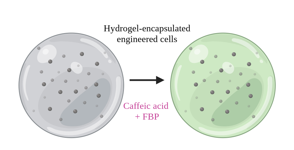
This work presents a caffeic acid and 5-CQA sensor based on the Fungal Bioluminescent Pathway (FBP), using Komagataella phaffii cells to express the FBP enzymes. The target diffuses through the hydrogel and activates the FBP pathway, resulting in light emission.
How would you use automation?
I would combine the hydrogel system with a microfluidic platform to automate sample handling and optical measurements. The system could control the introduction of the sample into the microfluidic chamber, regulate the flow and incubation time. An optical measurement step could then monitor the signal produced by the immobilized bacteria. Automation improves reproducibility by controlling parameters, while reducing manual handling. In the longer term, the platform could be used to test multiple samples and optimize the sensing parameters.
How much would you need to make, and how long would it take?
For an initial pilot-scale production, I would aim to manufacture approximately 1,000 biosensor units, each containing about 100 µL of bacteria-loaded hydrogel. This would require approximately 100 mL of hydrogel, including about 1 g of sodium alginate for a 1% w/v formulation. The main time requirement would be bacterial culture, which I estimate would take around 24 hours. Hydrogel preparation and sensor assembly could then be automated using a liquid-handling robot, requiring approximately 2–3 additional hours. Therefore, producing 1,000 sensing units would take approximately 26–27 hours, or about 1.5 days.
What don’t you know yet?
The main technical uncertainties are the stability and sensing performance of the immobilized Komagataella phaffii cells over time. I also need to determine how hydrogel composition affects biomarker diffusion and optical signal intensity. Finally, the scalability of the process needs to be validated.
References
[1] Torrez, V., Benavides-Frias, C., Jacobi, J., & Speranza, C. I. (2023). Ecological quality as a coffee quality enhancer. A review. Agronomy For Sustainable Development, 43(1), 19. https://doi.org/10.1007/s13593-023-00874-z
[2]Yang, S., Sarkar, S., Xie, X., Li, D., & Chen, J. (2023). Application of Optical Hydrogels in Environmental Sensing. Energy & Environment Materials, 7(3). https://doi.org/10.1002/eem2.12646
Part 1
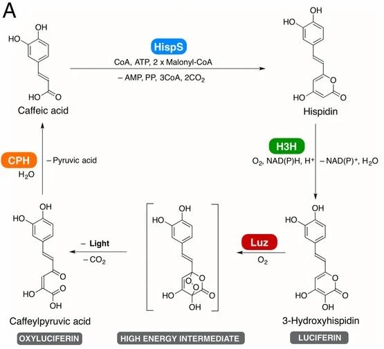
The idea is to detect caffeic acid by expressing the Fungal Bioluminescent Pathway FBP enzymes (hispidin synthase HispS, hispidin-3-hydroxylase H3H, luciferase, and caffeylpyruvate hydrolase CPH). Together, they convert caffeic acid into fungal luciferin. [1]
Figure 1. Proposed pathway of fungal luciferin biosynthesis and recycling. Caffeic acid is converted to hispidin by HispS and hydroxylated by H3H, yielding 3-hydroxyhispidin (fungal luciferin) [1]
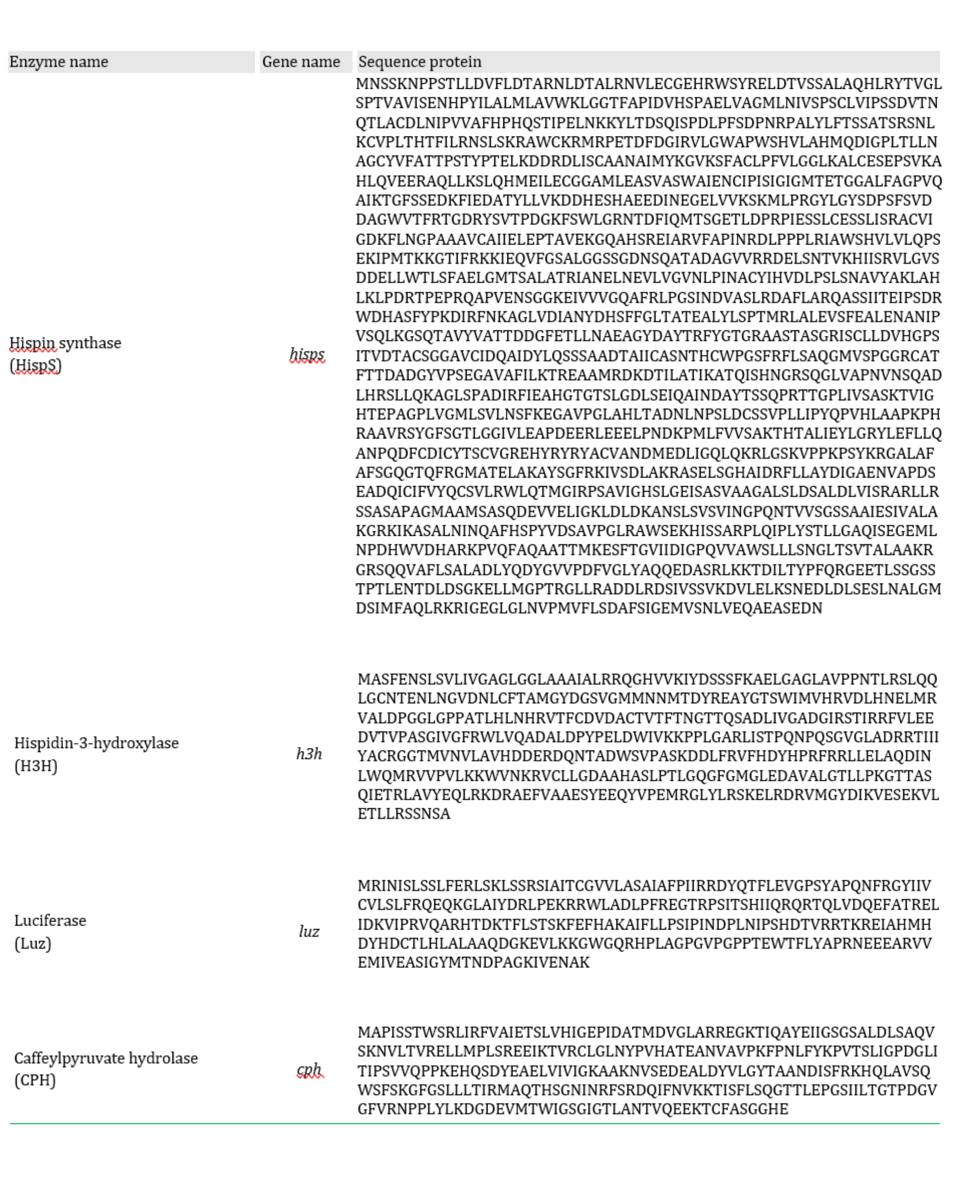
The sequence of enzymes that compose the FBP is described in the following table:
The chassis is Komagataella phaffii cells.
The FBP enzymes should be expressed in the cytoplasm.
References
[1] Kotlobay, A. A., Sarkisyan, K. S., Mokrushina, Y. A., Marcet-Houben, M., Serebrovskaya, E. O., Markina, N. M., Somermeyer, L. G., Gorokhovatsky, A. Y., Vvedensky, A., Purtov, K. V., Petushkov, V. N., Rodionova, N. S., Chepurnyh, T. V., Fakhranurova, L. I., Guglya, E. B., Ziganshin, R., Tsarkova, A. S., Kaskova, Z. M., Shender, V., . . . Yampolsky, I. V. (2018). Genetically encodable bioluminescent system from fungi. Proceedings Of The National Academy Of Sciences, 115(50), 12728-12732. https://doi.org/10.1073/pnas.1803615115
[2] Zamuner, C. K., Gomes, T. M. V., Bastos, E. L., Palkina, K. A., Yampolsky, I. V., Bechara, E. J. H., & Stevani, C. V. (2026). A Fungal Bioluminescent Pathway (FBP)-Based Yeast Biosensor for Caffeic Acid Quantification in Food and Beverages. ACS Sensors, 11(7), 5621-5634. https://doi.org/10.1021/acssensors.6c00051
A. COMPARE CELL-FREE REACTION COMPOSITIONS
Why does the concentration of potassium glutamate and magnesium glutamate differ between compositions? Which is more sensitive?
The concentrations of potassium glutamate and magnesium glutamate affect the cell-free reaction environment. Their optimal concentrations depend on the overall reaction formulation and the energy substrate being used. Jewett & Swartz (2004) reoptimized magnesium because PEP has a higher affinity for Mg2+ than pyruvate. PEP-based PANOx-SP used 20 mM Mg-Glu and 175 mM K-Glu, whereas pyruvate-based Cytomim used 8 mM Mg-Glu and 130 mM K-Glu
Magnesium glutamate is more sensitive to concentration changes. In Jewett & Swartz (2004) work, PANOx-SP changing Mg-Glu from 20 mM to 24 mM reduced relative protein expression from 100% to 73%, while K-Glu remained relatively stable around its optimum: 175 mM (100%), 200 (94%), and 225 mM (92%). In Cytomim, 8 mM gave the maximum yield, but 12 mM reduced it to 67% and 16 mM to 41%.
Explain how a PEP+NTP reaction formulation is different from a Ribose+NMP reaction formulation.
A PEP+NTP formulation supplies the cell-free system with nucleoside triphosphates (NTPs) such as ATP, GTP, CTP, and UTP. PEP serves as an energy phosphate donor to regenerate ATP, transferring its phosphate to ADP. For example, PANOx-SP (Jewett & Swartz, 2004) contains 33 mM PEP and 1.2 mM ATP plus 0.85 mM each of GTP, CTP, and UTP. This approach supplies transcription substrates, but PEP is relatively expensive, and its consumption generates inorganic phosphate.
In contrast, a Ribosome+ NMP formulation uses ribose together with nucleoside monophosphates (NMPs) such as AMP, CMP, GMP, and UMP. The lysate converts NMPs to NTPs. NMPs could support protein synthesis while reducing reagent costs.