Hello!! I’m Rishabh (or Rish), a first-year MS/PhD in the Molecular Machines group at MIT Media Lab building and validating AI methods for molecule design and drug discovery. I think AI has huge potential for making new biomedicine and therapeutics that go beyond human intuition, and HTBAA is exactly where I’d love to see just how much.
This site caricatures my journey holding a pipette … or at least getting a robot to do it for me ;)
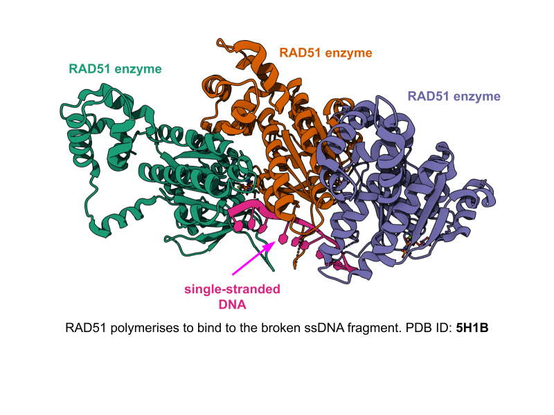
DNA randomly undergoes damage, and special dedicated Foci proteins are involved in DNA repair in the nucleus. I’d like to use deep learning methods to de novo design antagonist binders to RAD51, one such enzyme involved in this process (among other such proteins). If we can disrupt the DNA repeat mechanism, we can potentially use such binders to stop cancer cells from fixing their DNA and replicating.
I come from a computational (CS+Math) background and have worked in AI4Bio space for ~7 years now. I believe in the power of AI methods to go beyond human intuition when designing novel therapeutics and drugs. RAD51 is more abundant in cancer cells, making them a good target. I also am an aspiring structural biologist, and I’d like to approach this from a structural perspective because there the important protein-protein interface has a resolved structure in the PDB. I share more about the specific AI methods below. This idea of designing RAD51 antagonists with AI methods is novel according to a literature search with Claude.
What living system would you use?
I’d consider E.coli or yeast recombinant expression for protein-based binder design. Suppose I have a library of protein binder candidates (ie, their sequences), I’d start from DNA to synthesize the candidate sequence, insert this DNA into a living E.coli cell, and let the existing transcription+translation mechanism take care of protein design. Each DNA fragment carrying the instructions to become a unique binder candidate is attached to a circular plasmid, which also has a strong promoter that tells the cell’s transcription mechanism to amplify the specific protein’s synthesis, as well as an antibiotic resistance gene to prevent the plasmid from being destroyed by the cell.
The cell membranes undergo temporary shock, which makes them temporarily more permeable, allowing the plasmids to be placed into the E.coli cells. Soon after, we’d need to place the samples in liquid culture, which enables cell division. We’d also add a chemical trigger that tells the cells to synthesize a large quantity of our binder protein encoded in the plasmid. The cell’s ribosome translates the mRNA into our binders. The cells are then broken open to release everything inside, and we need to purify the proteins.
We’d finally then take the purified binders and pass them through purified RAD51 to screen them using a malachite green ATPase-inhibition assay. RAD51 uses ATP as fuel to power its DNA-repair filament activity, breaking each ATP into ADP + a free phosphate. The malachite green assay measures that released phosphate with a color change. More phosphate means a darker green on a plate reader. To screen the binders, I’d run RAD51 with and without each binder candidate. If a binder is disrupting RAD51’s function, less phosphate gets released, and the color stays lighter. So, lighter color across the library tells us which candidates are actually doing something.
How would you use automation?
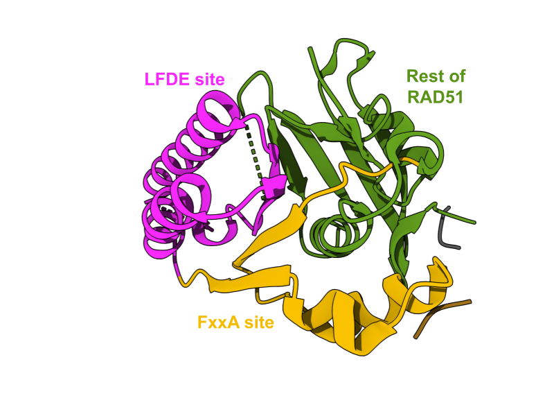
Before wet-lab validation, I’d have to curate a library of binders. Since I want to approach this from the structure-based perspective, I’d use open-source structure-based models like Complexa, RFDiffusion3, or BindCraft, which are deep learning methods for target-conditioned protein design (both sequence and structure). Here, RAD51 would be passed as an input and the model would generate new binder candidates. If parallelised on GPU, it’d take about a week to design a large enough library of binder candidates for RAD51. To in silico evaluate the quality of binders, I’d run them through the typical tools like AlphaFold3, ProteinMPNN (or its variants), and FoldSeek that measure how designable, novel, and diverse these protein binder candidates are. To ensure I’m not wasting compute on de novo binders, I’d condition RFDiffusion3 on known structural hotspots of RAD51 (eg: FxxA/LFDE pocket).
Once I have the filtered library, I’d have to use a synthesis vendor like Twist Bioscience to provide the initial DNA oligo-pool. Each binder can take up a well in a 96 or 384-well plate, where the arrayed transformation can be done by an OpenTron liquid handler robot. Robots can also be used to purify proteins, and put them through ATPase-inhibition assay as part of high-throughput screening of binders. Each binder is validated under the same reagent conditions and timing, making it easier to rank them all.
Lab automation would be great here because the deep learning models can generate thousands of binder candidates at high-throughput for RAD51. We’d ideally want to screen them at a similar pace as well, which is why using the liquid-handling OpenTron would help in a lab-in-the-loop design fashion.
How much would you need to make, and how long would it take?
Library design is bottlenecked by the inference throughput of RFDiffusion on a single GPU (eg: H100). Generating a single 100-150 residue binder with RFD3 takes 3-5 seconds. To computationally generate and evaluate ~100K designs, it will roughly take 5-6 days, with about ~20K being generated per day. To evaluate them would take another ~15 days using AF3, ProteinMPNN, and FoldSeek. I’d then filter to 100 most promising candidates using metrics like AF3’s pLDDT and ipTM, which is highly correlated with the true binding region of multimeric proteins. The wet-lab screening would take ~1 week to purify, express, and screen the 100 binders against RAD51. Expression and purification would take ~3 days and ATPase screening would take ~1 day.
Gene synthesis and transformation into E. coli, then expression and purification (~3 days total, using a yield of 8–10 mg/L for RFdiffusion3-designed proteins, which gives me the ~5 mg of total protein I calculated I’d need from well under a liter of culture), followed by ATPase-inhibition assays in the same day on an automated plate reader, since a single run handles up to 96 samples at once. Based on the hit rate of RFD3 of ~43%, I’d get 1-5 promising binders at the end.
What don’t you know yet?
The hit rate on RAD51 is currently unknown. My estimates are based on the performance of RFD3 and Complexa on other targets. The published RFdiffusion miniprotein study against SARS-CoV-2 targets found that computational confidence metrics (like ipTM) did not reliably predict which designs would actually bind. For instance, most candidates that passed filtering turned out to form unwanted oligomers. I don’t expect to finish this design campaign in one iteration. The bigger problem, which plagues the AI-for-binder design community in general, is engineering specificity into binders. I’d have to rely on computational tricks to guide RFD3-like models towards higher specificity (eg: inference-time steering, classifier guidance) as done in prior work. For such a lab-in-the-loop design pipeline, I’d assume it’d take a few iterations of designing binders before landing on any promising designs, using inspiration from success and failure cases in previous iterations.
I also foresee the bigger delivery problem of validating in vitro binders in the nucleus of living cells to be a challenge. The cell needs to cross the plasma membrane, cross the nuclear envelope (possibly with NLS tagging), and finally bind to free-floating RAD51. The designed binder could possibly be too big, so I’d have to consider RFD3 for miniprotein or cyclic peptide design instead of full-on protein binder design. RAD51 is also a dynamic molecule, staying in the repair foci region for ~200ms. This means our binders need to act in that window. The ATPase inhibition assay is a different isolated setting compared to the actual cellular environment the binder would have to act in. However, given this is exploratory work, seeing if we can even design binders of any kind for RAD51 would be a good first step, before thinking about safe delivery and passage into the nucleus.
Conducting an ATPase assay also doesn’t tell us the exact mechanism behind why we’d see an activity. Since a binder that reduces the ATPase activity of RAD51 is disrupting its normal catalytic cycle, we’d be unsure if it’s due to blocking the single-stranded DNA binding site, blocking the ATP binding pocket, or blocking the FxxA/LFDE site. It just tells us whether the binders are functionally good or not.
Week 3 HW: CFPS Coopetition
Copy and paste this section (section 3, A-F) into your website! Note, coopetition.htgaa.org is intended to support you as you answer the following questions.
Goal: using the collective intelligence of the HTBAA community and accessible AI systems, design novel CFPS reaction compositions for each of the three target proteins: sfGFP, PETase, and Reteplase to reduce the cost per gram of functional protein. Each design should be compatible with the experimental framework described above and should reflect the distinct expression and folding requirements of that protein. You can learn more about the details of the coopetition here.
A. COMPARE CELL-FREE REACTION COMPOSITIONS (20 minutes)
Why does the concentration of potassium glutamate and magnesium glutamate differ between compositions? Which is more sensitive?
Magnesium glutamate is a cofactor, so ribosomes, RNAP, and all ATP-using enzyme need it. It is more sensitive to potassium glutamate, which usually sets ionic strength and keeps the ribosome and translation machinery stable. It has to be present in bulk.
Explain how a PEP+NTP reaction formulation is different from a Ribose+NMP reaction formulation.
All cell-free reactions need a steady supply of ATP and other nuceleotide triphosphates. For PEP+NTP, the nucleotides added are already charged. PEP gives its phosphate to ADP using the lysate enzyme pyruvate kinase. However, it’s expensive but gives a fast start. For the Ribose+NMP reaction, the nucleotides are uncharged and cheaper. The lysate enzymes burn the sugar to make ATP, and other lysate enzymes add phosphates onto the NMPs. This gives it a slower start but lasts longer. Since it relies on the lysate’s own metabolism, there are more variables to tune in the lysate. For cell-free reactions, Ribose+NMP reactions are cheaper based on cost-per-gram optimisation.
Choose two cell-free reaction compositions from the literature, compare and contrast the choice of reagents and the concentration of reagents.
PANOx-SP and the coopetition baseline are built on the same core ingredients like lysate, HEPES buffer, glutamate salts, amino acids, an energy source, and nucleotides, but they have different purposes. PANOx-SP from Jewett & Swartz (2004) is a fixed recipe: it uses PEP as a direct energy source with NTPs, higher magnesium (about ~8 mM), and added cofactors and tRNA, all tuned for maximum protein yield.
The coopetition baseline is a lean starting point with fixed buffer, lysate, and DNA, leaving the remaining 11 µL open so you can choose cheaper energy sources (ribose, maltodextrin) and nucleotides (NMPs) and re-tune magnesium.
PANOx-SP optimizes for yield, while the coopetition optimizes for cost per gram.