No, I am not a Drosophila Melanogaster, I am a Medical Biotechnology student from Portugal.
I’m fascinated by the natural world and its molecules, its complexity and ingenuity: from plants to brains and everything in between.
I’ve been reflecting on how the future of scientific research and biopharmacological development could look like and be more democratic and community driven, not guided by profit only, while keeping safety as the main concern.
I imagine a future where an individual (or a small group of people) with access to remote labs and computational tools, including AI assisted research, can focus on a research topic or develop solutions that would otherwise be unexplored and that can eventually be shared, become the topic of institutional research, or capture funding to develop actual products.
I have an ambiguous relationship with computers. Do you know what I mean?
See the nav bar on the left to look through my work for How To
Biomanufacture Almost Anything (Fall 2026)!
Subsections of Rúben Gonçalves — HTBAA Fall 2026
Week 1 HW: Final Project Ideas
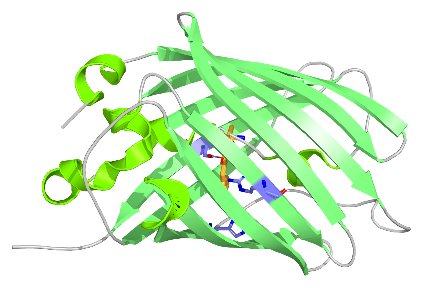
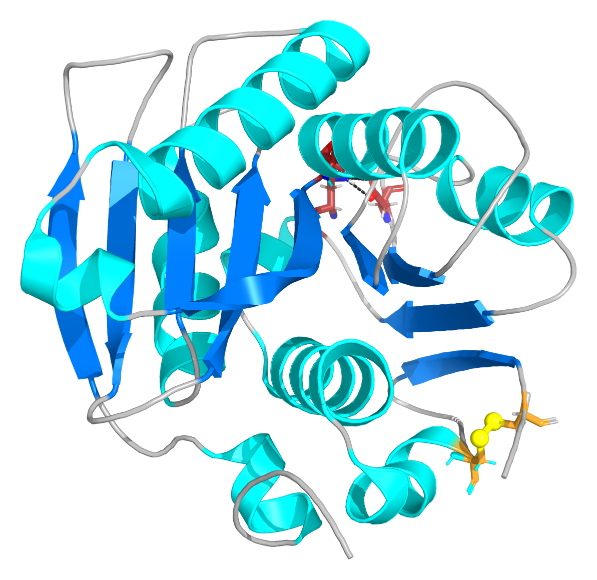
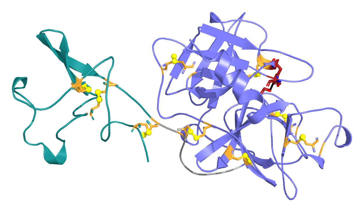
The cover image depicts the 3D molecular structure of the engineered plant defensin RsAFP2.
(1) What you would like to biomanufacture and why?
A leave-on topical hydrogel containing a re-engineered RsAFP2 plant defensin peptide to fight cutaneous fungal infections and restore skin microbiome balance.
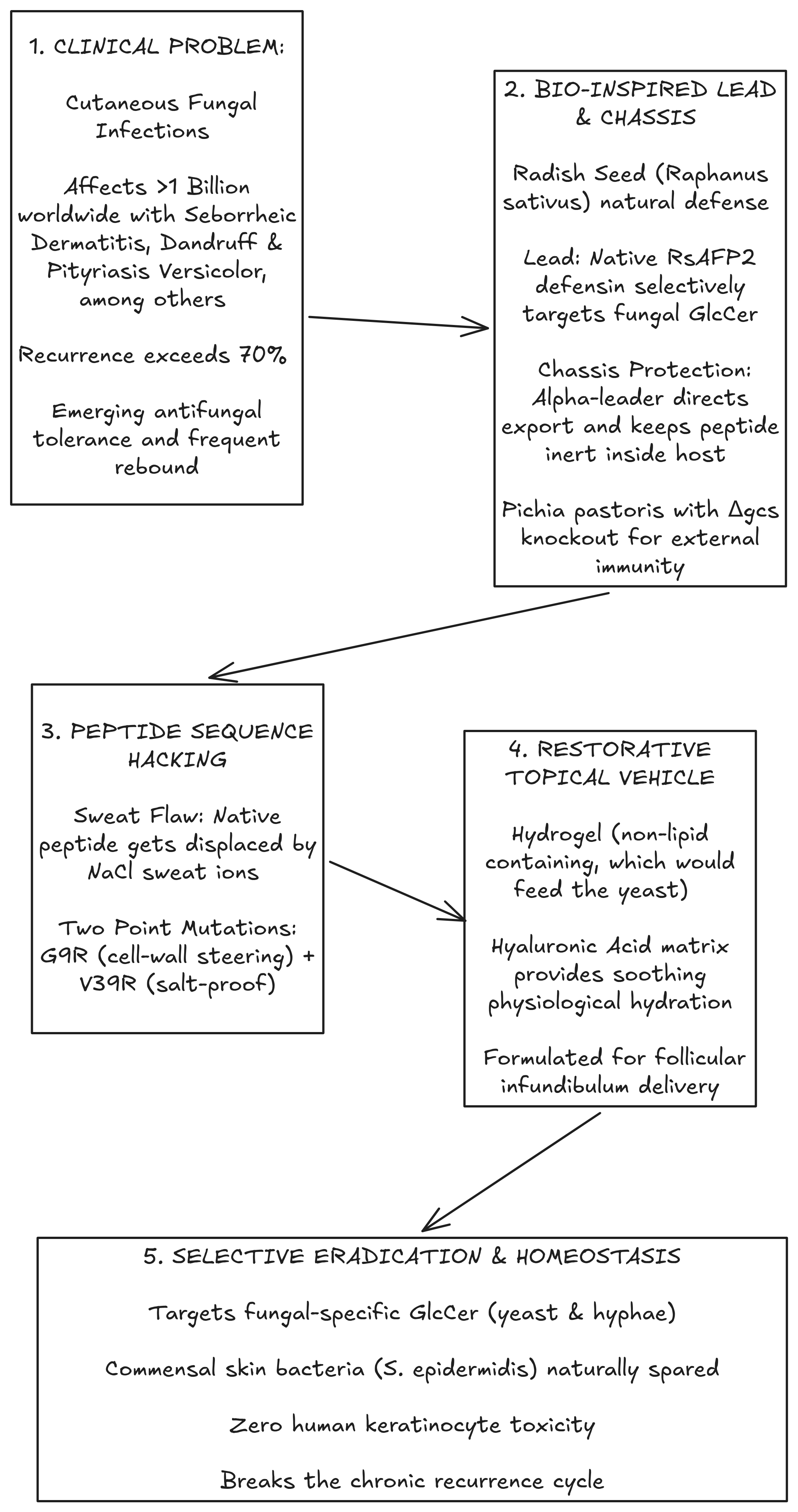
In the following diagram I present a quick overview of the rationale for the project followed by a more detailed description:
The idea for this project started with a condition that I see myself affected by and made me come up with the question: what would be a viable alternative to the current treatment methods that isn’t harsh on the skin and helps it reestablish balance instead of just fighting the infection?
This led me through an interesting journey of discovery about the skin, its interactions with the microbiome and what a safe biopharmaceutical intervention could look like.
I went on doing a literature review encompassing the etiology of these conditions and alternative biopharmaceutical approaches that could benefit from a biomanufacturing implementation.
Through this literature review I found out about a peptide, a plant defensin first isolated in 1992 from the common garden radish (Raphanus sativus L., family Brassicaceae), aiming for agricultural purposes, the RsAFP2 (Terras et al., 1992).
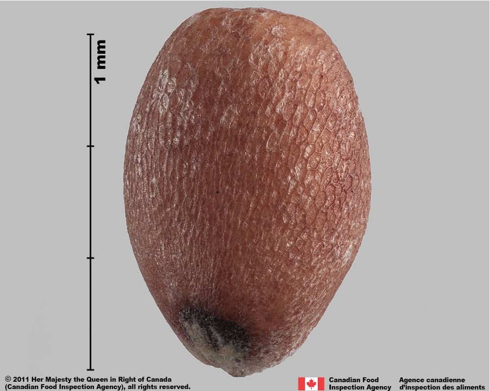
Figure: Seed of the common garden radish (Raphanus sativus L.), the natural source from which the antifungal defensin RsAFP2 was first isolated. I love the idea of leveraging a molecule from a plant seed to create a re-engineered human therapeutic.
My initial idea is about developing a hydrogel containing a re-engineered version of this plant defensin that reduces the yeast overgrowth in the skin, while not disrupting the beneficial microflora and has no detrimental effects on the skin. In the future, other components like urea and/or probiotics could be added to the final product, so that it leads to a more thorough skin health rebalance.
Virtually all human fungal pathogens including Malassezia, Candida, and dermatophytes (ringworm) depend on the exact same cell-membrane receptor: fungal glucosylceramide (GlcCer). Because this specific branched lipid is completely absent in human cells and beneficial bacteria, our engineered defensin acts as a selective fungal therapeutic with zero mammalian toxicity (Thevissen et al., 2004).
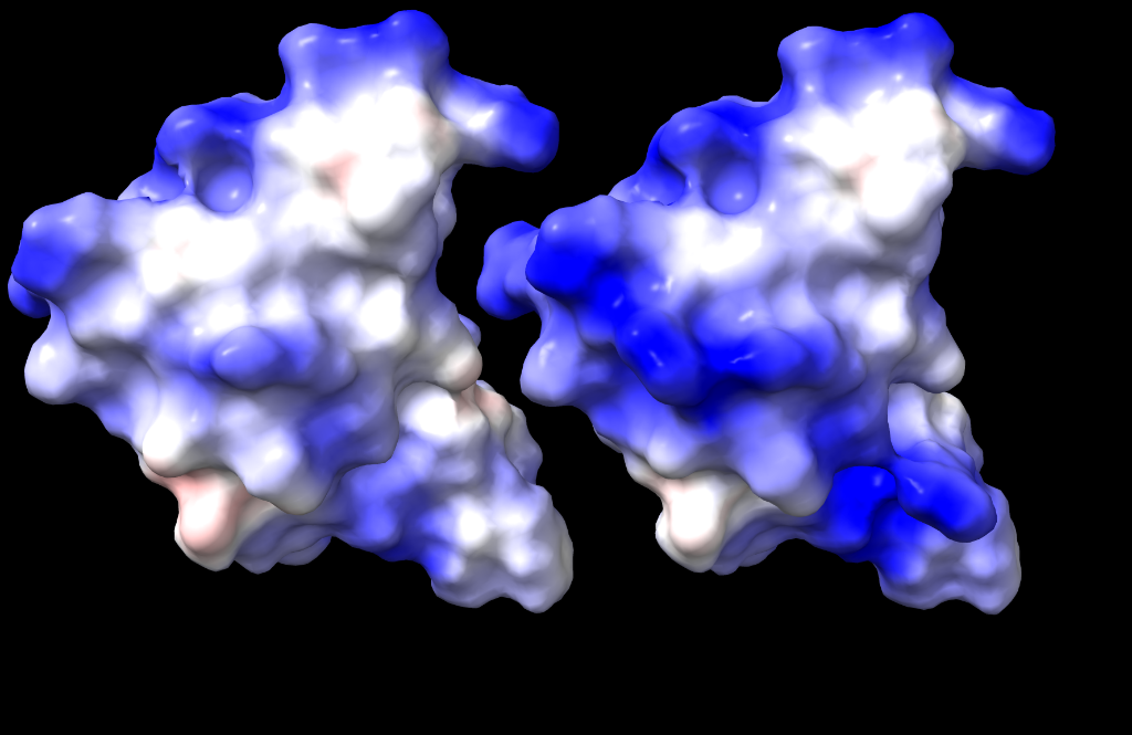
For optimal results, we would be tailoring the wild-type peptide specifically for the human cutaneous environment, applying two point mutations documented in the literature (G9R/V39R; Pandey et al., 2020): basically we are hacking the wild-type peptide to optimize for skin conditions, making it more salt-tolerant and electrostatically targeted.
Figure: Electrostatic surface potential comparison between wild-type RsAFP2 (left) and our engineered G9R/V39R double mutant (right). The expanded deep-blue surface indicates an increased positive electrostatic charge, enhancing attraction toward negatively charged fungal membranes and preventing ionic deactivation by sweat (rendered using UCSF ChimeraX).
This is also an exercise in how an individual can leverage scientific research to solve a personal problem and eventually work together with others that share the same interest: a different way of developing health solutions that is not dependent on a company’s initiative. Obviously this also poses questions about safety and regulation, which I find will become more and more important as biotechnological experiments become accessible outside regulated institutions.
Safety
Different studies have characterized RsAFP2 as safe for human cells:
Tavares et al. (2008): Specifically tested RsAFP2 against human cells using an automated Lactate Dehydrogenase (LDH) release assay. RsAFP2 caused zero LDH leakage, completely inert toward mammalian cells.
Terras et al. (1992) and Thevissen et al. (2004): Demonstrated 0% hemolytic activity against human red blood cells even at concentrations >500 μg/mL.
Future directions
adding extra components that help the skin recover and avoid recurrence (e.g. urea, probiotics);
after the engineered peptide proves effective for our use case (skin dysbiosis), we could develop different delivery systems for using in the scalp or against vaginal candidiasis, where it’s also very important not to disrupt the beneficial bacterial flora;
chromogenic feedback, transforming the product into a theragnostic that gives the user an immediate reading of the treatment’s effectiveness;
grafting an Arg-Arg-Arg-Trp (RRRW) motif into the defensin’s flexible β2–β3 loop (γ-core). In plant defensin studies, this cationic-hydrophobic patch accelerates fungal membrane permeabilization by over 300% (Sagaram et al., 2011). This would have to be screened for production viability and safety, as described in the automation section;
eventual translation to internal use, producing an antifungal that has no hepatotoxicity, in contrast to current drugs in the market.
We need an eukaryotic chassis for specific folding that wouldn’t be possible in a bacterial one and we don’t need any mammalian cell specific machinery, hence this choice.
Because wild-type Pichia pastoris is a fungus whose plasma membrane contains glucosylceramide, expressing an antifungal defensin in a wild-type fungal host would result in autotoxicity. For this reason, manufacturing requires a two-stage engineering sequence:
Host Immunization: we disrupt the Pichia glucosylceramide synthase gene (GCS, locus PAS_chr4_0775) using a transient, marker-free CRISPR/Cas9 plasmid. The resulting ΔgcsPichia pastoris mutant produces no glucosylceramide and is completely immune to defensin-mediated lysis (confirmed by Thevissen et al., 2004).
API Expression Vector Integration: the immune Δgcs host is transformed with the linearized expression vector pPICZαA-RsAFP2[G9R/V39R] for stable single-crossover integration into the chromosomal AOX1 locus.
(3) How would you use automation?
This is an area that I hope to improve as the course proceeds and I get more familiar with the available automation methods and their possible uses.
Through an AI guided exploration where I provided the AI with the course content and available automation systems along with the project details, it suggested some uses, from which I selected what seems to me a sensible use for automation during the biomanufacturing process which I describe below (the text has been manually edited by me).
Example using the Opentrons OT-2 for testing formulation phase-behavior and solubility:
RsAFP2 (our peptide) has a +7 positive charge. Hyaluronic acid (the hydrogel polymer) is an anionic polyglycosaminoglycan covered in negative carboxylic acid groups (-COO⁻).
At certain pH and salt levels, strongly positive peptides and strongly negative polymers can bind together and form cloudy precipitates or “clumps” (polyelectrolyte complexation).
The automated screen: we would be using the Opentrons robot to run a combinatorial 96-well grid:
pH gradient: 4.0, 4.5, 4.8, 5.2, 5.5, 6.0, 6.5
Salt gradient: 0, 25, 50, 100, 150 mM NaCl
Polymer: 0.5%, 1.0%, 1.5% Hyaluronic acid
We would then measure absorbance at 405 nm to find the exact formulation window where the gel remains 100% crystal-clear and the peptide is free to diffuse and kill yeast.
Another possible use
I also find this use for automation really interesting: as mentioned in the “future directions” section, there is another peptide modification (γ-core RRRW grafting) that could be used to improve the antifungal activity but that has never been tested for use in humans (as far as I could find). An automated safety pipeline could be implemented that verifies if it doesn’t compromise host safety:
Opentrons OT-2 automated 96-well HaCaT human keratinocyte cytotoxicity screen (CellTiter-Glo viability and LDH release assays).
This would establish a developability gate to confirm if the high selectivity of RsAFP2 for fungal glucosylceramide is preserved with zero mammalian membrane permeabilization.
(4) How much would you need to make, and how long would it take?
I used AI to come up with the estimations and calculations below, using the articles mentioned in the bibliography as references (“Clinical scale and dosing standards” and “Living system and Biomanufacturing automation” sections). I will iterate on them as I become more familiar with the methods. All the text was manually edited by me but I must clarify that I am not familiar with all the processes that are mentioned.
(a) How much?
For a 4-week clinical treatment course for 10,000 patients (10,000×50 mL tubes of 0.1% topical hydrogel): (Borda and Wikramanayake, 2015; Long and Finlay, 1991):
Calculations
Mass of pure RsAFP2[G9R/V39R] active peptide needed per tube:
Mass per tube = 50 mL × 1.0 mg/mL = 50 mg = 0.050 grams of pure RsAFP2
Total active product needed for 10,000 patients:
Total Active Peptide Required = 10,000 patients × 0.050 g/patient = 500 grams (0.50 kg)
(b) How long?
The overall workflow divides into two distinct automated phases: initial prototyping and the 500-gram production campaign:
Prototyping Automation vs. Production Automation
Prototyping phase (2–3 weeks, upfront R&D): High-throughput micro-scale screening using the Opentrons OT-2 to screen colonies, optimize media in 96-well plates, and test formulation compatibility (μL to mL scale; Asin-Garcia et al., 2025).
Production phase (3 weeks, manufacturing campaign): Pilot-scale manufacturing of the 500 g using a SCADA-controlled 500 L Bioreactor with automated Process Analytical Technology (PAT; Cereghino and Cregg, 2000; Petzold and Mukhopadhyay, 2025): closed-loop DO-stat methanol feeding, mass-flow gas blending, and automated Tangential Flow Filtration (TFF).
Bioreactor volume and time calculation
Target pure peptide: 500 g
Expected fermentation titer: 1.5 g/L in Pichia pastoris broth
Downstream recovery: ~70% (yielding ~1.0 g/L pure peptide)
Total broth required: 500 g ÷ 1.0 g/L = 500 L broth
To produce 500 L with adequate headroom for aeration and foaming, we run two 250 L batches in a 500 L pilot vessel (working at 50% capacity).
Manufacturing Timeline
Batch strategy: Run two consecutive 250 L batches in a 500 L pilot vessel
Production manufacturing duration = (2×8 days) + 5 days = 21 days (≈3 weeks) (Total initial campaign duration including upfront prototyping: ≈5–6 weeks)
5. What don’t you know yet?
The fact that we are dealing with a biopharmaceutical project means that we would need a lot of careful testing to validate the action (some of it could probably be done through automation, as clarified above) and safety. The ultimate validation would be testing it on human skin and measure its effect on infected skin: I am not familiar with the process but that’s something I will explore in the meanwhile.
Being pretty much a beginner when it comes to biomanufacturing, I can’t still define exactly what is the best way to implement the fabrication process of such a product. Hopefully as the course advances, things will become more clear. For this proposal I am using some ideas retrieved through AI (always using reliable sources as references) and these should be considered “temporary”.
I also need to dive deeper on how to calculate and establish the right posology and treatment guidelines.
AI use:
I’ve been experimenting with an AI assisted research environment, using Google Antigravity 2.0 with the Gemini Flash 3.8 model. The agent is connected to external scientific literature sources (using the Consensus MCP server as well as other sources through web-search) so that I can more easily find relevant articles related to the project.
I have been having good results with finding relevant literature but the AI is still quite naïve with some of its statements and overly optimistic about some of the ideas I present to it. For this reason, I use it to gather information (which really speeds up the process) and as a brainstorming companion, while always checking the sources as the ultimate source of truth.
For the proposal, I prompted the AI with my original “problem” and asked it to find possible solutions that could be implemented through biomanufacturing. I then went on asking for details about specific parts of the project, gathering relevant articles that I accessed directly and keep as a reference locally for me and for the AI agent itself.
All the information is then kept in the form of a local “wiki” that I access as an Obsidian vault.
AI was also used to generate the clean citation list.
I still prefer to write the final text myself, and that’s what I did for this proposal.
Bibliography
Foundational Plant Defensin and Biology:
Terras, F. R., Schoofs, H. M., De Bolle, M. F., Van Leuven, F., Rees, S. B., Vanderleyden, J., Cammue, B. P., and Broekaert, W. F. (1992). Analysis of two novel classes of plant antifungal proteins from radish (Raphanus sativus L.) seeds.Journal of Biological Chemistry, 267(22), 15301–15309. DOI: 10.1016/s0021-9258(19)49534-3
Thevissen, K., Warnecke, D. C., François, I. E., Leipelt, M., Heinz, E., Ott, C., Zähringer, U., Thomma, B. P., Ferket, K. K., and Cammue, B. P. (2004). Defensins from insects and plants interact with fungal glucosylceramides: Pichia pastoris Δgcs mutants are highly resistant to defensin-mediated permeabilization.Journal of Biological Chemistry, 279(6), 3900–3905. DOI: 10.1074/jbc.M311165200
Tavares, P. M., Thevissen, K., Cammue, B. P., François, I. E., Barreto-Bergter, E., Taborda, C. P., Marques, A. F., Rodrigues, M. L., and Nimrichter, L. (2008). In vitro activity of the antifungal plant defensin RsAFP2 against Candida isolates and its in vivo efficacy in prophylactic murine models of candidiasis.Antimicrobial Agents and Chemotherapy, 52(12), 4522–4525. DOI: 10.1128/AAC.00448-08
Pandey, B., Tyagi, C., Prajapati, G. K., Mishra, A. K., Hashem, A., Alqarawi, A. A., and Abd_Allah, E. F. (2020). Analysis of mutations of defensin protein using accelerated molecular dynamics simulations.PLoS ONE, 15(11), e0241679. DOI: 10.1371/journal.pone.0241679
Sagaram, U. S., Pandurangi, R., Kaur, J., Smith, T. J., and Shah, D. M. (2011). Structure-activity determinants in antifungal plant defensins MsDef1 and MtDef4 with different modes of action against Fusarium graminearum.PLoS ONE, 6(4), e18550. DOI: 10.1371/journal.pone.0018550
Clinical Scale and Dosing Standards:
Borda, L. J., and Wikramanayake, T. C. (2015). Seborrheic dermatitis and dandruff: a comprehensive review.Journal of Clinical and Investigative Dermatology, 3(2), 10. DOI: 10.13188/2373-1044.1000019 | PMCID: PMC4852869
Long, C. C., and Finlay, A. Y. (1991). The finger-tip unit—a new practical measure.Clinical and Experimental Dermatology, 16(6), 444–447. DOI: 10.1111/j.1365-2230.1991.tb01232.x
Living System and Biomanufacturing Automation:
Cereghino, J. L., and Cregg, J. M. (2000). Heterologous protein expression in the methylotrophic yeast Pichia pastoris.FEMS Microbiology Reviews, 24(1), 45–66. DOI: 10.1111/j.1574-6976.2000.tb00532.x
Petzold, C. J., and Mukhopadhyay, A. (2025). From bench to biofactory: high-throughput technologies and automated workflows to accelerate biomanufacturing.Current Opinion in Biotechnology, 94, 103320. DOI: 10.1016/j.copbio.2025.103320
Asin-Garcia, E., Fawcett, Z., Batianis, C., et al. (2025). A snapshot of biomanufacturing and the need for enabling research infrastructure.Trends in Biotechnology, 43(5), 1000–1014. DOI: 10.1016/j.tibtech.2024.10.014
Project Follow-Up
Here I plan to add new findings/ideas for my final project.
Alternative Project Ideas
These are some alternative ideas I had for the final project:
Relaxing Sound Chewing Gum
A chewing gum that produces relaxing sounds conducted via bones. The more relaxed the chewing rythm, the more relaxing the sounds become… Be careful, don’t fall asleep or you can swallow it. Actually, not a problem (I guess) - it’s not plastic!
Just so that we can stay even more relaxed, let’s add some antimicrobial components and some viral interceptors to it.
100% Biodegradable! Degrades harmlessly into natural fatty acid monomers in compost, soil, or marine environments within weeks.
Oral Biotherapeutic MSG / Glutamate Neutralizer
Problem
While monosodium glutamate (MSG) is generally recognized as safe for the majority of the population, a subset of sensitive individuals (me included) experiences adverse reactions (“MSG symptom complex”): diaphoresis, severe flushing, chest tightness, palpitations, and throbbing headaches within 15–30 minutes of ingesting free glutamate.
The supposed physiological mechanism: unlike protein-bound glutamate (which is digested and absorbed slowly), free MSG dissolved in soups or sauces causes a rapid, massive spike in portal and systemic blood plasma glutamate concentration, overstimulating peripheral glutamate receptors and autonomic ganglia.
The (hypothetical) solutions bellow are AI generated, using current scientific literature as a reference:
Mechanism: GAD is a pyridoxal 5’-phosphate (PLP)-dependent enzyme that decarboxylates L-glutamate into GABA, the mammalian nervous system’s primary inhibitory, calming neurotransmitter.
Engineering: Directed evolution of bacterial GAD (from Lactobacillus brevis or Escherichia coli) to resist pepsin/trypsin proteolysis and operate across the acidic-to-neutral pH gradient of the stomach and upper duodenum.
Outcome: Converts excess dietary excitatory glutamate into calming GABA directly inside the stomach lumen before it can enter the bloodstream.
Solution 2
Non-Absorbable Molecular Scavenger Proteins (The “Glutamate Sponge”)
Engineered derivatives of the bacterial periplasmic glutamate/aspartate-binding protein (GltI / PEB1a).
Formulated as an insoluble, non-absorbable crosslinked polymer or edible hydrogel. It acts as an oral ion-exchange sponge, binding free glutamate ions with nanomolar affinity (Kd < 100 nM) and carrying them through the stool, completely blunting the plasma Cmax surge.
(or stop addind MSG to food)
Week 2 HW: DNA Design
Part 0: Set up in Kernel
ruben_goncalves/goncalves-antifungal_peptide
Part 1: Define your expression target
What molecule are you expressing?
I’m expressing an engineered plant peptide: the RsAFP2[G9R/V39R].
What is the sequence?
The amino acid sequence for the wild-type defensin Rs-AFP2 from Raphanus sativus was obtained from UniProtKB (Accession: P30230, Entry: DEF1_RAPSA; https://www.uniprot.org/uniprotkb/P30230/entry):
In the UniProt Feature viewer the first 29 AAs are identified as a plant signal sequence, which we need to remove as later we are adding the yeast signal peptide:
Where does the product need to go?
It needs to be secreted (extracellular). The peptide is targeted co-translationally into the endoplasmic reticulum lumen using the S. cerevisiae αα-mating factor pre-pro leader peptide.
Part 2: Design your genetic parts
For defining the parts that need to be included in the plasmid, apart from the peptide sequence, the “pPICZ A” Pichia pastoris vector was used as a reference (retrieved from https://www.snapgene.com/plasmids/yeast_plasmids/pPICZ_A ).
Figure: Reference plasmid map of pPICZα A (3,593 bp; Invitrogen/SnapGene).
Each of the necessary components was individually created as a part in Kernel. The sequence for each of the parts was extracted from the .dna file retrieved from the address above.
The sequence for the RsAFP2 plant peptide was retrived from the European Nucleotide Archive (https://www.ebi.ac.uk/ena/browser/api/embl/U18556.1?lineLimit=1000). Several edits had to be done to achieve the DNA coding sequence for the mature 51-amino-acid engineered defensin:
removal of the DNA bases (1–41): plant’s 5’ UTR;
removal of the DNA bases (42–128): plant’s native signal peptide;
removal of the DNA bases (285–457): plant’s 3’ UTR and poly(A) tail;
engineered point mutations (G9R and V39R).
Parts created in Kernel:
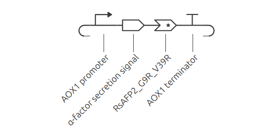
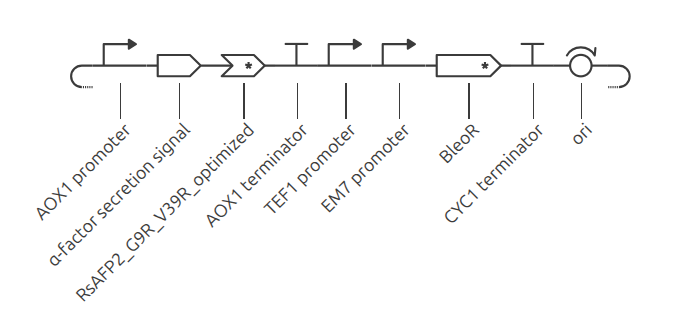
AOX1 promoter Type: promoter Ultra-strong, methanol-inducible eukaryotic promoter from Pichia pastoris (Invitrogen pPICZαA) driving high-titer defensin expression upon glycerol depletion, harboring a unique SacI linearization site (bp 208) for Campbell chromosomal integration. Sequence (939 bp): GATCTAACATCCAAAGACGAAAGGTTGAATGAAACCTTTTTGCCATCCGACATCCACAGGTCCATTCTCACACATAAGTGCCAAACGCAACAGGAGGGGATACACTAGCAGCAGACCGTTGCAAACGCAGGACCTCCACTCCTCTTCTCCTCAACACCCACTTTTGCCATCGAAAAACCAGCCCAGTTATTGGGCTTGATTGGAGCTCGCTCATTCCAATTCCTTCTATTAGGCTACTAACACCATGACTTTATTAGCCTGTCTATCCTGGCCCCCCTGGCGAGGTTCATGTTTGTTTATTTCCGAATGCAACAAGCTCCGCATTACACCCGAACATCACTCCAGATGAGGGCTTTCTGAGTGTGGGGTCAAATAGTTTCATGTTCCCCAAATGGCCCAAAACTGACAGTTTAAACGCTGTCTTGGAACCTAATATGACAAAAGCGTGATCTCATCCAAGATGAACTAAGTTTGGTTCGTTGAAATGCTAACGGCCAGTTGGTCAAAAAGAAACTTCCAAAAGTCGGCATACCGTTTGTCTTGTTTGGTATTGATTGACGAATGCTCAAAAATAATCTCATTAATGCTTAGCGCAGTCTCTCTATCGCTTCTGAACCCCGGTGCACCTGTGCCGAAACGCAAATGGGGAAACACCCGCTTTTTGGATGATTATGCATTGTCTCCACATTGTATGCTTCCAAGATTCTGGTGGGAATACTGCTGATAGCCTAACGTTCATGATCAAAATTTAACTGTTCTAACCCCTACTTGACAGCAATATATAAACAGAAGGAAGCTGCCCTGTCTTAAACCTTTTTTTTTATCATCATTATTAGCTTACTTTCATAATTGCGACTGGTTCCAATTGACAAGCTTTTGATTTTAACGACTTTTAACGACAACTTGAGAAGATCAAAAAACAACTAATTATTCGAAACG
α-factor secretion signal Type: peptide-cds (+start, -stop) Bipartite pre-pro secretion leader from Saccharomyces cerevisiaeMFα1 (Invitrogen pPICZαA) directing the nascent peptide co-translationally into the oxidizing ER lumen for 4-disulfide bond formation, terminating in a dibasic Lys-Arg motif for scarless Golgi Kex2 endoprotease cleavage. Sequence (267 bp): ATGAGATTTCCTTCAATTTTTACTGCTGTTTTATTCGCAGCATCCTCCGCATTAGCTGCTCCAGTCAACACTACAACAGAAGATGAAACGGCACAAATTCCGGCTGAAGCTGTCATCGGTTACTCAGATTTAGAAGGGGATTTCGATGTTGCTGTTTTGCCATTTTCCAACAGCACAAATAACGGGTTATTGTTTATAAATACTACTATTGCCAGCATTGCTGCTAAAGAAGAAGGGGTATCTCTCGAGAAAAGAGAGGCTGAAGCT
RsAFP2_G9R_V39R_optimizedType: peptide-cds (-start, +stop) De novo engineered plant defensin coding sequence (based on UniProt P30230 / PDB 2N2R) harboring G9R and V39R substitutions, codon-harmonized for Saccharomyces cerevisiae as the closest organism to Pichia pastoris available and terminated with tandem TAA stop codons. Sequence (156 bp): CAAAAATTGTGTCAAAGACCATCTAGAACTTGGTCTGGTGTTTGTGGTAATAATAATGCTTGTAAAAATCAATGTATTAGATTGGAAAAAGCTAGACATGGTTCTTGTAATTATAGATTTCCAGCTCATAAATGTATTTGTTATTTTCCATGTTAA
AOX1 terminator Type: terminator Native Pichia pastorisAOX1 transcription terminator and polyadenylation signal (Invitrogen pPICZαA) ensuring high mRNA transcript stability, efficient 3’ cleavage, and prevention of transcriptional read-through into downstream units. Sequence (247 bp): TCAAGAGGATGTCAGAATGCCATTTGCCTGAGAGATGCAGGCTTCATTTTTGATACTTTTTTATTTGTAACCTATATAGTATAGGATTTTTTTTGTCATTTTGTTTCTTCTCGTACGAGCTTGCTCCTGATCAGCCTATCTCGCAGCTGATGAATATCTTGTGGTAGGGGTTTGGGAAAATCATTCGAGTTTGATGTTTTTCTTGGTATTTCCCACTCCTCTTCAGAGTACAGAAGATTAAGTGAGA
TEF1 promoter Type: promoter Robust, constitutive RNA Polymerase II promoter from Saccharomyces cerevisiae Translation Elongation Factor 1 (Invitrogen pPICZαA) driving continuous expression of the Zeocin resistance marker in yeast without requiring chemical inducers. Sequence (387 bp): AATGTTTCTACTCCTTTTTTACTCTTCCAGATTTTCTCGGACTCCGCGCATCGCCGTACCACTTCAAAACACCCAAGCACAGCATACTAAATTTTCCCTCTTTCTTCCTCTAGGGTGTCGTTAATTACCCGTACTAAAGGTTTGGAAAAGAAAAAAGAGACCGCCTCGTTTCTTTTTCTTCGTCGAAAAAGGCAATAAAAATTTTTATCACGTTTCTTTTTCTTGAAATTTTTTTTTTTAGTTTTTTTCTCTTTCAGTGACCTCCATTGATATTTAAGTTAATAAACGGTCTTCAATTTCTCAAGTTTCAGTTTCATTTTTCTTGTTCTATTACAACTTTTTTTACTTCTTGTTCATTAGAAAGAAAGCATAGCAATCTAATCTAAG
EM7 promoter Type: promoter Minimal synthetic prokaryotic promoter (Invitrogen pPICZαA) providing constitutive σ70-driven expression of the BleoR resistance gene in Escherichia coli during plasmid propagation, functioning in an orthogonal tandem arrangement with the upstream TEF1 promoter. Sequence (48 bp):
GTTGACAATTAATCATCGGCATAGTATATCGGCATAGTATAATACGAC
BleoR Type: cds (+start, +stop) Dominant antibiotic resistance gene from Streptoalloteichus hindustanus (sh ble, Invitrogen pPICZαA) encoding a 13.7 kDa acidic protein that stoichiometrically binds and neutralizes Zeocin in a 1:1 complex, conferring chemical selection in both bacteria and yeast. Sequence (375 bp): ATGGCCAAGTTGACCAGTGCCGTTCCGGTGCTCACCGCGCGCGACGTCGCCGGAGCGGTCGAGTTCTGGACCGACCGGCTCGGGTTCTCCCGGGACTTCGTGGAGGACGACTTCGCCGGTGTGGTCCGGGACGACGTGACCCTGTTCATCAGCGCGGTCCAGGACCAGGTGGTGCCGGACAACACCCTGGCCTGGGTGTGGGTGCGCGGCCTGGACGAGCTGTACGCCGAGTGGTCGGAGGTCGTGTCCACGAACTTCCGGGACGCCTCCGGGCCGGCCATGACCGAGATCGGCGAGCAGCCGTGGGGGCGGGAGTTCGCCCTGCGCGACCCGGCCGGCAACTGCGTGCACTTCGTGGCCGAGGAGCAGGACTGA
CYC1 terminator Type: terminator Native Saccharomyces cerevisiae Cytochrome c-1 transcription terminator and polyadenylation signal (Invitrogen pPICZαA) directing 3’ transcript processing of the BleoR mRNA in yeast, insulating the adjacent bacterial origin of replication. Sequence (248 bp): TCATGTAATTAGTTATGTCACGCTTACATTCACGCCCTCCCCCCACATCCGCTCTAACCGAAAAGGAAGGAGTTAGACAACCTGAAGTCTAGGTCCCTATTTATTTTTTTATAGTTATGTTAGTATTAAGAACGTTATTTATATTTCAAATTTTTCTTTTTTTTCTGTACAGACGCGTGTACGCATGTAACATTATACTGAAAACCTTGCTTGAGAAGGTTTTGGGACGCTCGAAGGCTTTAATTTGC
ori Type: origin-of-replication High-copy prokaryotic replicon derived from plasmid pUC19/ColE1 (Invitrogen pPICZαA) recruiting E. coli DNA Polymerase III on the reverse strand to maintain 500–700 plasmid copies per cell for rapid, high-yield cloning and miniprep harvesting. Sequence (589 bp): TTTCCATAGGCTCCGCCCCCCTGACGAGCATCACAAAAATCGACGCTCAAGTCAGAGGTGGCGAAACCCGACAGGACTATAAAGATACCAGGCGTTTCCCCCTGGAAGCTCCCTCGTGCGCTCTCCTGTTCCGACCCTGCCGCTTACCGGATACCTGTCCGCCTTTCTCCCTTCGGGAAGCGTGGCGCTTTCTCAATGCTCACGCTGTAGGTATCTCAGTTCGGTGTAGGTCGTTCGCTCCAAGCTGGGCTGTGTGCACGAACCCCCCGTTCAGCCCGACCGCTGCGCCTTATCCGGTAACTATCGTCTTGAGTCCAACCCGGTAAGACACGACTTATCGCCACTGGCAGCAGCCACTGGTAACAGGATTAGCAGAGCGAGGTATGTAGGCGGTGCTACAGAGTTCTTGAAGTGGTGGCCTAACTACGGCTACACTAGAAGGACAGTATTTGGTATCTGCGCTCTGCTGAAGCCAGTTACCTTCGGAAAAAGAGTTGGTAGCTCTTGATCCGGCAAACAAACCACCGCTGGTAGCGGTGGTTTTTTTGTTTGCAAGCAGCAGATTACGCGCAGAAAAAAAGGATCTCAA
Part 3: Assemble your construct
(Descriptions are AI generated from reference material)
1. Linear Expression Cassette (RsAFP2_G9R_V39R_Cassette)
Description (1,609 bp): Minimal eukaryotic expression cassette driving secretion of the defensin in Pichia pastoris. It joins the inducible AOX1 promoter, the α-factor secretion signal, the mature RsAFP2[G9R/V39R] coding sequence, and the AOX1 terminator into a single in-frame 423 bp open reading frame (141 AA) with zero frameshifts and an ochre stop codon (TAA).
Description (3,256 bp): Complete recombinant shuttle vector embedding the 1,609 bp expression cassette within the pPICZαA backbone. It provides the pUC origin for high-copy cloning in E. coli and the dual TEF1/EM7 promoter driving Zeocin resistance (BleoR), harboring a unique SacI site at bp 208 for linearization and chromosomal integration into Pichia pastoris.
Part 4
Codon-optimization:
The peptide sequence was codon-optimized using the Kernel optimization tool for Saccharomyces cerevisiae as this was the the closest organism to Pichia pastoris available.
GC content: from 42.9% to 29.5%. Shifted to AT-rich yeast preference;
30 of 52 codons swapped (57.7%).
Why does codon usage matter for expression in this organism?(validated AI generated text)
Although the genetic code is redundant, organisms exhibit strong species-specific codon bias that mirrors the intracellular abundance of their matching tRNA pools. Expressing native plant (Raphanus sativus) cDNA directly in yeast frequently leads to ribosome stalling, translational pausing, and mRNA degradation due to the scarcity of tRNAs for plant-preferred codons. Codon optimization resolves this by systematically substituting rare codons with high-frequency synonymous yeast codons (favoring AT-rich triplets like CAA, AAA, and TCT), which accelerates translation elongation and maximizes recombinant peptide yield without altering the final amino acid sequence. Saccharomyces cerevisiae was selected as the optimization host in Kernel because its tRNA availability and codon preferences closely mirror those of Pichia pastoris.
BLAST:
(I asked AI for the steps required for the analysis)
Database: Non-redundant protein sequences (nr) Algorithm: blastp (protein-protein BLAST)
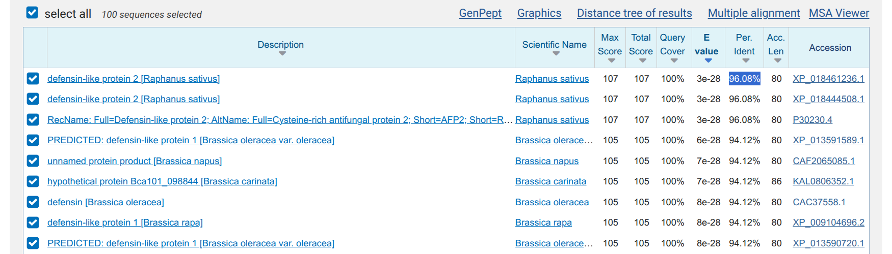
Inserted the engineered peptide aminoacid sequence: QKLCQRPSRTWSGVCGNNNACKNQCIRLEKARHGSCNYRFPAHKCICYFPC
Results: While being a novel engineered variant, as expected, the CDS is similar to existing proteins, as just two point mutations were made to the original radish plant defensin. The closest match is exactly the “defensin-like protein 2 [Raphanus sativus]” with 96.08%.
Figure: NCBI blastp results showing defensin-like protein 2 (Raphanus sativus) as the top match (96.08% identity).
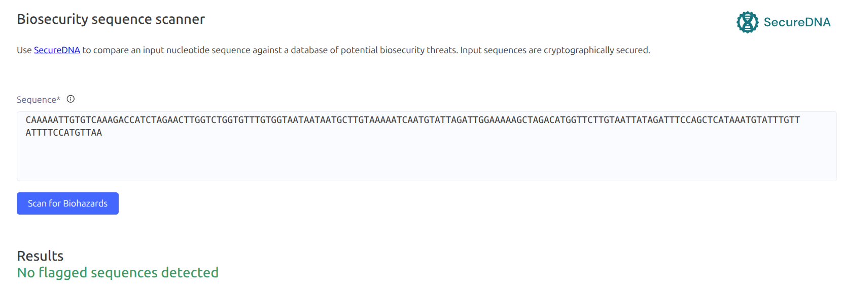
Biosecurity sequence scanner:
The scanner evaluates genetic sequences against international regulatory databases for:
Dual-Use Toxins: High-consequence protein toxins and virulence factors (e.g., ricin, botulinum, diphtheria toxin).
Pandemic Hazards: Sequences of concern that present severe biological safety or biosecurity risks prior to commercial DNA synthesis.
The engineered peptide sequence shows no flags when run throught the biosecurity scanner:
Part 5: From design to build
(this part of the text was AI generated against the reference literature and its still under review)
How would you build it?
Strategy: I would order only the 156 bp coding sequence as a double-stranded synthetic gene fragment (e.g., IDT gBlock or Twist fragment) and insert it into a commercial, linearized pPICZαA vector using Gibson Assembly (Gibson et al., 2009).
Why this choice:
Synthesizing the entire ~3.6 kb plasmid from scratch makes no economic or practical sense: the vector backbone is an inexpensive commodity, and only the 156 bp defensin payload is novel.
Gibson assembly joins overlapping DNA ends in a simple 1-hour isothermal reaction (using a 5’ exonuclease, DNA polymerase, and DNA ligase). By designing ~30 bp overlaps matching the vector ends, the fragment inserts seamlessly right after the secretion signal without leaving unwanted restriction scars.
What does synthesis constrain?
Chemical synthesis error rates: Oligo synthesis has an inherent error rate per base that accumulates with length. For a short ~200 bp fragment (including terminal overlaps), direct synthesis is fast and cheap, and simple colony picking followed by Sanger sequencing is sufficient to verify an error-free clone.
Error correction (MutS): If assembling longer multikilobase genes or using crude oligo pools, enzymatic error correction using the mismatch-binding protein MutS (Carr et al., 2004) would be employed to remove error-containing molecules (reducing error rates by >15-fold).
Manufacturability check:
Our codon-optimized sequence passed all vendor complexity checks: 29.5% GC content, no extreme GC/AT patches, no repetitive hairpins, and no homopolymers longer than 4 bp.
All internal cut sites for key cloning enzymes (EcoRI, NotI, XhoI) and the SacI linearization site were domesticated.
Prototype vs. scale
Prototyping (Benchtop Testing): To test the idea, we only need to build 1–2 constructs (e.g., wild-type vs. G9R/V39R) using individually synthesized gene fragments assembled by Gibson and amplified in E. coli minipreps.
Production Scale: In biomanufacturing, DNA is not the final product. The sequence-verified plasmid is integrated once into the Pichia chromosome to establish a clonal Master Cell Bank (MCB). Subsequent manufacturing scales entirely through yeast fermentation, not repeated DNA synthesis.
High-throughput variant libraries: Chip-synthesized oligo pools (Tian et al., 2004; Kosuri et al., 2010; Kong et al., 2007) would only be needed if we were scaling the design search to screen hundreds of defensin mutants in parallel.
Part 6: What don’t you know yet?
(this part of the text was AI generated against the reference literature and its still under review)
Here are the 3 biggest biological unknowns and bets in the construct, and how to test them:
1. Titer & Biomanufacturing Yield
The Risk: In HW1, our manufacturing plan aims for ~1 g/L of pure peptide from a 500 L bioreactor. However, published yields for plant defensins secreted by Pichia in shake flasks are typically much lower (10–65 mg/L; Almeida et al., 2001; Cabral et al., 2003). Reaching gram-per-liter titers is possible, but requires substantial clone screening and bioprocess tuning.
How to find out: Screen multiple transformed Pichia colonies for gene copy number and quantify secreted peptide by RP-HPLC. Then run a benchtop fed-batch fermentation to compare the methanol-induced AOX1 promoter against a constitutive GAP promoter.
The Risk: Secretion relies on yeast proteases to remove the α-factor leader. While Kex2 cleaves efficiently at -Lys-Arg-, the secondary enzyme Ste13 (which trims the Glu-Ala repeats) is notoriously inefficient during high expression (Cabral et al., 2003). Incomplete trimming leaves extra amino acids on the N-terminus (+400 Da for EAEA), which can disrupt folding or increase cation sensitivity.
How to find out: Analyze the culture supernatant with intact-mass LC-MS to verify whether the peptide has the exact mature mass or carries extra residues.
Next design variant: If trimming is incomplete, remove the EAEA spacer so Kex2 cleaves directly before the mature defensin sequence.
3. Activity of the Double Mutant in Sweat Salinity
The Risk: The G9R and V39R mutations were originally identified in plant defense against crop fungi (Fusarium) (De Samblanx et al., 1997) and modeled computationally (Pandey et al., 2020). However, the combined double mutant has not yet been experimentally tested against human skin pathogens (Candida albicans and Malassezia) under cutaneous salt concentrations (20–100 mM NaCl).
How to find out: Run a broth microdilution assay (CLSI standard for Candida, and lipid-supplemented media for Malassezia) measuring Minimum Inhibitory Concentration (MIC) across a titration of NaCl (0 to 100 mM) plus Ca²⁺/Mg²⁺.
AI
AI (Gemini 3.8 Flash) was used for general guidance and text generation where indicated, using peer-reviewed articles and curated databases as reference. It was also used to retrieve, verify, and format the bibliographic citations.
Terras, F. R., Schoofs, H. M., De Bolle, M. F., Van Leuven, F., Rees, S. B., Vanderleyden, J., Cammue, B. P., and Broekaert, W. F. (1992). Analysis of two novel classes of plant antifungal proteins from radish (Raphanus sativus L.) seeds.Journal of Biological Chemistry, 267(22), 15301–15309. DOI: 10.1016/S0021-9258(19)49534-3
De Samblanx, G. W., Goderis, I. J., Thevissen, K., Raemaekers, R., Fant, F., Borremans, F., Acland, D. P., and Broekaert, W. F. (1997). Mutational analysis of a plant defensin from radish (Raphanus sativus L.) reveals two adjacent sites important for antifungal activity.Journal of Biological Chemistry, 272(2), 1171–1179. DOI: 10.1074/jbc.272.2.1171
Thevissen, K., Warnecke, D. C., François, I. E., Leipelt, M., Heinz, E., Ott, C., Zähringer, U., Thomma, B. P., Ferket, K. K., and Cammue, B. P. (2004). Defensins from insects and plants interact with fungal glucosylceramides: Pichia pastoris Δgcs mutants are highly resistant to defensin-mediated permeabilization.Journal of Biological Chemistry, 279(6), 3900–3905. DOI: 10.1074/jbc.M311165200
Pandey, B., Tyagi, C., Prajapati, G. K., Mishra, A. K., Hashem, A., Alqarawi, A. A., and Abd_Allah, E. F. (2020). Analysis of mutations of defensin protein using accelerated molecular dynamics simulations.PLoS ONE, 15(11), e0241679. DOI: 10.1371/journal.pone.0241679
Recombinant Expression & Protease Processing in Pichia pastoris:
Almeida, M. S., Cabral, K. M., Zingali, R. B., and Kurtenbach, E. (2001). cDNA cloning and heterologous expression of functional cysteine-rich antifungal protein Psd1 in the yeast Pichia pastoris.Archives of Biochemistry and Biophysics, 395(2), 199–207. DOI: 10.1006/abbi.2001.2564
Cabral, K. M., Almeida, M. S., Valente, A. P., Almeida, F. C., and Kurtenbach, E. (2003). Production of the active antifungal Pisum sativum defensin 1 (Psd1) in Pichia pastoris: overcoming the inefficiency of the STE13 protease.Protein Expression and Purification, 31(1), 115–122. DOI: 10.1016/s1046-5928(03)00136-0
Assigned Course Readings:
Gibson, D. G., Young, L., Chuang, R. Y., Venter, J. C., Hutchison, C. A. 3rd, and Smith, H. O. (2009). Enzymatic assembly of DNA molecules up to several hundred kilobases.Nature Methods, 6(5), 343–345. DOI: 10.1038/nmeth.1318
Carr, P. A., Park, J. S., Lee, Y. J., Yu, T., Zhang, S., and Jacobson, J. M. (2004). Protein-mediated error correction for de novo DNA synthesis.Nucleic Acids Research, 32(20), e162. DOI: 10.1093/nar/gnh160
Kosuri, S., Eroshenko, N., Leproust, E. M., Super, M., Way, J., Li, J. B., and Church, G. M. (2010). Scalable gene synthesis by selective amplification of DNA pools from high-fidelity microchips.Nature Biotechnology, 28(12), 1295–1299. DOI: 10.1038/nbt.1716
Tian, J., Gong, H., Sheng, N., Zhou, X., Gulari, E., Gao, X., and Church, G. (2004). Accurate multiplex gene synthesis from programmable DNA microchips.Nature, 432(7020), 1050–1054. DOI: 10.1038/nature03151
Kong, D. S., Carr, P. A., Chen, L., Zhang, S., and Jacobson, J. M. (2007). Parallel gene synthesis in a microfluidic device.Nucleic Acids Research, 35(8), e61. DOI: 10.1093/nar/gkm121
Week 3 HW: Cell-Free Protocols
This is a first draft with AI generated content that is still under review.
A. COMPARE CELL-FREE REACTION COMPOSITIONS
1. Why does the concentration of potassium glutamate and magnesium glutamate differ between compositions?
The optimal concentrations of potassium glutamate and magnesium glutamate differ across cell-free protein synthesis (CFPS) formulations due to several biochemical and biophysical factors:
Substrate Affinity and Ion Chelation:
Formulations using high-energy phosphorylated substrates like phosphoenolpyruvate (PEP) (e.g., PANOx-SP) require higher magnesium concentrations (~16–20 mM) because PEP has a high binding affinity for magnesium and chelates Mg²⁺ ions.
Formulations utilizing pyruvate (Cytomim) or non-phosphorylated energy sources like nucleoside monophosphates (NMPs) and glucose (RFopt) do not chelate Mg²⁺ to the same extent, requiring lower added magnesium glutamate concentrations (~7–8 mM).
Charge Neutralization and Ribosomal Function:
Cations balance the net negative charge of nucleic acid phosphate backbones (tRNA, mRNA, plasmid DNA), ATP/NTPs, and anionic proteins.
Potassium (K⁺) is the primary intracellular monovalent cation in E. coli, while glutamate (Glu⁻) is the principal organic anion produced during osmotic stress. High potassium glutamate concentrations are needed to replicate the cytoplasmic ionic environment and maintain favorable protein–nucleic acid interactions without inhibiting translation.
Phosphate Byproduct Accumulation:
Consumption of PEP liberates high levels of inorganic phosphate (Pi), which can chelate free Mg²⁺ ions and inhibit transcription/translation if magnesium is not kept elevated. Pyruvate and NMP-based systems generate minimal free phosphate, reducing the required Mg²⁺ buffer.
Which is more sensitive?
Magnesium glutamate is significantly more sensitive to concentration changes than potassium glutamate.
Magnesium Glutamate (High Sensitivity):
Protein expression follows a very sharp, narrow optimum curve for Mg²⁺.
In the Cytomim system, shifting magnesium glutamate by just 4 mM above or below its 8 mM optimum drops protein yields to 62% (at 4 mM) and 67% (at 12 mM), collapsing further to 41% at 16 mM.
Similarly, in PANOx-SP, dropping magnesium glutamate from 20 mM down to 8 mM cuts protein yield nearly in half (54%).
Potassium Glutamate (High Tolerance):
Potassium glutamate displays a broad optimal plateau.
In PANOx-SP, yields remain above 92% of maximum across a wide concentration span from 150 mM to 225 mM.
In Cytomim, potassium glutamate maintains 88% to 95% of maximum yield anywhere between 100 mM and 200 mM.
2. Explain how a PEP+NTP reaction formulation is different from a Ribose+NMP reaction formulation.
A PEP+NTP reaction formulation (such as PANOx or PANOx-SP) and a Ribose+NMP reaction formulation (such as RFopt or minimal cell-free formulations) represent two fundamentally different strategies for powering cell-free protein synthesis (CFPS).
The primary differences between the two systems span energy substrates, metabolic homeostasis, cation requirements, reaction kinetics, and overall cost:
1. Energy Substrates & Nucleotide Inputs
PEP+NTP Formulations: Rely on adding pre-synthesized nucleoside triphosphates (ATP, CTP, GTP, UTP) alongside phosphoenolpyruvate (PEP) as a sacrificial, high-energy secondary phosphate donor to drive ATP regeneration.
Ribose+NMP Formulations: Eliminate PEP and NTPs entirely or partially. Instead, they supply cheaper nucleoside monophosphates (AMP, CMP, GMP, UMP) or ribose combined with nucleobases (such as guanine or adenine). The cell extract’s active endogenous salvage pathways and central metabolic networks synthesize and convert these lower-cost inputs into functional NTPs in situ.
2. Byproduct Accumulation & Metabolic Homeostasis
PEP+NTP Formulations: The enzymatic degradation and consumption of PEP liberates substantial amounts of inorganic phosphate (Pi), leading to an accumulation of up to ~30 mM phosphate in batch reactions. This high Pi concentration eventually inhibits protein synthesis machinery and poisons the reaction.
Ribose+NMP Formulations: Because no sacrificial phosphorylated energy substrate is added, free inorganic phosphate is not liberated. Phosphate levels remain low and stable (~10–15 mM), preserving metabolic equilibrium throughout the reaction.
3. Cation (Mg²⁺) Requirements
PEP+NTP Formulations: PEP exhibits a high binding affinity for divalent cations and actively chelates free Mg²⁺ ions. Combined with the high Pi accumulation, these reactions require significantly elevated magnesium glutamate concentrations (~16–20 mM) to sustain translation.
Ribose+NMP Formulations: Lacking PEP chelation and excess Pi, these systems require substantially less added magnesium glutamate (~7–8.8 mM).
4. ATP Dynamics & Oxygen Sensitivity
PEP+NTP Formulations: Generate a rapid, early surge of ATP within the first 30 minutes, but ATP levels collapse to near zero as PEP is consumed and Pi accumulates, terminating protein synthesis early.
Ribose+NMP Formulations: Maintain steady-state ATP levels (~250 µM) over extended batch durations (20+ hours). Because energy regeneration relies on active central carbon metabolism and oxidative phosphorylation rather than chemical phosphate transfer, these formulations are highly sensitive to dissolved oxygen flux and surface area-to-volume ratios.
5. Cost & Economic Viability
PEP+NTP Formulations: Extremely expensive. PEP alone accounts for ~40% of small-molecule reagent costs, pushing total reagent costs to $3,000–$4,500 per liter of CFE.
Ribose+NMP Formulations: Reduce small-molecule reagent costs by ~95% to 99% (down to ~$143/L CFE or $26–$60 per gram of protein), making large-scale biomanufacturing and point-of-use applications economically realistic.
3. Choose two cell-free reaction compositions from the literature, compare and contrast the choice of reagents and the concentration of reagents.
Comparing Two Cell-Free Formulations: PANOx-SP vs. RFopt
To answer this question, we compare two landmark E. coli cell-free protein synthesis (CFPS) formulations:
PANOx-SP (Jewett & Swartz 2004): The classic, high-performance benchmark that powers protein synthesis using expensive, high-energy chemical donors.
RFopt (Olsen et al. 2026): A modern, low-cost formulation that replaces expensive additives with simple sugars and lets the cell extract’s natural enzymes do the recycling.
Reagent & Concentration Comparison Table
Category
Reagent Component
PANOx-SP (Classic)
RFopt (Low-Cost)
Why They Differ
Fuel / Energy
Phosphoenolpyruvate (PEP)
33 mM
0 mM
PANOx-SP uses an expensive high-energy chemical donor; RFopt eliminates it entirely.
D-Ribose + Glucose
0 mM
50 mM + 10 mM
RFopt uses simple, cheap sugars that feed into natural cellular metabolism.
RFopt adds cheaper monophosphate building blocks and re-energizes them in situ.
Essential Salts
Magnesium Glutamate
20 mM
8 mM
PANOx-SP needs extra magnesium because PEP breakdown traps free magnesium ions.
Potassium Glutamate
175 mM
362 mM
RFopt doubles potassium to keep ribosomes and recycling enzymes active.
Buffering & Helpers
HEPES Buffer (pH 7.5)
0–57 mM
75 mM
RFopt uses a stronger buffer to prevent sugar breakdown from making the broth too acidic.
Sodium Oxalate
2.7 mM
0 mM
Added in PANOx-SP to stop enzymes from wasting PEP; unneeded in RFopt.
Inorganic Phosphate
0 mM
15 mM
RFopt adds phosphate as raw material to turn sugar and NMPs into ATP.
Nicotinamide (Vitamin B3)
0 mM
4 mM
Added in RFopt to keep the cell’s natural energy-carrying cofactors stable.
Purified Add-ins
tRNA, CoA, NAD+, Folinic Acid
All 4 added
None added (0 mM)
RFopt relies on the natural background levels already present in the cell extract.
Protein Building Blocks
Amino Acids
2 mM each
5 mM each
RFopt adds higher concentrations so the reaction does not run out of fuel during long runs.
Final Result
Yield & Specific Cost
1.1 g/L ($4,083/g)
2.4 g/L ($60/g)
RFopt doubles the yield at a 68-fold lower cost.
Key Comparisons
1. Energy & Building Blocks: “Pre-made” vs. “Recycled”
PANOx-SP works like buying pre-charged batteries: it feeds high-energy PEP and fully assembled NTPs. This is fast and reliable, but extremely expensive (> $100/L in nucleotides alone).
RFopt works like using a solar charger: it feeds simple sugars (Ribose and Glucose) and raw, uncharged building blocks (NMPs). Enzymes already present in the bacterial lysate use the sugar to recharge NMPs back into active NTPs continuously.
2. Magnesium Concentration: The “Clogging” Problem
Ribosomes need free magnesium (Mg2+) to hold their subunits together.
In PANOx-SP, when PEP is consumed, it releases free phosphate waste. This waste reacts with magnesium to form chalk-like, insoluble precipitates (Mg3(PO4)2), stealing free magnesium from the ribosomes. Therefore, PANOx-SP must start with a very high magnesium concentration (20 mM).
In RFopt, the recycling pathway constantly reuses phosphate, preventing buildup. Because free magnesium is not depleted, RFopt only needs a gentle, physiological level (8 mM).
3. Potassium Concentration: Balancing the Salt Environment
RFopt uses more than double the potassium glutamate (362 mM vs. 175 mM).
Monovalent potassium acts like an electrostatic shield on RNA backbones. Because RFopt runs at lower magnesium, it increases potassium to keep ribosomes structured and support the active flow of metabolic enzymes.
4. Additives & Buffers: Preventing Waste vs. Managing Acid
PANOx-SP includes sodium oxalate as a targeted “brake” to stop a specific bacterial enzyme (pps) from burning through expensive PEP without making ATP.
RFopt does not use PEP, so it omits oxalate. Instead, because fermenting sugars generates mild organic acids, RFopt uses a higher buffer concentration (75 mM HEPES) to keep the pH neutral, alongside a bit of phosphate and nicotinamide to sustain the recycling engine.
5. Complex Cofactors
PANOx-SP supplements four expensive purified components: transfer RNA (tRNA), Coenzyme A (CoA), NAD+, and folinic acid.
RFopt proves these purchases are unnecessary: if the cell lysate is prepared without harsh dialysis, sufficient quantities of these cofactors naturally carry over, slashing the total reaction cost.
Bottom Line
PANOx-SP delivers high-energy parts for a fast reaction (1–2 hours), but quickly stops due to phosphate accumulation and high component cost.
RFopt supplies simple sugars, extra amino acids (5 mM each), and higher buffer capacity, sustaining steady synthesis for over 10 hours to achieve higher yields at a fraction of the cost.
B. DESIGN ONE REACTION COMPOSITION FOR EACH OF THE THREE PROTEINS
1. Reaction compositions
For this part of the homework I collaborated with Beatriz Mesquita and Ada Yazan (both colleagues from the Bioclub Tokyo node) so that each of us could focus on gathering information about each of the proteins and what would be the most important aspects to take into account when designing compositions for each of them. The following compositions are based on Beatriz’s findings for sfGFP and Ada’s for PETase. I focused on gathering information on Reteplase.
My main strategy for the first submission was: start with a baseline and then make changes based on my and my colleagues’ findings for each of the proteins.
Baseline: Started with the reference plate sfGFP Ribose baseline.
Modifications:
Extended energy regeneration: To avoid early energy depletion, I added maltodextrin (600 nL) and sodium pyruvate (200 nL) with potassium oxalate (150 nL) to sustain prolonged ATP synthesis without free phosphate buildup.
Translation stability: Added spermidine (100 nL) to stabilize translation elongation complexes.
Ionic tuning: Lowered potassium glutamate slightly (1675 nL) to keep optimal ionic strength alongside the added energy salts.
Baseline: Started with the reference plate PETase PEP baseline.
Modifications:
Added redox buffer: The reference baseline had no glutathione. PETase (TfCut2)needs an oxidizing environment for its disulfide bond (Cys241–Cys259). I added oxidized glutathione (350 nL GSSG) and reduced glutathione (100 nL GSH) (~3.5:1 ratio) to establish the necessary redox poise.
Maintained isomerase: Kept DsbC (25 nL) to correct mispaired disulfides.
Increased energy: Boosted PEP to 4000 nL to meet translation demands.
Baseline: Started with the reference plate Reteplase PEP baseline.
Modifications:
Lowered DsbC to the catalytic plateau: Lowered DsbC from the excessive 5000 benchmark level down to 200 nL (1.0 uM). This eliminates the massive reagent cost penalty while still supplying sufficient isomerase activity for disulfide shuffling.
Added antioxidant protection: Added catalase (125 nL) to scavenge harmful hydrogen peroxide (H2O2) generated during continuous disulfide formation.
Enhanced thiol exchange: Supplemented free cysteine (225 nL) and adjusted glutathione (475 nL GSSG / 200 nL GSH) to support dynamic disulfide rearrangement across Reteplase’s 9 disulfide bonds (18 cysteines).
Hybrid energy system: Reduced PEP to 2300 nL and added glucose (725 nL) and sodium pyruvate (550 nL) to sustain longer protein synthesis while mitigating phosphate buildup.
2. Suggested new reagent
Glycine betaine
In E. coli, glycine betaine acts as a potent chemical chaperone that directly stabilizes native protein backbones and specifically stimulates the disaggregation and refolding kinetics of the endogenous DnaK/DnaJ/GrpE and ClpB chaperone network. Supplementing betaine prevents misfolding and aggregation of complex target proteins in cell-free extract.
Google Antigravity (Gemini 3.8 Flash) and Google Notebook.
Bibliography
Cai, Q., Hanson, J. A., Steiner, A. R., Tran, C., Masikat, M. R., Chen, R., Zawada, J. F., Sato, A. K., Hallam, T. J., & Yin, G. (2015). A simplified and robust protocol for immunoglobulin expression in Escherichia coli cell-free protein synthesis systems. Biotechnology Progress, 31(3), 823–831. https://doi.org/10.1002/btpr.2082
Fu, C., Shi, W., Li, J., You, S., Qi, W., Peng, X., & Wang, M. (2026). A cell-free platform for the high-efficiency synthesis and screening of PET hydrolase. Journal of Biotechnology, 417, 101–107. https://doi.org/10.1016/j.jbiotec.2026.05.012
Hunt, A. C., Rasor, B. J., Seki, K., Ekas, H. M., Warfel, K. F., Karim, A. S., & Jewett, M. C. (2025). Cell-free gene expression: Methods and applications. Chemical Reviews, 125(1), 91–149. https://doi.org/10.1021/acs.chemrev.4c00116
Hwang, C., Sinskey, A. J., & Lodish, H. F. (1992). Oxidized redox state of glutathione in the endoplasmic reticulum. Science, 257(5076), 1496–1502. https://doi.org/10.1126/science.1523409
Jewett, M. C., & Swartz, J. R. (2004). Mimicking the Escherichia coli cytoplasmic environment activates long-lived and efficient cell-free protein synthesis. Biotechnology and Bioengineering, 86(1), 19–26. https://doi.org/10.1002/bit.20026
Knapp, K. G., Goerke, A. R., & Swartz, J. R. (2007). Cell-free synthesis of proteins that require disulfide bonds using glucose as an energy source. Biotechnology and Bioengineering, 97(4), 901–908. https://doi.org/10.1002/bit.21296
Olsen, M. L., Copeland, C. E., Sundberg, C. A., Aw, R., Shaver, Z. M., Rao, G., Swartz, J. R., Karim, A. S., & Jewett, M. C. (2026). Design-driven optimization of low-cost reagent formulations for reproducible and high-yielding cell-free gene expression. Nature Communications, 17, Article 3478. https://doi.org/10.1038/s41467-026-69605-8
Schafer, F. Q., & Buettner, G. R. (2001). Redox environment of the cell as viewed through the redox state of the glutathione disulfide/glutathione couple. Free Radical Biology & Medicine, 30(11), 1191–1212. https://doi.org/10.1016/s0891-5849(01)00480-4
Yin, G., & Swartz, J. R. (2004). Enhancing multiple disulfide bonded protein folding in a cell-free system. Biotechnology and Bioengineering, 86(2), 188–195. https://doi.org/10.1002/bit.10827


_A_reference_map.png)