Week 3 HW: Cell-Free Protocols
This is a first draft with AI generated content that is still under review.
A. COMPARE CELL-FREE REACTION COMPOSITIONS
1. Why does the concentration of potassium glutamate and magnesium glutamate differ between compositions?
The optimal concentrations of potassium glutamate and magnesium glutamate differ across cell-free protein synthesis (CFPS) formulations due to several biochemical and biophysical factors:
Substrate Affinity and Ion Chelation:
- Formulations using high-energy phosphorylated substrates like phosphoenolpyruvate (PEP) (e.g., PANOx-SP) require higher magnesium concentrations (~16–20 mM) because PEP has a high binding affinity for magnesium and chelates Mg²⁺ ions.
- Formulations utilizing pyruvate (Cytomim) or non-phosphorylated energy sources like nucleoside monophosphates (NMPs) and glucose (RFopt) do not chelate Mg²⁺ to the same extent, requiring lower added magnesium glutamate concentrations (~7–8 mM).
Charge Neutralization and Ribosomal Function:
- Cations balance the net negative charge of nucleic acid phosphate backbones (tRNA, mRNA, plasmid DNA), ATP/NTPs, and anionic proteins.
- Potassium (K⁺) is the primary intracellular monovalent cation in E. coli, while glutamate (Glu⁻) is the principal organic anion produced during osmotic stress. High potassium glutamate concentrations are needed to replicate the cytoplasmic ionic environment and maintain favorable protein–nucleic acid interactions without inhibiting translation.
Phosphate Byproduct Accumulation:
- Consumption of PEP liberates high levels of inorganic phosphate (Pi), which can chelate free Mg²⁺ ions and inhibit transcription/translation if magnesium is not kept elevated. Pyruvate and NMP-based systems generate minimal free phosphate, reducing the required Mg²⁺ buffer.
Which is more sensitive?
Magnesium glutamate is significantly more sensitive to concentration changes than potassium glutamate.
Magnesium Glutamate (High Sensitivity):
- Protein expression follows a very sharp, narrow optimum curve for Mg²⁺.
- In the Cytomim system, shifting magnesium glutamate by just 4 mM above or below its 8 mM optimum drops protein yields to 62% (at 4 mM) and 67% (at 12 mM), collapsing further to 41% at 16 mM.
- Similarly, in PANOx-SP, dropping magnesium glutamate from 20 mM down to 8 mM cuts protein yield nearly in half (54%).
Potassium Glutamate (High Tolerance):
- Potassium glutamate displays a broad optimal plateau.
- In PANOx-SP, yields remain above 92% of maximum across a wide concentration span from 150 mM to 225 mM.
- In Cytomim, potassium glutamate maintains 88% to 95% of maximum yield anywhere between 100 mM and 200 mM.
2. Explain how a PEP+NTP reaction formulation is different from a Ribose+NMP reaction formulation.
A PEP+NTP reaction formulation (such as PANOx or PANOx-SP) and a Ribose+NMP reaction formulation (such as RFopt or minimal cell-free formulations) represent two fundamentally different strategies for powering cell-free protein synthesis (CFPS).
The primary differences between the two systems span energy substrates, metabolic homeostasis, cation requirements, reaction kinetics, and overall cost:
1. Energy Substrates & Nucleotide Inputs
- PEP+NTP Formulations: Rely on adding pre-synthesized nucleoside triphosphates (ATP, CTP, GTP, UTP) alongside phosphoenolpyruvate (PEP) as a sacrificial, high-energy secondary phosphate donor to drive ATP regeneration.
- Ribose+NMP Formulations: Eliminate PEP and NTPs entirely or partially. Instead, they supply cheaper nucleoside monophosphates (AMP, CMP, GMP, UMP) or ribose combined with nucleobases (such as guanine or adenine). The cell extract’s active endogenous salvage pathways and central metabolic networks synthesize and convert these lower-cost inputs into functional NTPs in situ.
2. Byproduct Accumulation & Metabolic Homeostasis
- PEP+NTP Formulations: The enzymatic degradation and consumption of PEP liberates substantial amounts of inorganic phosphate (Pi), leading to an accumulation of up to ~30 mM phosphate in batch reactions. This high Pi concentration eventually inhibits protein synthesis machinery and poisons the reaction.
- Ribose+NMP Formulations: Because no sacrificial phosphorylated energy substrate is added, free inorganic phosphate is not liberated. Phosphate levels remain low and stable (~10–15 mM), preserving metabolic equilibrium throughout the reaction.
3. Cation (Mg²⁺) Requirements
- PEP+NTP Formulations: PEP exhibits a high binding affinity for divalent cations and actively chelates free Mg²⁺ ions. Combined with the high Pi accumulation, these reactions require significantly elevated magnesium glutamate concentrations (~16–20 mM) to sustain translation.
- Ribose+NMP Formulations: Lacking PEP chelation and excess Pi, these systems require substantially less added magnesium glutamate (~7–8.8 mM).
4. ATP Dynamics & Oxygen Sensitivity
- PEP+NTP Formulations: Generate a rapid, early surge of ATP within the first 30 minutes, but ATP levels collapse to near zero as PEP is consumed and Pi accumulates, terminating protein synthesis early.
- Ribose+NMP Formulations: Maintain steady-state ATP levels (~250 µM) over extended batch durations (20+ hours). Because energy regeneration relies on active central carbon metabolism and oxidative phosphorylation rather than chemical phosphate transfer, these formulations are highly sensitive to dissolved oxygen flux and surface area-to-volume ratios.
5. Cost & Economic Viability
- PEP+NTP Formulations: Extremely expensive. PEP alone accounts for ~40% of small-molecule reagent costs, pushing total reagent costs to $3,000–$4,500 per liter of CFE.
- Ribose+NMP Formulations: Reduce small-molecule reagent costs by ~95% to 99% (down to ~$143/L CFE or $26–$60 per gram of protein), making large-scale biomanufacturing and point-of-use applications economically realistic.
3. Choose two cell-free reaction compositions from the literature, compare and contrast the choice of reagents and the concentration of reagents.
Comparing Two Cell-Free Formulations: PANOx-SP vs. RFopt
To answer this question, we compare two landmark E. coli cell-free protein synthesis (CFPS) formulations:
- PANOx-SP (Jewett & Swartz 2004): The classic, high-performance benchmark that powers protein synthesis using expensive, high-energy chemical donors.
- RFopt (Olsen et al. 2026): A modern, low-cost formulation that replaces expensive additives with simple sugars and lets the cell extract’s natural enzymes do the recycling.
Reagent & Concentration Comparison Table
| Category | Reagent Component | PANOx-SP (Classic) | RFopt (Low-Cost) | Why They Differ |
|---|---|---|---|---|
| Fuel / Energy | Phosphoenolpyruvate (PEP) | 33 mM | 0 mM | PANOx-SP uses an expensive high-energy chemical donor; RFopt eliminates it entirely. |
| D-Ribose + Glucose | 0 mM | 50 mM + 10 mM | RFopt uses simple, cheap sugars that feed into natural cellular metabolism. | |
| RNA Building Blocks | NTPs (ATP, CTP, GTP, UTP) | 1.2 mM / 0.85 mM | 0 mM | PANOx-SP adds fully built, expensive triphosphates directly. |
| NMPs (AMP, CMP, GMP, UMP) | 0 mM | 3.0 mM / 2.15 mM | RFopt adds cheaper monophosphate building blocks and re-energizes them in situ. | |
| Essential Salts | Magnesium Glutamate | 20 mM | 8 mM | PANOx-SP needs extra magnesium because PEP breakdown traps free magnesium ions. |
| Potassium Glutamate | 175 mM | 362 mM | RFopt doubles potassium to keep ribosomes and recycling enzymes active. | |
| Buffering & Helpers | HEPES Buffer (pH 7.5) | 0–57 mM | 75 mM | RFopt uses a stronger buffer to prevent sugar breakdown from making the broth too acidic. |
| Sodium Oxalate | 2.7 mM | 0 mM | Added in PANOx-SP to stop enzymes from wasting PEP; unneeded in RFopt. | |
| Inorganic Phosphate | 0 mM | 15 mM | RFopt adds phosphate as raw material to turn sugar and NMPs into ATP. | |
| Nicotinamide (Vitamin B3) | 0 mM | 4 mM | Added in RFopt to keep the cell’s natural energy-carrying cofactors stable. | |
| Purified Add-ins | tRNA, CoA, NAD+, Folinic Acid | All 4 added | None added (0 mM) | RFopt relies on the natural background levels already present in the cell extract. |
| Protein Building Blocks | Amino Acids | 2 mM each | 5 mM each | RFopt adds higher concentrations so the reaction does not run out of fuel during long runs. |
| Final Result | Yield & Specific Cost | 1.1 g/L ($4,083/g) | 2.4 g/L ($60/g) | RFopt doubles the yield at a 68-fold lower cost. |
Key Comparisons
1. Energy & Building Blocks: “Pre-made” vs. “Recycled”
- PANOx-SP works like buying pre-charged batteries: it feeds high-energy PEP and fully assembled NTPs. This is fast and reliable, but extremely expensive (> $100/L in nucleotides alone).
- RFopt works like using a solar charger: it feeds simple sugars (Ribose and Glucose) and raw, uncharged building blocks (NMPs). Enzymes already present in the bacterial lysate use the sugar to recharge NMPs back into active NTPs continuously.
2. Magnesium Concentration: The “Clogging” Problem
- Ribosomes need free magnesium (Mg2+) to hold their subunits together.
- In PANOx-SP, when PEP is consumed, it releases free phosphate waste. This waste reacts with magnesium to form chalk-like, insoluble precipitates (Mg3(PO4)2), stealing free magnesium from the ribosomes. Therefore, PANOx-SP must start with a very high magnesium concentration (20 mM).
- In RFopt, the recycling pathway constantly reuses phosphate, preventing buildup. Because free magnesium is not depleted, RFopt only needs a gentle, physiological level (8 mM).
3. Potassium Concentration: Balancing the Salt Environment
- RFopt uses more than double the potassium glutamate (362 mM vs. 175 mM).
- Monovalent potassium acts like an electrostatic shield on RNA backbones. Because RFopt runs at lower magnesium, it increases potassium to keep ribosomes structured and support the active flow of metabolic enzymes.
4. Additives & Buffers: Preventing Waste vs. Managing Acid
- PANOx-SP includes sodium oxalate as a targeted “brake” to stop a specific bacterial enzyme (pps) from burning through expensive PEP without making ATP.
- RFopt does not use PEP, so it omits oxalate. Instead, because fermenting sugars generates mild organic acids, RFopt uses a higher buffer concentration (75 mM HEPES) to keep the pH neutral, alongside a bit of phosphate and nicotinamide to sustain the recycling engine.
5. Complex Cofactors
- PANOx-SP supplements four expensive purified components: transfer RNA (tRNA), Coenzyme A (CoA), NAD+, and folinic acid.
- RFopt proves these purchases are unnecessary: if the cell lysate is prepared without harsh dialysis, sufficient quantities of these cofactors naturally carry over, slashing the total reaction cost.
Bottom Line
- PANOx-SP delivers high-energy parts for a fast reaction (1–2 hours), but quickly stops due to phosphate accumulation and high component cost.
- RFopt supplies simple sugars, extra amino acids (5 mM each), and higher buffer capacity, sustaining steady synthesis for over 10 hours to achieve higher yields at a fraction of the cost.
B. DESIGN ONE REACTION COMPOSITION FOR EACH OF THE THREE PROTEINS
1. Reaction compositions
For this part of the homework I collaborated with Beatriz Mesquita and Ada Yazan (both colleagues from the Bioclub Tokyo node) so that each of us could focus on gathering information about each of the proteins and what would be the most important aspects to take into account when designing compositions for each of them. The following compositions are based on Beatriz’s findings for sfGFP and Ada’s for PETase. I focused on gathering information on Reteplase.
My main strategy for the first submission was: start with a baseline and then make changes based on my and my colleagues’ findings for each of the proteins.
sfGFP Reaction Composition
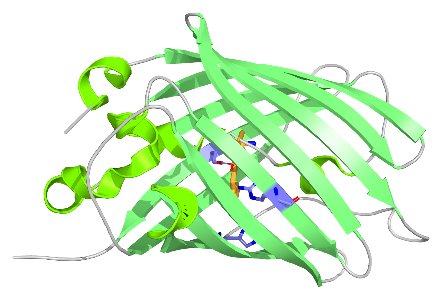
- Platform Link:
https://coopetition.htgaa.org/cfps?design=29ym3na6546ki2n - Sample ID:
ruben_goncalves_2026-10-05_sfgfp_v1.1
JSON
Rationale
- Baseline: Started with the reference plate sfGFP Ribose baseline.
- Modifications:
- Extended energy regeneration: To avoid early energy depletion, I added maltodextrin (600 nL) and sodium pyruvate (200 nL) with potassium oxalate (150 nL) to sustain prolonged ATP synthesis without free phosphate buildup.
- Translation stability: Added spermidine (100 nL) to stabilize translation elongation complexes.
- Ionic tuning: Lowered potassium glutamate slightly (1675 nL) to keep optimal ionic strength alongside the added energy salts.
PETase Reaction Composition
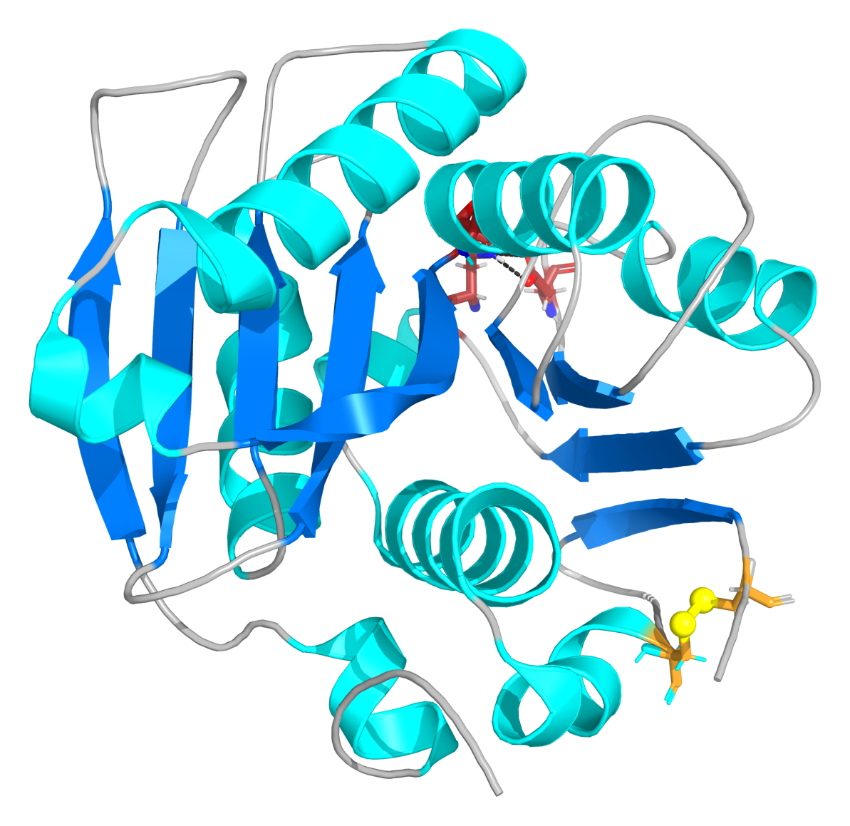
- Platform Link:
https://coopetition.htgaa.org/cfps?design=87u97tdicf6kke7 - Sample ID:
ruben_goncalves_2026-10-05_petase_v1
JSON
Rationale
- Baseline: Started with the reference plate PETase PEP baseline.
- Modifications:
- Added redox buffer: The reference baseline had no glutathione. PETase (TfCut2)needs an oxidizing environment for its disulfide bond (Cys241–Cys259). I added oxidized glutathione (350 nL GSSG) and reduced glutathione (100 nL GSH) (~3.5:1 ratio) to establish the necessary redox poise.
- Maintained isomerase: Kept DsbC (25 nL) to correct mispaired disulfides.
- Increased energy: Boosted PEP to 4000 nL to meet translation demands.
Reteplase Reaction Composition
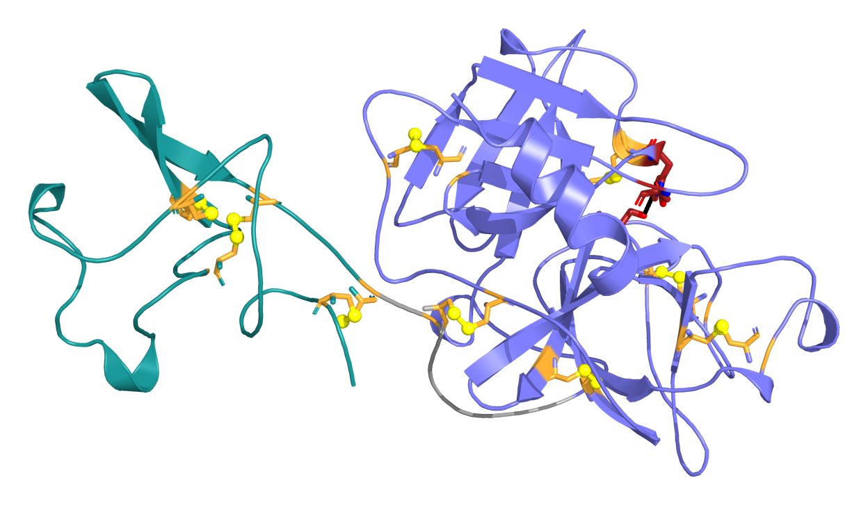
- Platform Link:
https://coopetition.htgaa.org/cfps?design=k0oc48m0mssy5um - Sample ID:
ruben_goncalves_2026-10-05_reteplase_v1
JSON
Rationale
- Baseline: Started with the reference plate Reteplase PEP baseline.
- Modifications:
- Lowered DsbC to the catalytic plateau: Lowered DsbC from the excessive 5000 benchmark level down to 200 nL (1.0 uM). This eliminates the massive reagent cost penalty while still supplying sufficient isomerase activity for disulfide shuffling.
- Added antioxidant protection: Added catalase (125 nL) to scavenge harmful hydrogen peroxide (H2O2) generated during continuous disulfide formation.
- Enhanced thiol exchange: Supplemented free cysteine (225 nL) and adjusted glutathione (475 nL GSSG / 200 nL GSH) to support dynamic disulfide rearrangement across Reteplase’s 9 disulfide bonds (18 cysteines).
- Hybrid energy system: Reduced PEP to 2300 nL and added glucose (725 nL) and sodium pyruvate (550 nL) to sustain longer protein synthesis while mitigating phosphate buildup.
2. Suggested new reagent
Glycine betaine
In E. coli, glycine betaine acts as a potent chemical chaperone that directly stabilizes native protein backbones and specifically stimulates the disaggregation and refolding kinetics of the endogenous DnaK/DnaJ/GrpE and ClpB chaperone network. Supplementing betaine prevents misfolding and aggregation of complex target proteins in cell-free extract.
https://pubmed.ncbi.nlm.nih.gov/11517217/
C. ASIMOV KERNEL
In progress!
Use of AI
Google Antigravity (Gemini 3.8 Flash) and Google Notebook.
Bibliography
Cai, Q., Hanson, J. A., Steiner, A. R., Tran, C., Masikat, M. R., Chen, R., Zawada, J. F., Sato, A. K., Hallam, T. J., & Yin, G. (2015). A simplified and robust protocol for immunoglobulin expression in Escherichia coli cell-free protein synthesis systems. Biotechnology Progress, 31(3), 823–831. https://doi.org/10.1002/btpr.2082
Fu, C., Shi, W., Li, J., You, S., Qi, W., Peng, X., & Wang, M. (2026). A cell-free platform for the high-efficiency synthesis and screening of PET hydrolase. Journal of Biotechnology, 417, 101–107. https://doi.org/10.1016/j.jbiotec.2026.05.012
Hunt, A. C., Rasor, B. J., Seki, K., Ekas, H. M., Warfel, K. F., Karim, A. S., & Jewett, M. C. (2025). Cell-free gene expression: Methods and applications. Chemical Reviews, 125(1), 91–149. https://doi.org/10.1021/acs.chemrev.4c00116
Hwang, C., Sinskey, A. J., & Lodish, H. F. (1992). Oxidized redox state of glutathione in the endoplasmic reticulum. Science, 257(5076), 1496–1502. https://doi.org/10.1126/science.1523409
Jewett, M. C., & Swartz, J. R. (2004). Mimicking the Escherichia coli cytoplasmic environment activates long-lived and efficient cell-free protein synthesis. Biotechnology and Bioengineering, 86(1), 19–26. https://doi.org/10.1002/bit.20026
Knapp, K. G., Goerke, A. R., & Swartz, J. R. (2007). Cell-free synthesis of proteins that require disulfide bonds using glucose as an energy source. Biotechnology and Bioengineering, 97(4), 901–908. https://doi.org/10.1002/bit.21296
Olsen, M. L., Copeland, C. E., Sundberg, C. A., Aw, R., Shaver, Z. M., Rao, G., Swartz, J. R., Karim, A. S., & Jewett, M. C. (2026). Design-driven optimization of low-cost reagent formulations for reproducible and high-yielding cell-free gene expression. Nature Communications, 17, Article 3478. https://doi.org/10.1038/s41467-026-69605-8
Schafer, F. Q., & Buettner, G. R. (2001). Redox environment of the cell as viewed through the redox state of the glutathione disulfide/glutathione couple. Free Radical Biology & Medicine, 30(11), 1191–1212. https://doi.org/10.1016/s0891-5849(01)00480-4
Yin, G., & Swartz, J. R. (2004). Enhancing multiple disulfide bonded protein folding in a cell-free system. Biotechnology and Bioengineering, 86(2), 188–195. https://doi.org/10.1002/bit.10827