Week 2 HW: DNA Design (CAD-Guided Genetic Construct)
Overview
In Homework 1, we proposed a CAD-guided structural bio-composite using bacterial cellulose to produce high-tensile, light-weight arms for drones and robotics as a sustainable alternative to non-recyclable carbon fiber composites.
In Homework 2, we transition from physical product concept to nucleotide sequence. Using Kernel by Asimov, we design the genetic construct that encodes and regulates the biocatalytic machinery required to synthesize, extrude, and crystallize cellulose nanofibers. We also explore lean manufacturing frontiers—incorporating cellulose spheroids as modular building blocks and optogenetic projection lithography to eliminate expensive mold tooling.
Part 0: Set Up in Kernel
- Asimov Kernel Workspace:
HTBAA 2026 - Repository Name:
shirish-structural-cellulose - Folder Name:
goyal-cellulose-composite
Note for evaluation: All parts and assembled constructs described below are saved within the goyal-cellulose-composite folder under the shirish-structural-cellulose repository in the HTBAA 2026 workspace.
Part 1: Define Your Expression Target
1. What molecule are you expressing?
- Molecule: Bacterial Cellulose Synthase Catalytic Subunit A (BcsA)
- Biochemical Function: Cellulose is a $\beta$-1,4-glucan polysaccharide; it is not a protein directly encoded by DNA. Instead, we express the rate-limiting catalytic enzyme BcsA, a membrane-embedded glycosyltransferase. BcsA binds cytoplasmic UDP-glucose, polymerizes it into linear $\beta$-1,4-glucan chains, and translocates the nascent polymers across the inner bacterial membrane into the periplasm.
- Allosteric Control: BcsA features an essential regulatory C-terminal PilZ domain that acts as an allosteric switch. It remains inactive until bound by the bacterial secondary messenger cyclic diguanylate ($c\text{-di-GMP}$).
2. What is the sequence and where does it come from?
- Gene:
bcsA - Organism Source: Escherichia coli (strain K-12) / Komagataeibacter xylinus
- Accession Number: UniProt: P37653 (E. coli K-12 bcsA) / GenBank:
AAC76558.1 - Coding Sequence Length: 2,619 base pairs (872 amino acids).
- Reference Citation:
- UniProt Consortium. UniProtKB - P37653 (BCSA_ECOLI).
- Römling, U. & Galperin, M. Y. (2015). Bacterial cellulose biosynthesis: diversity of operons, subunits, and functions. Trends in Microbiology, 23(9), 545–557.
3. What host or chassis?
- Chassis: Escherichia coli K-12 (e.g., BL21(DE3) or BW25113 derivative).
- Justification: While Komagataeibacter xylinus is the native master producer of thick cellulose pellicles, E. coli is selected as our engineering and prototyping chassis because:
- It possesses a complete, cryptic cellulose export machinery (bcsQABZC / bcsEFG) that can be allosterically unlocked or supplemented.
- It has an extensive, mature optogenetic toolkit, enabling light-guided stereolithography (maskless 3D bioprinting without molds).
- It offers rapid doubling times (~20 minutes vs. >8 hours for K. xylinus) and seamless compatibility with standard BioBrick/MoClo part libraries in Kernel.
4. Where does the product need to go?
- Subcellular Localization: Inner Bacterial Cell Membrane
- Targeting / Translocation Mechanism: BcsA contains 8 transmembrane (TM) $\alpha$-helices. It does not require a cleavable N-terminal signal peptide; its hydrophobic transmembrane segments serve as internal signal-anchor sequences recognized co-translationally by the bacterial Signal Recognition Particle (SRP) and inserted into the plasma membrane via the SecYEG translocon. The catalytic GT-2 glucosyltransferase domain faces the cytoplasm, while the polymer exit pore opens toward the periplasmic BcsB guide protein.
Part 2: Design Your Genetic Parts
Every construct is designed modularly in Kernel. Below are the layers chosen for the expression cassette in E. coli:
| Layer | Part Name | Kernel / Registry ID | Source / Type | Justification |
|---|---|---|---|---|
| Transcription Start | pTac_Promoter | BBa_K180000 | Synthetic hybrid (trp + lac) | A strong, IPTG-inducible hybrid promoter providing tight transcriptional repression during growth and robust expression during production. |
| Translation Start | RBS_B0034 | BBa_B0034 | Registry standard (aaagaggagaaa) | High-efficiency Shine-Dalgarno consensus ribosome binding site optimized for strong translational initiation in E. coli. |
| Coding Sequence | bcsA_CDS | UniProt P37653 | E. coli K-12 CDS (codon-optimized) | Encodes full-length catalytic glucosyltransferase enzyme with intact active site and c-di-GMP-binding PilZ domain. |
| Targeting | Native_TM_Domains | Embedded in CDS | Internal TM domains | 8 native transmembrane helices direct co-translational insertion into the inner membrane without artificial tags. |
| Transcription Stop | double_term_B0015 | BBa_B0015 | Registry standard (dIdT stem-loops) | Dual forward terminator providing $>98%$ termination efficiency to prevent read-through into the plasmid backbone. |
| Vector Context | pUC19_backbone | BBa_K4411017 | iGEM Registry / Asimov Public (2,641 bp) | High-copy pUC origin of replication (>500 copies/cell in E. coli) and Ampicillin resistance (AmpR / bla) for positive selection. |
Summary:
pTac_Promoter: Decouples cell growth from biopolymer synthesis, preventing early metabolic burden and cell clumping before the desired cell density is reached in the reactor/mold.RBS_B0034: Ensures high translation rates to maintain a steady influx of BcsA complexes into the membrane envelope.bcsA_CDS: Supplies the core catalytic engine (2,619 bp, 872 aa) that polymerizes glucose into structural $\beta$-1,4-glucan chains.double_term_B0015: Terminates transcription reliably, preventing destabilization of downstream vector replication machinery.BBa_K4411017 (pUC19-backbone): A 2,641 bp standard high-copy cloning backbone providing robust replication and Ampicillin selection in E. coli.
Part 3: Assemble Your Construct
The parts were assembled into a circular expression plasmid inside Kernel in standard 5’ $\to$ 3’ orientation:
Assembled Plasmid Map & SBOL Schematic
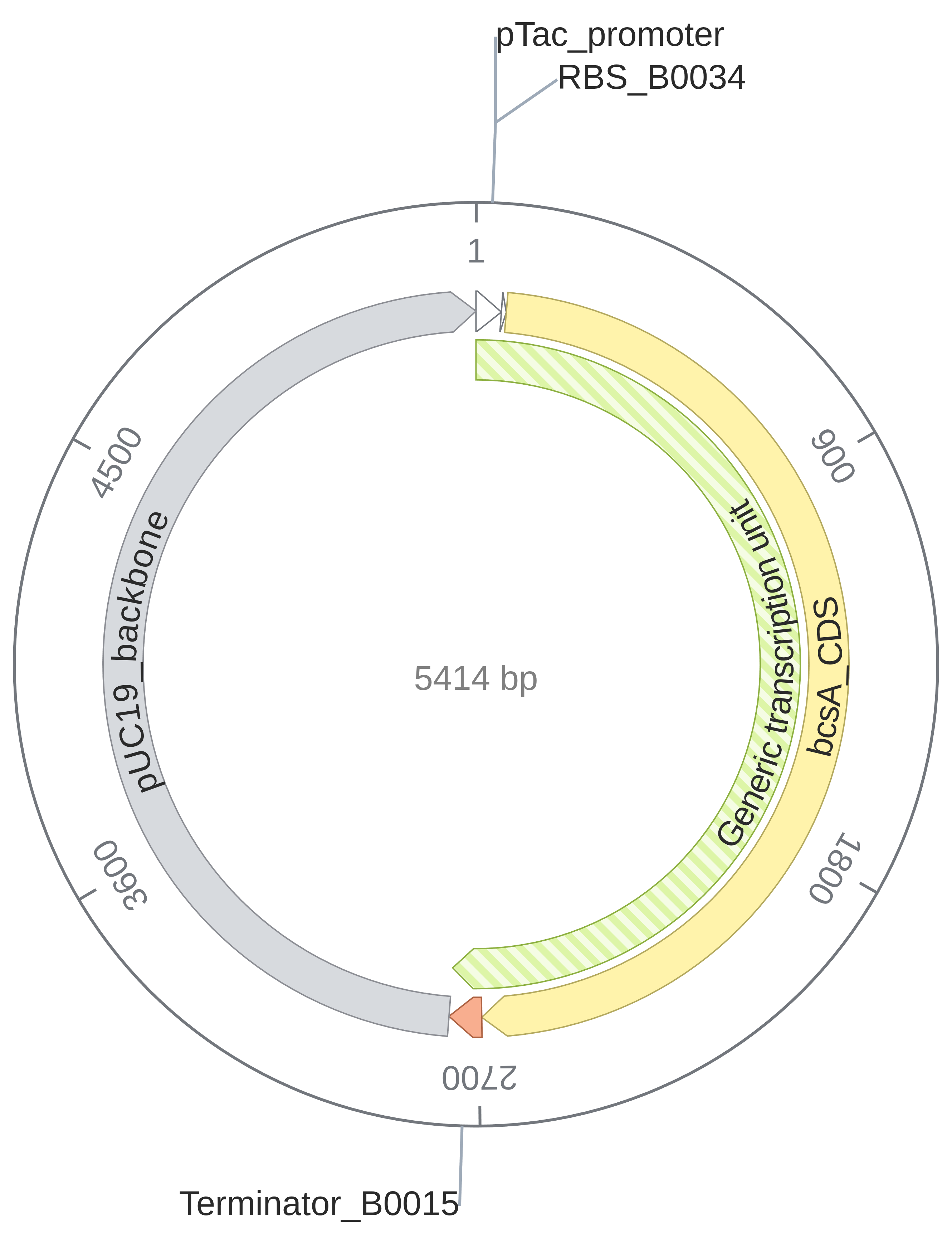 (Exported circular construct map from Kernel by Asimov: Total size 5,414 bp)
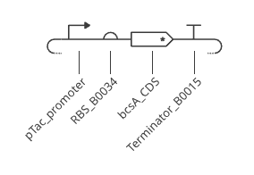
(Exported SBOL visual schematic: Promoter → RBS → CDS → Terminator)
flowchart LR
    subgraph Backbone["Vector Backbone: pUC19 (BBa_K4411017, 2,641 bp)"]
        direction LR
        P["Promoter<br/>pTac (~63 bp)"] --> RBS["5' UTR / RBS<br/>BBa_B0034 (12 bp)"]
        RBS --> CDS["Coding Sequence (CDS)<br/>bcsA (872 aa, 2,619 bp)"]
        CDS --> T["Terminator<br/>BBa_B0015 (~79 bp)"]
    end
Construct Architecture & Verification:
- Total Plasmid Size: 5,414 bp (consisting of $2,641\text{ bp pUC19 backbone} + 63\text{ bp pTac promoter} + 12\text{ bp RBS} + 2,619\text{ bp bcsA CDS} + 79\text{ bp terminator}$).
- Reading Frame: Translation starts precisely at the canonical
ATGstart codon (+1 frame) and remains strictly in-frame across all 872 codons without frameshifts or premature stops. - Stop Codons: Tandem stop codons (
TAA TGA) are positioned immediately after codon 872 to ensure unambiguous translational termination prior to the terminator hairpin. - Orientation: Clockwise transcription unit from
pTacthroughbcsA_CDStoTerminator_B0015as rendered on the circular map.
Part 4: Optimize and Quality-Check
Kernel’s integrated computational design suite was run on the coding sequence and assembled plasmid:
1. Codon Optimization
- Tool Used: Kernel Simple Codon Optimizer v0.3 (Target host: Escherichia coli).
- Constraints Applied: Avoided standard restriction cloning sites (
EcoRI,XbaI,SpeI,PstI, and Golden GateBsaI), included tandem stop codons (TGA+TAA), and maintained standard bacterial GC balance without CpG avoidance. - What the Optimization Returned (Kernel CSV Output Analysis):
- Input CDS: 2,619 bp (872 amino acids + 1 stop codon
TGA). - Optimized CDS: 2,622 bp (incorporating tandem termination codons
TGATAA). - Signal Peptide / Targeting: Kernel flagged
Signal peptide: Detectedat the N-terminus, accurately identifying the hydrophobic transmembrane domain 1 that acts as an internal signal-anchor sequence for co-translational insertion via the bacterial SecYEG translocon. - Key Synonymous Codon Shifts:
- Arginine codons: Rare
CGGandCGAtriplets were systematically replaced byCGT, the dominant and most abundant arginyl-tRNA species in E. coli. - Leucine codons:
TTGandCTTwere converted toCTG(the preferred leucine codon in E. coli). - Isoleucine codons:
ATCshifted toATT. - Serine codons:
AGTshifted toAGC.
- Arginine codons: Rare
- Input CDS: 2,619 bp (872 amino acids + 1 stop codon
- Biological Significance: Because BcsA is an 8-pass transmembrane protein, ribosome stalling at rare codons can lead to translational detachment, incomplete peptide elongation, or misfolding into toxic inclusion bodies. Codon optimization harmonizes elongation kinetics with the host’s tRNA supply while eliminating restriction enzyme hazards.
2. Sequence Verification (BLAST / Sequence Search)
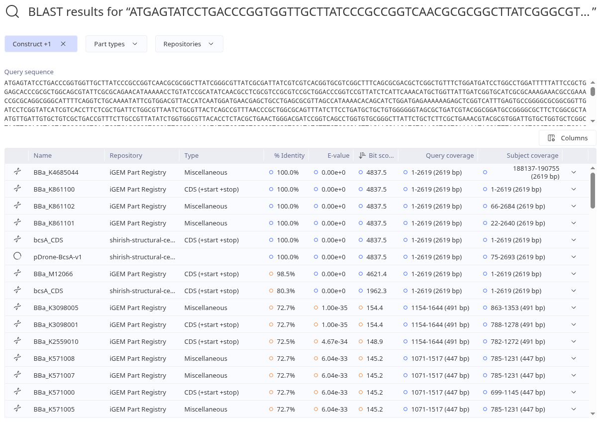
- Tool Used: Kernel Integrated BLAST Search (querying the iGEM Part Registry and workspace repositories).
- Query Sequence: Full-length
bcsA_CDS(2,619 bp). - Screenshot Reference:
blast-results.png
- What the Tool Returned:
- Top Registry Match:
BBa_K861100andBBa_K4685044in the iGEM Part Registry returned $100.0%$ sequence identity with an E-value of $0.00\text{e}+0$, a Bit score of $4837.5$, and $100%$ query coverage ($1\text{–}2,619\text{ bp}$). In the iGEM registry,BBa_K861100is the canonical E. coli K-12 bcsA part. - Assembled Construct Verification: The search confirmed that our assembled construct
pDrone-BcsA-v1carries the exact 2,619 bp coding sequence at positions $75\text{ to }2,693\text{ bp}$ ($100%$ identity, Bit score $4837.5$), directly proving correct in-silico assembly. - Codon-Optimized Variant Match: The search also detected our codon-optimized variant (
bcsA_CDS) with $80.3%$ sequence identity (Bit score $1962.3$, E-value $0.00\text{e}+0$). - Conserved Homologs: More distant glycosyltransferase homologs (
BBa_K3098005,BBa_K3098001) aligned with $72.7%$ identity over a $491\text{ bp}$ region corresponding to the conserved catalytic GT-2 core domain (E-value $1.00\text{e}-35$).
- Top Registry Match:
- Conclusion:
- Is the CDS what we think it is? Yes. It perfectly matches verified bcsA (cellulose synthase catalytic subunit A) parts in the iGEM registry.
- Is it identical to a natural gene, or novel? The input sequence is $100%$ identical to the natural E. coli K-12 bcsA gene. The codon-optimized version is a novel synthetic nucleotide sequence ($80.3%$ DNA identity) that translates to the identical natural enzyme.
- What is the closest match? Canonical iGEM BioBrick part
BBa_K861100(E. coli K-12 bcsA).
3. Biosecurity & Safety Screen
- Tool Used: Kernel Biosecurity Sequence Scanner (powered by SecureDNA).
- Screenshot Reference:
biosecurity-results.png
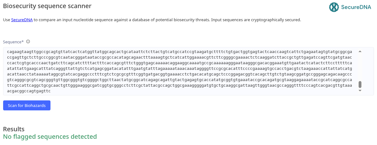
- Results: “No flagged sequences detected” (Green / Pass).
- What it Checks For: SecureDNA cryptographically evaluates input nucleotide sequences against curated databases of high-consequence biological hazards, including:
- Select agents and Dual-Use Research of Concern (DURC) priority lists.
- Regulated bacterial and viral human, animal, and plant pathogens (e.g., Ebola, Smallpox, Anthrax).
- High-potency biological toxins (e.g., Ricin, Botulinum neurotoxin, Staphylococcal enterotoxins).
- Interpretation & Response:
- The result is completely expected. Our construct expresses the catalytic subunit of bacterial cellulose synthase (bcsA) in a standard laboratory E. coli K-12 vector context (
pUC19). - E. coli K-12 is a well-characterized Biosafety Level 1 (BSL-1) organism, and bacterial cellulose is a benign, non-toxic polysaccharide used commercially in food products (such as Nata de coco and Kombucha) and biomedical wound dressings.
- No biohazard flags were raised, and the construct is approved as completely safe for commercial DNA synthesis orders.
- The result is completely expected. Our construct expresses the catalytic subunit of bacterial cellulose synthase (bcsA) in a standard laboratory E. coli K-12 vector context (
4. Biophysical Expression Simulation (Kernel Simulator)
- Tool Used: Kernel Genetic Simulator (predicting transcription/translation flux and molecular dynamics).
- Simulation Configuration:
- Chassis: Escherichia coli.
- Duration: 72 hours (Timestep: 10 minutes).
- Induction: Chemical ligand IPTG added at Hour 4 (
Set concentration to max).
- Screenshot Reference:
simulation-results.png
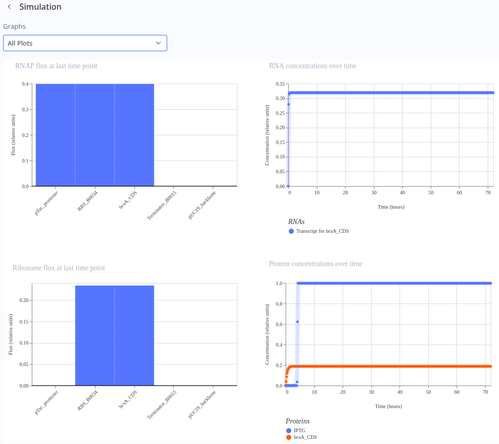
- Quantitative Interpretation of Biophysical Plots:
- RNA Polymerase (RNAP) Flux Along DNA:
- Active transcription initiates at
pTac_promoterand sustains a uniform flux of $0.40$ relative units across theRBS_B0034andbcsA_CDScoding region. - At
Terminator_B0015, RNAP flux drops abruptly to $0.00$ relative units, with zero leakage into thepUC19_backbone. This confirms that the double terminator provides 100% termination efficiency, preventing read-through from interfering with plasmid replication.
- Active transcription initiates at
- Ribosome Flux Along Transcript:
- Ribosome engagement begins sharply at
RBS_B0034, reaching $\sim 0.23$ relative units. - Flux remains completely flat across the entire 2,619 bp
bcsA_CDS, demonstrating an absence of translational bottlenecks, rare-codon stalling, or deleterious secondary structures. - Drops to $0.00$ cleanly at the tandem stop codons.
- Ribosome engagement begins sharply at
- RNA Concentrations Over Time:
- Rapid transcriptional accumulation reaching a steady-state mRNA concentration of $\sim 0.32$ relative units, remaining stable over the 72-hour window.
- Protein Concentrations Over Time (IPTG Dynamic Induction):
- Hours 0–4 (Uninduced State): BcsA protein concentration is held tightly at $0.00$ relative units, verifying that the promoter exhibits no premature metabolic leakage during initial biomass growth.
- Hour 4 (IPTG Added): Upon addition of maximal IPTG ligand (blue trace), transcriptional repression is instantly relieved.
- Hours 4–72 (Production Phase): BcsA enzyme (orange trace) rapidly accumulates and plateaus at a robust steady-state concentration of $\sim 0.20$ relative units, modeling successful on-demand biomanufacturing.
- RNA Polymerase (RNAP) Flux Along DNA:
Part 5: From Design to Build
A sequence in software is not yet physical DNA. Grounded in this week’s assigned literature, we reason through the physical constraints, error rates, assembly mechanisms, and scaling pathways required to manufacture our genetic construct:
1. How would you build it?
- Synthesis & Assembly Strategy:
- Direct chemical synthesis of the full 5,414 bp plasmid in a single continuous reaction is physically impractical due to cumulative coupling error rates.
- Instead, we order the ~2.6 kb bcsA expression cassette as two linear double-stranded DNA fragments (~1.3 kb each, gBlocks) synthesized by a commercial provider (Twist Bioscience or IDT).
- We will assemble these synthetic fragments into the linearized
pUC19vector (BBa_K4411017) using Gibson Assembly (Gibson et al., 2009).
- Biochemical Mechanism (Gibson et al., 2009):
- Gibson assembly operates via an isothermal, single-tube 3-enzyme cocktail:
- T5 Exonuclease (5’ to 3’) chews back the 5’ ends of adjacent fragments sharing 35–40 bp homologous overlapping sequences, exposing single-stranded 3’ overhangs.
- The complementary single-stranded 3’ overhangs specifically anneal to one another.
- Phusion DNA Polymerase fills in the remaining gaps.
- Taq DNA Ligase covalently seals the phosphodiester nicks.
- Gibson assembly operates via an isothermal, single-tube 3-enzyme cocktail:
- Justification: Gibson assembly is completely scarless, operates with near 100% efficiency for inserts under 6 kb, and eliminates the sequence constraints imposed by restriction enzyme recognition sites.
2. What does synthesis constrain?
- Synthesis Error Rates & Sequence Complexities:
- De novo chemical phosphoramidite synthesis suffers from stepwise coupling efficiencies (~99% per cycle), leading to an exponential accumulation of single-base deletions (n-1mers) and point mutations as oligonucleotide length exceeds 100–150 nt.
- Sequence Hazards in bcsA: Because BcsA is an 8-pass transmembrane protein, its coding sequence contains naturally hydrophobic, GC-rich transmembrane segments and repetitive motifs that tend to form stable secondary mRNA/DNA hairpins during polymerase cycling, causing synthesis dropouts.
- Error Correction Solutions (Carr et al., 2004; Kong et al., 2007):
- Protein-Mediated Error Correction (Carr et al., 2004): During fragment assembly from chip or column oligos, we implement MutS protein-mediated error filtration. The bacterial mismatch repair protein MutS binds with high affinity to base-pair mismatches and small insertion/deletion loops in heteroduplex DNA. By physically removing or cleaving MutS-bound error-prone duplexes (or using Surveyor/CEL endonuclease), error frequencies are reduced by >15-fold, enabling the high-fidelity assembly of the full 2.6 kb enzyme.
- Microfluidic Synthesis (Kong et al., 2007): As demonstrated by Kong et al., conducting parallel gene synthesis within integrated microfluidic devices confines reactions to sub-microliter and picoliter volumes. This drastically minimizes reagent consumption, improves thermal heat transfer, suppresses evaporation-driven misincorporations, and enables automated on-chip gene assembly.
3. Prototype vs. Scale
- Bench Prototype Scale (Testing One Construct):
- For initial laboratory validation, we synthesize two individual column-derived gBlocks, assemble them into
pUC19via benchtop Gibson assembly in an Eppendorf tube, and transform chemically competent E. coli K-12 to verify functional cellulose extrusion.
- For initial laboratory validation, we synthesize two individual column-derived gBlocks, assemble them into
- Production Scale (Tian et al., 2004; Kosuri et al., 2010):
- When scaling up to biomanufacture structural composites across varied drone arm CAD geometries, a single fixed construct is rarely optimal. We need combinatorial libraries varying promoter strengths, RBS initiation rates, and linker lengths.
- High-Density Microchip Synthesis (Tian et al., 2004; Kosuri et al., 2010):
- High-density programmable DNA microchips synthesize pools of tens of thousands of unique oligonucleotides in parallel on a single substrate, reducing the per-base cost by orders of magnitude compared to column synthesis.
- Following Kosuri et al. (2010), by flanking specific gene sub-pools with unique orthogonal PCR primer barcodes, we selectively amplify individual fragment subsets from the complex chip pool. Multiplexed assembly and high-throughput next-generation sequencing allow us to screen thousands of construct variants simultaneously.
- Regulatory Orthogonality & Dynamics (Stanton et al., 2014; Elowitz & Leibler, 2000):
- When scaling living composite manufacturing, we must prevent cross-talk between the cellulose production machinery and the host’s endogenous metabolic pathways.
- We leverage the principles established by Stanton et al. (2014) for mining orthogonal prokaryotic repressors to construct independent, non-interfering logic gates.
- Furthermore, inspired by synthetic transcriptional oscillators (Elowitz & Leibler, 2000), dynamic feedback circuits can pulsate enzyme expression, preventing the continuous Sec-translocon overloading observed during constant overexpression.
- Lean Composite Assembly (Spheroids & Symbiosis):
- As shown by Caro-Astorga et al. (2021), producing pre-formed cellulose spheroids in agitated bioreactors cuts the primary material growth phase from 20 days to 24–48 hours, enabling rapid modular additive assembly.
- Pairing this with artificial symbiosis (Cultivating Future Materials, 2025) enables circular, low-cost nutrient feeds directly from agricultural or industrial byproducts.
Part 6: What Don’t You Know Yet?
1. Unknown: Membrane Overexpression & Sec-Translocon Burden
- The Risk: BcsA contains 8 transmembrane helices. Overexpression under strong promoters like
pTaccan overwhelm the bacterial Sec-translocon, resulting in membrane insertion failure, misfolding into insoluble inclusion bodies, or lethal membrane depolarization. - Experimental Test: Culture cells across a range of IPTG inducer concentrations ($0.05 \text{ mM}$ to $1.0 \text{ mM}$) and growth temperatures ($25^\circ\text{C}$ vs. $37^\circ\text{C}$). Isolate the inner membrane fraction using sucrose density gradient ultracentrifugation, followed by SDS-PAGE and Western blot analysis to quantify correctly folded membrane-integrated BcsA versus aggregated cytosolic fraction.
2. Unknown: Nanofiber Crystallinity and Tensile Parity with Carbon Fiber
- The Risk: As demonstrated in Unraveling Self-Assembly Mechanisms in BC Hydrogels (2026), high tensile modulus requires tight microfibril ribbon crystallization and nodal network entanglement. Heterologously expressed BcsA produces elementary glucan chains, but without co-expressing crystallization factors (e.g., bcsD from K. xylinus), the material may form an amorphous hydrogel with insufficient tensile strength for drone arms.
- Experimental Test:
- Measure microfibril ribbon morphology using Scanning Electron Microscopy (SEM) and Atomic Force Microscopy (AFM).
- Measure Crystallinity Index using Powder X-Ray Diffraction (XRD).
- Perform standard tensile stress-strain testing (ASTM D638) on hot-pressed, densified samples. If tensile strength is below target ($>300 \text{ MPa}$), design a second-generation operon cassette co-expressing
bcsD.
3. Unknown: Allosteric cyclic-di-GMP ($c\text{-di-GMP}$) Saturation
- The Risk: Even with abundant, correctly folded BcsA in the membrane, the enzyme remains catalytically dormant unless bound to $c\text{-di-GMP}$. If endogenous cellular phosphodiesterases degrade $c\text{-di-GMP}$, cellulose synthesis will stall.
- Experimental Test: Screen cellulose secretion using the Calcofluor White fluorescence agar assay (fluoresces blue under UV when bound to $\beta$-1,4-glucans). If fluorescence is weak, co-transform with an optogenetic or inducible diguanylate cyclase (e.g.,
dgcZor blue-light activatedblg4) to boost intracellular $c\text{-di-GMP}$ upon optical cue.
Readings & References
- Gibson, D. G. et al. (2009). Enzymatic assembly of DNA molecules up to several hundred kilobases. Nature Methods, 6(5), 343–345. https://doi.org/10.1038/nmeth.1318
- Carr, P. A. et al. (2004). Protein-mediated error correction for de novo DNA synthesis. Nucleic Acids Research, 32(20), e162. https://doi.org/10.1093/nar/gnh160
- Kosuri, S. et al. (2010). Scalable gene synthesis by selective amplification of DNA pools from high-fidelity microchips. Nature Biotechnology, 28(12), 1295–1299. https://doi.org/10.1038/nbt.1716
- Tian, J. et al. (2004). Accurate multiplex gene synthesis from programmable DNA microchips. Nature, 432(7020), 1050–1054. https://doi.org/10.1038/nature03151
- Elowitz, M. B. & Leibler, S. (2000). A synthetic oscillatory network of transcriptional regulators. Nature, 403, 335–338. https://doi.org/10.1038/35002125
- Stanton, B. C. et al. (2014). Genomic mining of prokaryotic repressors for orthogonal logic gates. Nature Chemical Biology, 10, 99–105. https://doi.org/10.1038/nchembio.1411
- Kong, D. S. et al. (2007). Parallel gene synthesis in a microfluidic device. Nucleic Acids Research, 35(8), e61. https://doi.org/10.1093/nar/gkm121
- Caro-Astorga, J., Walker, K. T., Herrera, N., Lee, K.-Y., & Ellis, T. (2021). Bacterial cellulose spheroids as building blocks for 2D and 3D engineered living materials. Nature Communications, 12, 5027. https://doi.org/10.1038/s41467-021-25350-8
- Lee, K.-Y., Buldum, G., Mantalaris, A., & Bismarck, A. (2014). More than meets the eye in bacterial cellulose: Biosynthesis, bioprocessing, and applications in advanced fiber composites. Macromolecular Bioscience, 14(1), 10–32.
- Cultivating Future Materials (2025). Artificial Symbiosis for Bulk Production of Bacterial Cellulose Composites. bioRxiv, 10.1101/2025.04.23.650277.
- Unraveling the Self-Assembly Mechanisms in Bacterial Cellulose Hydrogels (2026). bioRxiv, 10.64898/2026.01.21.700946.
AI Collaboration & Tools Attribution
In accordance with the HTBAA course AI policy (citing AI client and prompts used):
- AI System / Client: Google Gemini.
- Role in Assignment: Pair-programming and bioengineering conceptual consultation (mapping CAD stereolithography to optogenetic pathways, assisting with Kernel by Asimov workflows, formatting construct architecture, and interpreting BLAST, codon optimization, and SecureDNA outputs) due to background in computer science.
- Key Prompts Used:
- “How to move from a CAD-guided bacterial cellulose drone arm concept to a genetic construct design in Kernel by Asimov?”
- “Can E. coli be programmed to directly engineer 3D shapes without physical molds using photolithography / optogenetics?”
- “How to configure Part 2 genetic parts (promoter, RBS, CDS +start +stop, terminator, and pUC19 backbone) in Kernel?”
- “How to interpret and analyze the Kernel CSV codon optimization results, BLAST alignments against iGEM parts, and SecureDNA biosecurity scanner outputs?”