See the nav bar on the left to look through my work for How To
Biomanufacture Almost Anything (Fall 2026)!
Subsections of <Sukie Wang> — HTBAA Fall 2026
Week 1 HW: Final Project Ideas
What you would like to biomanufacture and why?
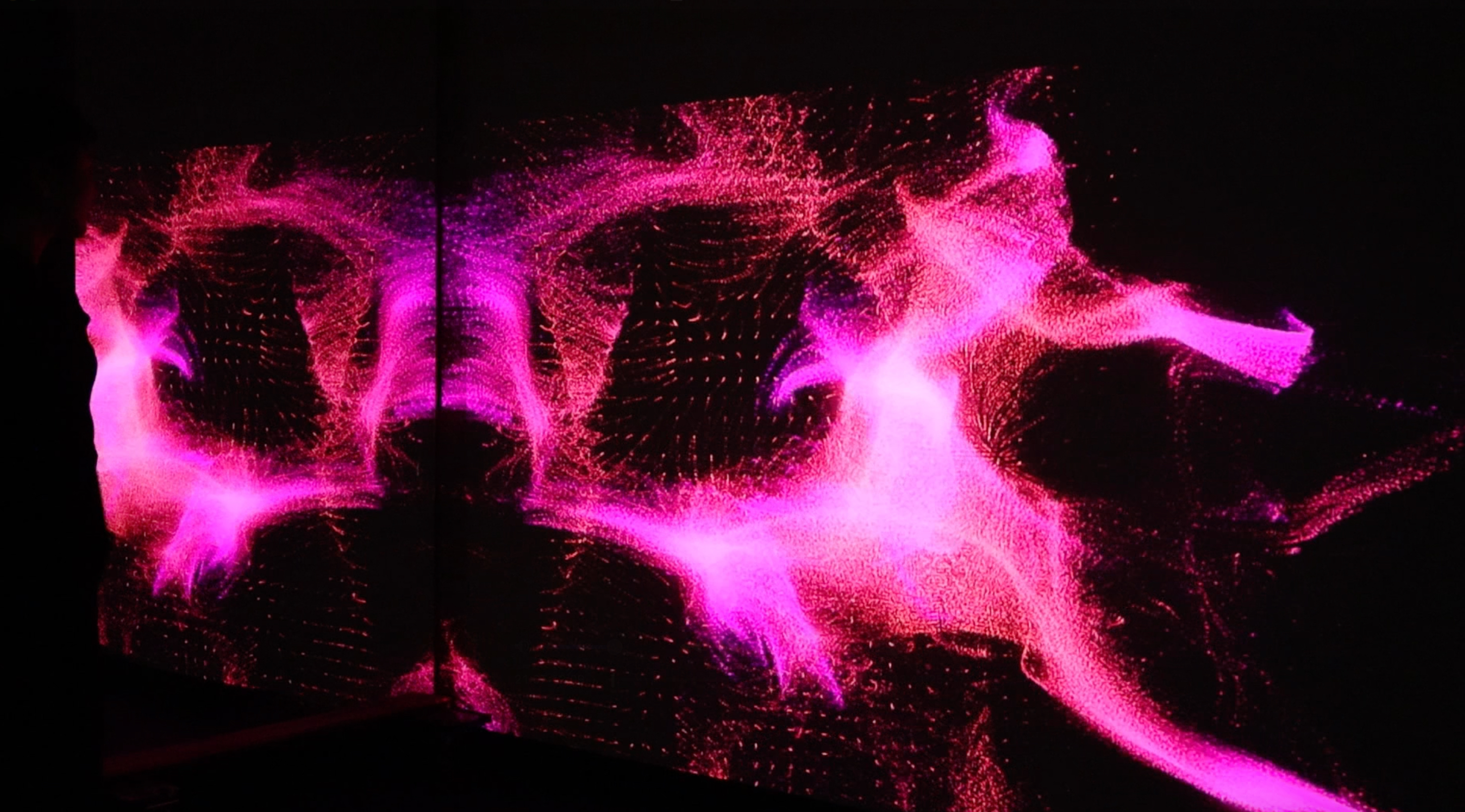
I would like to biomanufacture a wearable made from bacterial cellulose where color, thickness, transparency and surface pattern are influenced by a person’s brain activity.
I am interested in this idea because many of the brain to computer interfaces translate brain activity into numbers, images and sound but not really something you can feel. With this idea, I hope to explore what happens when brain activity instead influences a material that grows slowly and can be touched.
What living system would you use?
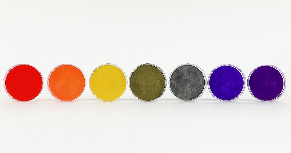
From my preliminary research, I would be using bacterial cellulose and/or microalgae that then would be dyed by machine based on the brain wave. I have found that aerobic bacteria will convert sugars in a growth medium which is then into nanoscale cellulose fibers. In this process the fibers will accumulate into a physical material.
From my initial assesment, I believe the system will incorporate the following:
Cellulose producing bacteria
A scaffold or mold where oxygen can pass through
Pigment producting bacteria or microorganism
How would you use automation?
I would be using EEG processing, machine learning, digital fabrication and automated liquid handling in this process. The start of the process would be an EEG device recording brainwave of a participent during a short activity. A computational system would then extract features from the data to create the pattern and color of the growing environment. With the pattern produced, a liquid handling robot will dispense bacterial cultures and different concentrations of growth medium into multiwell plates or small culture chambers based on the data previously collected.
How much would you need to make, and how long would it take?
With it being a wearable/art piece, I’m estimating it to be the size of a forearm, which would be in the size:
Length : 12in
Avg. Forearm Length: 9.5in
Some calculations supported by Chat GPT:
The required surface area would be:
12\ \x{in} \times 9.5\ \x{in} = 114\ \x{in}^2
In addition to adding approximately 20% extra material for seams, overlap, shrinkage, trimming, and unsuccessful regions:
114\ \x{in}^2 \times 1.20 = 136.8\ \x{in}^2
I would therefore need to manufacture approximately 137 square inches of finished bacterial-cellulose material.
For an initial estimate, I will assume that the dried bacterial-cellulose material has a target areal mass of approximately 0.026 grams per square inch. This is equivalent to approximately 40 grams per square meter.
The total amount of dry bacterial cellulose required would therefore be:
137\ \x{in}^2 \times 0.026\ \x{g/in}^2
= 3.56\ \x{g}
What don’t you know yet?
Honestly there is still so much I need to know but I think some of the main questions would be the following:
How can EEG creating meaningful material pattern and is it the most effective way?
How precise the calculation need to be to fully reflect the pattern design by machine?
Will the method for growing the most effective and can it be controlled?
Is it safe to wear such material?
Week 2 HW: DNA Design
Part 1 : Expression Target
I would express MscL (large conductance mechanosensitive channel) from E. coli as the primary mechanosensing protein. MscL is a membrane protein that opens in response to mechanical tension in the cell membrane, which allows mechanical stimulation to be converted into an intracellular ionic signal. In my proposed idea, a person’s physiological state would be measured electronically and translated by a machine into controlled fluid flow and shear stress. The goal is to use MscL as the first biological interface through which cells can sense this mechanically encoded human information.
For an initial proof of concept, MscL mediated mechanosensing will eventually be coupled to a visible reporter which allows for different mechanical inputs to produce measurable cellular outputs. In later iterations, it can be replaced with a protein or other pathway that produces a visible and persistent change on a textile scaffold. The E. coli MscL protein, as of now, is encoded by mscL (UniProt P0A742; RefSeq NP_417749.1) and is a 136-amino-acid inner-membrane protein.
Part 2 : Genetic Parts
Part type
Short name
Sequence / Source
Justification
Promoter
J23110
Sequence from the iGEM Registry / Kernel standard part library
J23110 is a constitutive bacterial promoter that allows continuous transcription of mscL in E. coli without requiring an external chemical inducer.
Ribosome Binding Site (RBS)
B0034
Sequence from the iGEM Registry / Kernel standard part library
B0034 is a commonly used bacterial RBS that provides efficient translation initiation in E. coli.
Coding Sequence (CDS)
mscL
E. coli K-12 mscL; UniProt P0A742; RefSeq NP_417749.1
mscL encodes the large conductance mechanosensitive channel MscL, which responds to membrane tension and serves as the mechanosensing protein in the proposed interface.
Targeting / Signal peptide
None
Not required
MscL is an integral inner membrane protein whose native transmembrane domains direct membrane localization, so an additional secretion signal is unnecessary.
Terminator
B0015
Sequence from the iGEM Registry / Kernel standard part library
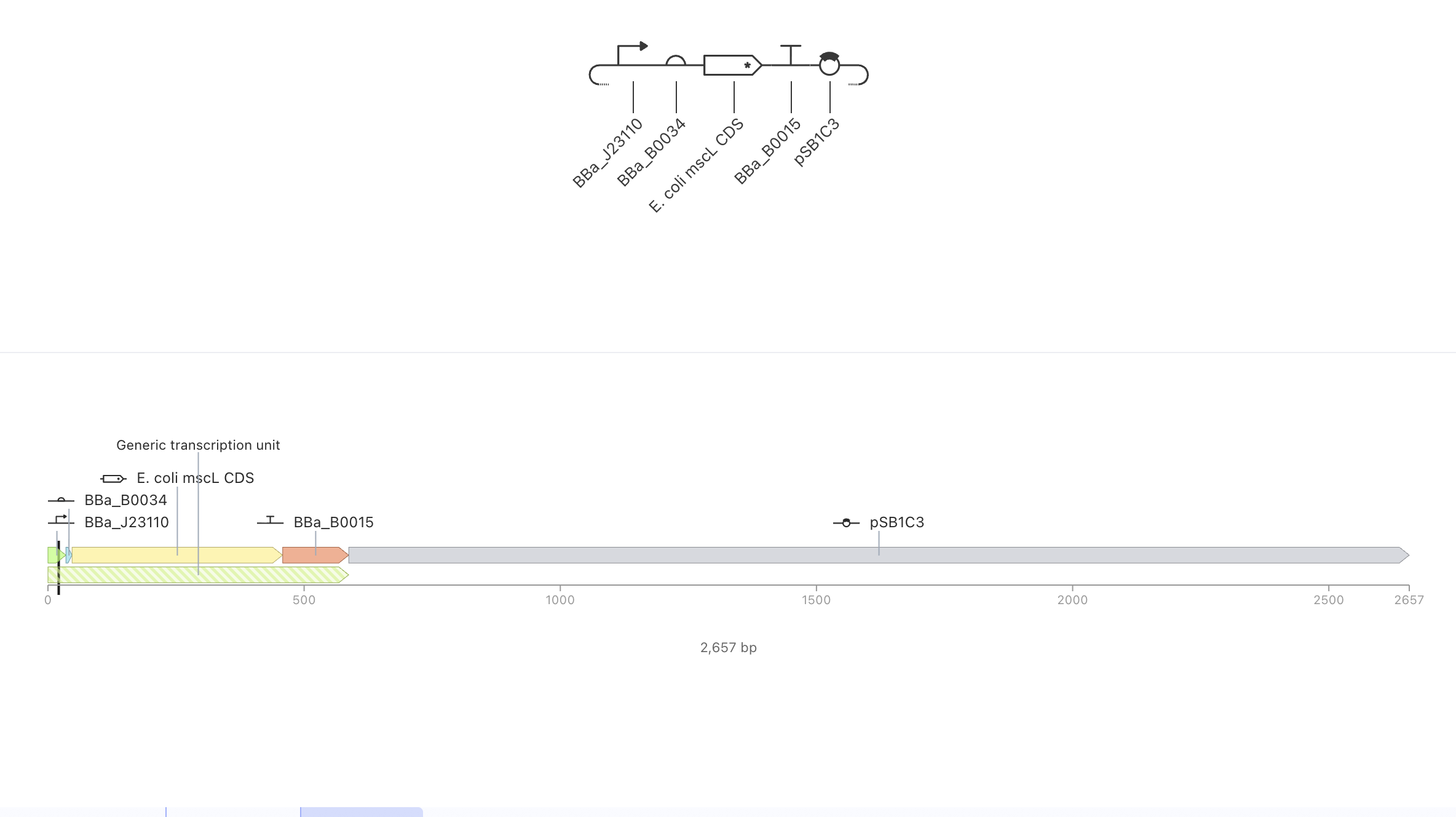
B0015 provides transcription termination downstream of mscL, producing a defined expression cassette.
Vector backbone
pSB1C3
iGEM standard plasmid backbone; use existing Kernel/backbone sequence
pSB1C3 provides an E. coli-compatible origin of replication and chloramphenicol-resistance selection for maintaining the construct.
Part 3 : Assembled Construct
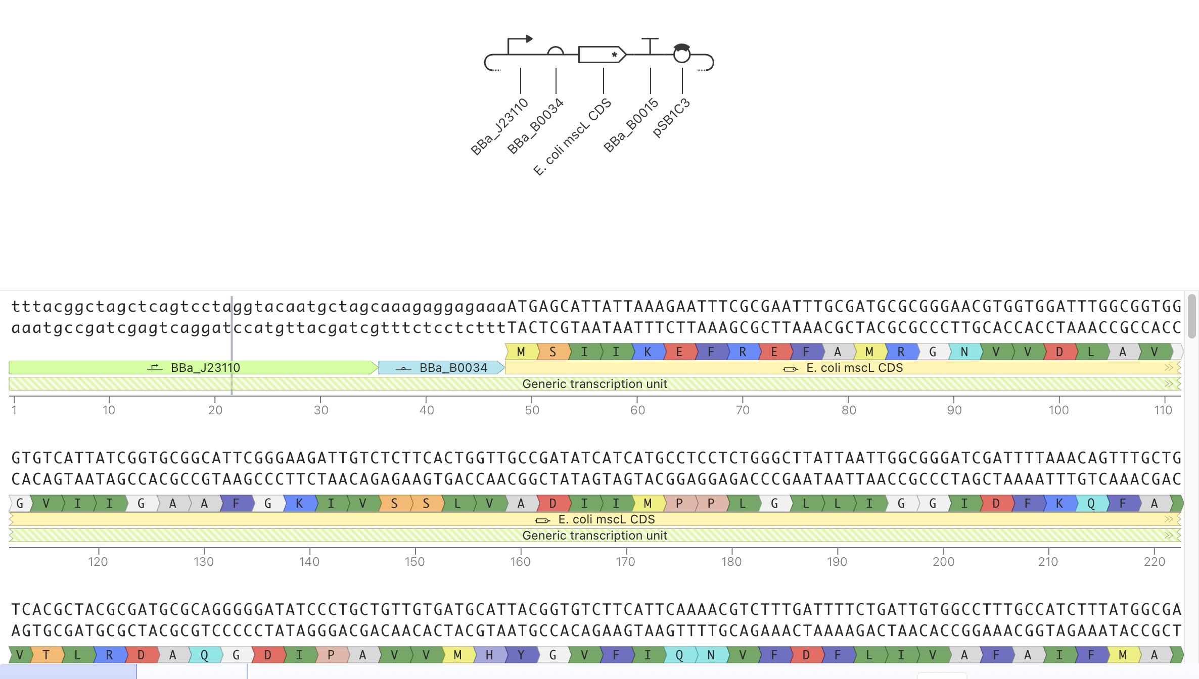
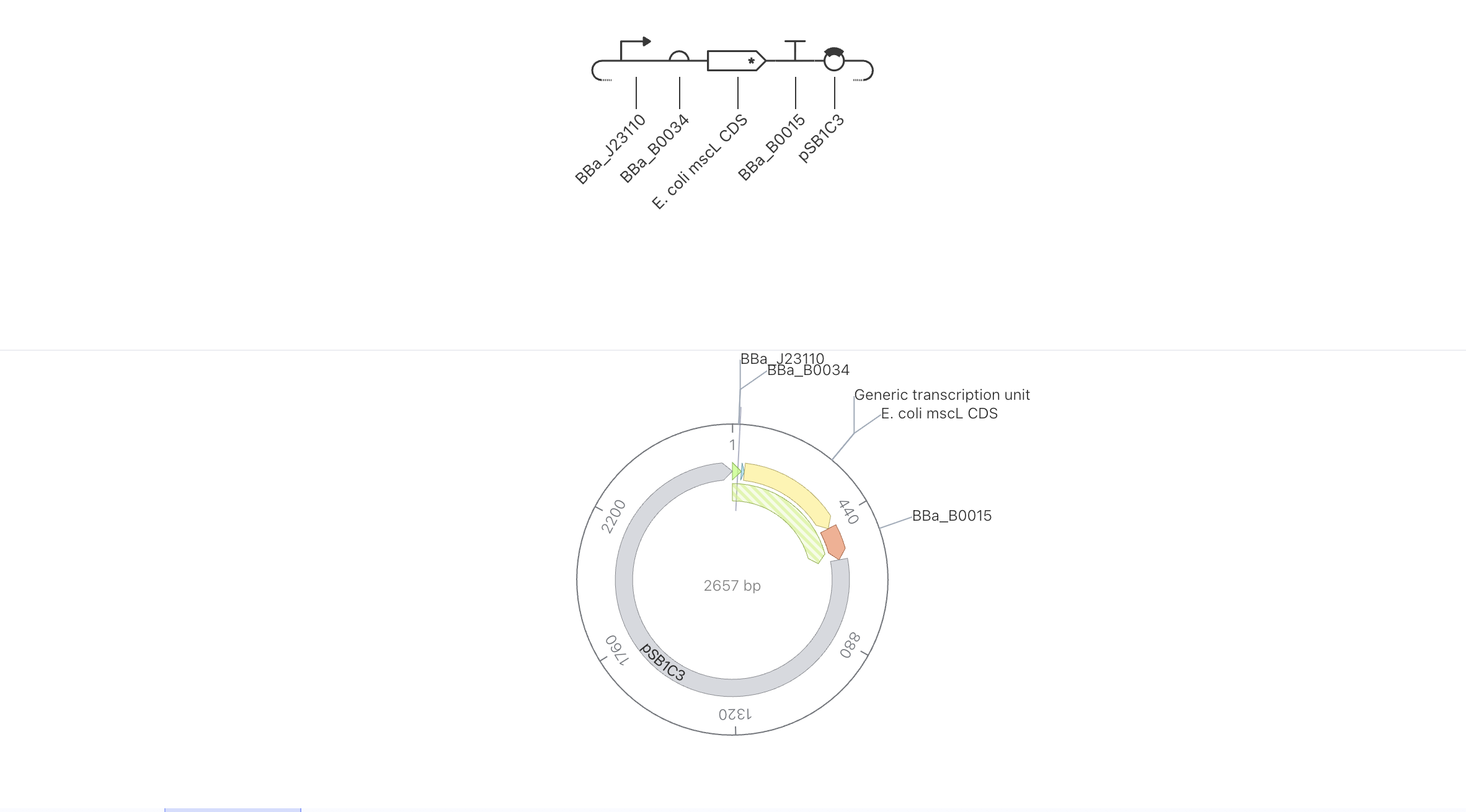
Kernel file has been saved under name Wang - Hw2 DNA Design
Part 4 : Optimize & Quality Check
Condon Optimizer
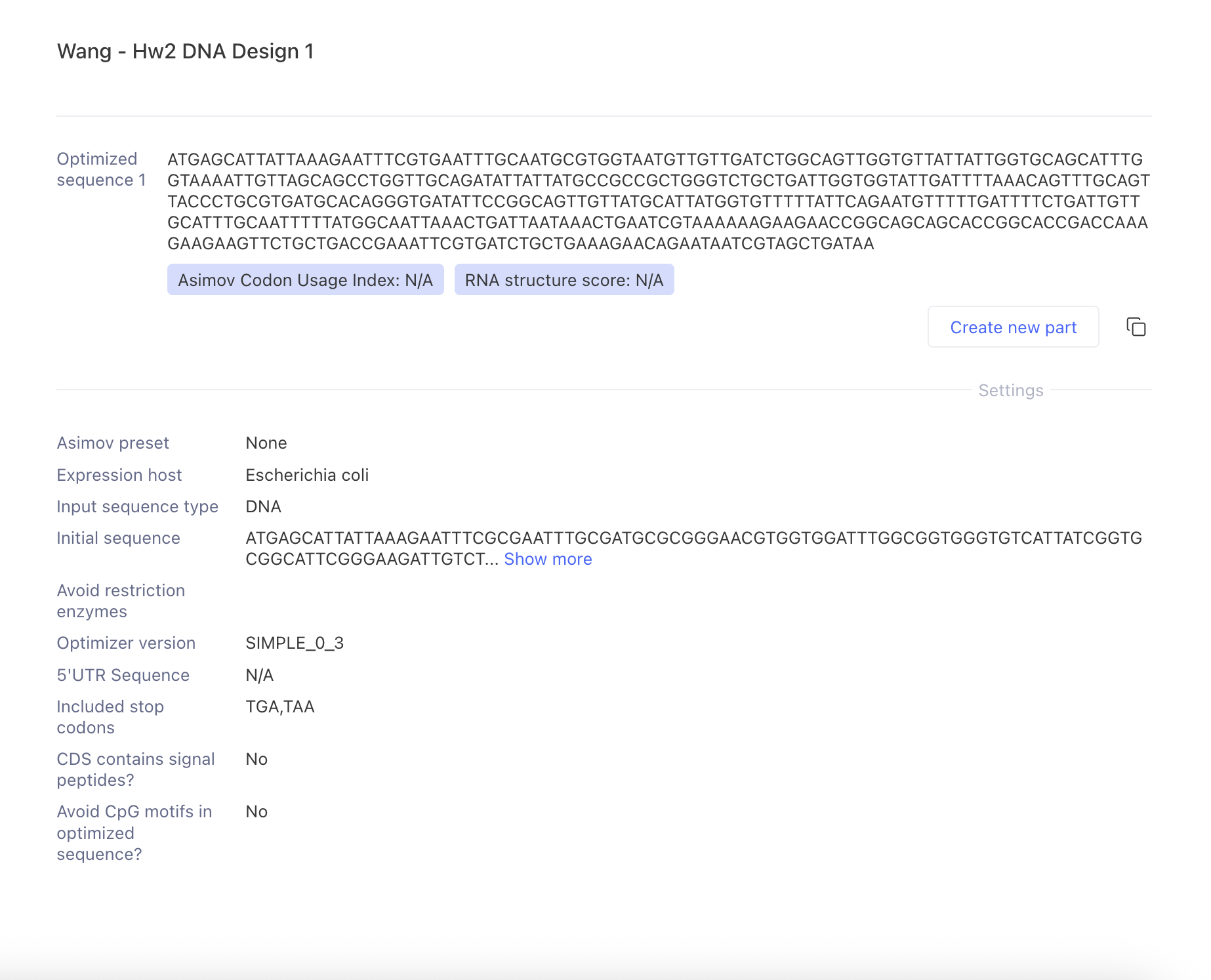
I have codon optimized the mscL coding sequence for expression in Escherichia coli using Kernel’s SIMPLE_0_3 optimizer. The original sequence was the native mscL gene from E. coli K-12, but Kernel still redesigned synonymous codons according to its E. coli expression parameters. These nucleotide changes are intended to improve compatibility with host codon usage while preserving the MscL amino-acid sequence. I did not apply additional restriction-enzyme or motif-avoidance constraints, allowing me to evaluate codon usage independently. Kernel did not report a Codon Usage Index or RNA structure score for this run (both were listed as N/A).
Sequence Search
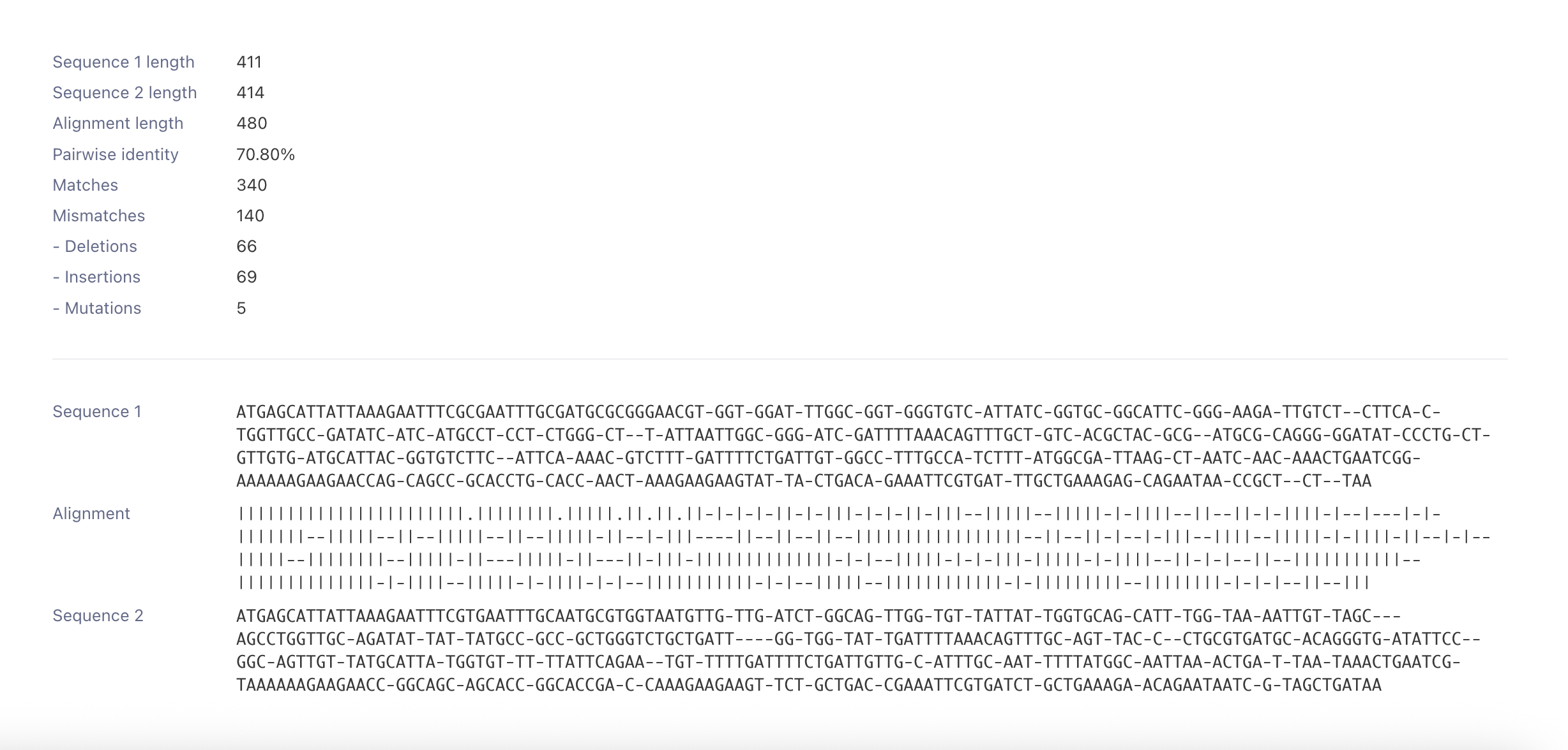
The alignment between the native and Kernel optimized mscL sequences showed substantial nucleotide level redesign. The native sequence was 411 bp, while the optimized sequence was 414 bp, and the pairwise nucleotide identity was 70.80%. This indicates that Kernel changed many codons during optimization rather than making only a few nucleotide substitutions. Because codon optimization is intended to alter DNA codon usage while preserving the encoded protein, I would verify the translated amino acid sequence before using the optimized CDS experimentally.
Security Search
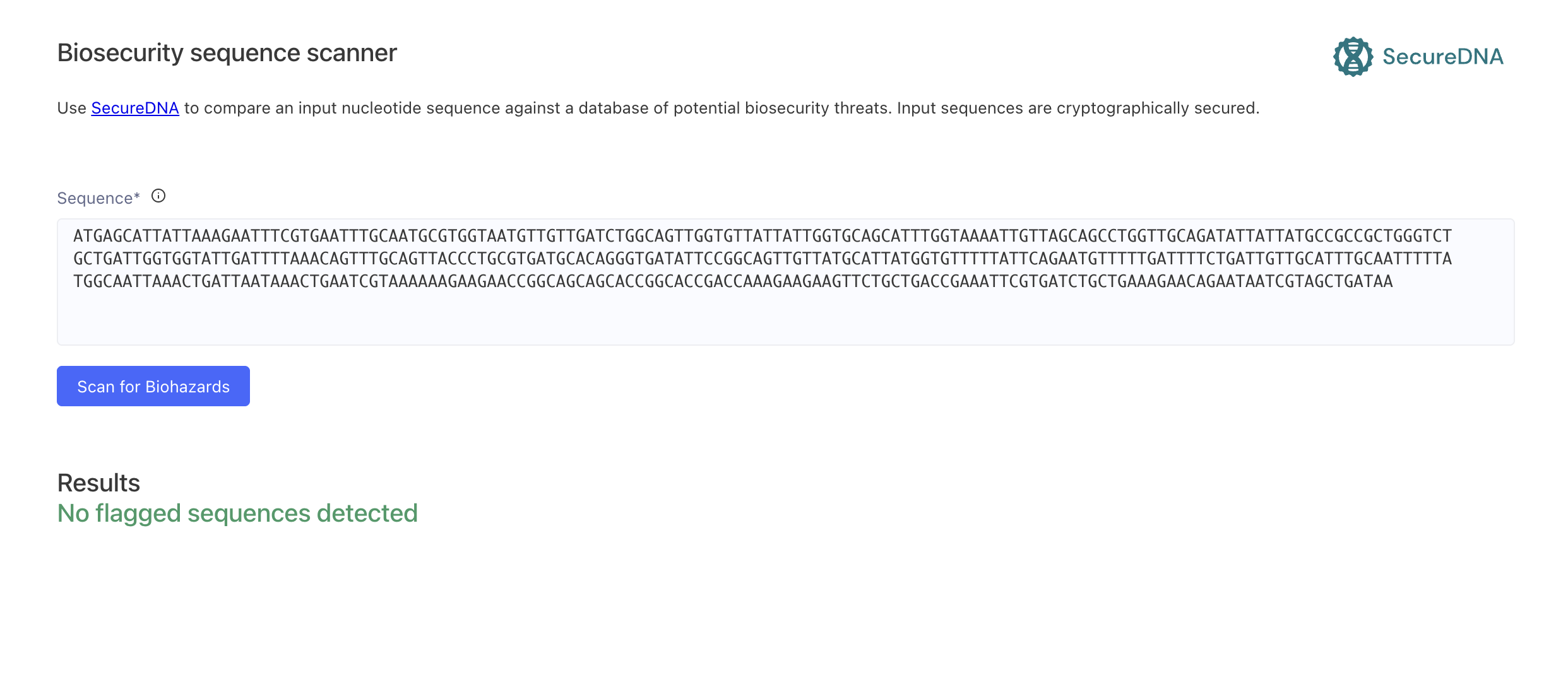
I ran the new mscL sequence through Kernel’s SecureDNA Biosecurity Sequence Scanner, which screens nucleotide sequences against a database of potential biosecurity threats. The scan returned “No flagged sequences detected.” This indicates that the MscL coding sequence did not trigger any biosecurity flags in the screening system, so no sequence changes were required based on this result.
Part 5 : Design to Build
How to build?
I would start with the mscL expression cassette as synthetic DNA and assemble it into the existing pSB1C3 backbone. With the mscL CDS being only 411 bp and the complete expression cassette is relatively small, creating it as one DNA fragment may be more practical and effective than producing it from a large pool of short oligonucleotides. I would use Gibson Assembly to join the synthetic insert and plasmid backbone using overlapping ends. The reading mentioned that overlapping DNA fragments can assemble seamlessly in a single isothermal reaction, allowing this method to be effective for inserting a small synthetic expression cassette into an existing vector.
What does synthesis constrain?
The synthesized construct would need sequence verification before being used. Through the reading, it is to my understanding that mismatched binding proteins such as MutS can selectively remove error containing products and substantially improve the fidelity of de novo DNA synthesis.
My construct, I believe, it is comparatively shorter and does not intentionally contain long repetitive regions, so I’m hoping it to provide me less space for constrain. However, if errors or problematic sequence regions are produced, the cassette could potentially be synthesized in smaller fragments, error corrected or sequence verified which can be assembled in the end.
Prototype vs. scale.
For my idea in a prototype form, I believe commercial synthesis followed by assembly into pSB1C3 is enough. In large scale production, it would be more difficult as it may require producing a library of MscL variants or different mechanosensing circuits instead of repeating one identical construct. In that situation, oligonucleotide pool or microchip based synthesis could become practical. Through the reading, I was inspired that complex pools of chip synthesized oligonucleotides can be selectively amplified and assembled into many genes in paralle as using error correction strategies to address the higher error rates associated with pooled synthesis.
Part 6 : Questions
Will externally applied shear stress actually activate MscL?
If MscL will, how will the activation perform a visible output?
Is constitutive MscL expression at the right level?
Use of AI
I have used ChatGPT in support of explaination of sequence and DNA construct.
Week 3 HW: Cell-Free Protocols
A. COMPARE CELL-FREE REACTION COMPOSITIONS
Why does the concentration of potassium glutamate and magnesium glutamate differ between compositions? Which is more sensitive?
a. Compositions
I believe that magnesium and potassium glutamate differ for different reasons.
Magnesium differs because of the energy substrate where PEP, and the inorganic phosphate it generatesm, combinds and sequesters Mg². Due to such, the PEP based systems is in need of a high total magnesium to keep enough avaliable Mg²⁺ for the ribosomes. Pyruvate binds magnesium weakly and produces almost no phosphate. Because of this, the optimal avaliable magnesium level is reached with even less than half the total magnesium.
Potassium glutamate on the other hand, differs because of ionic/charge balance. As K⁺ and glutamate balance the charge from nucleic acid phosphates, it also recreated a cell alike ionic strength where protein nucleic acid interactions are favored. In the designed system, PEP, HEPES and PEG were removed while acetate was replaced with glutamate salts in addition to the magnesium and ammonium being lowered, it naturally return the ionic environment which mimic the E. coli cytoplasm.
b. Sensitivity
Based on the same reading (Jewett & Swartz) and their optimization studies in both the PANOx-SP (with PEP), I believe that magnesium is more sensitive than potassium. Through data they have presented in their paper (Table II.), magnesium glutamate has performed a sharp peak, a shift 4 mM from the optimum have decreased the harvest to about 62–67% of maximum. Whereas potassium glutamate had a broad optimum where it was able to keep about 95% of maximum yield even when it has increased to 200 mM.
Explain how a PEP+NTP reaction formulation is different from a Ribose+NMP reaction formulation.
I believe that the core difference is how NTP and energy are being supplied to each reaction formulation.
In the PEP+NTP formulation (based on Jewett & Swartz, 2004) the four nucleoside triphosphates (ATP, GTP, CTP and UTP) are being added directly, so RNA polymerase can use them directly. With PEP being a high energy phosphorylated secondary energy substrate, it is regenerating ATP by donating its phosphate to ADP. This reaction formulation is higher in cost as the PEP consumption liberates inhibitory inorganic phosphate and drives the pH down.
In the Ribose+NMP reaction formulation (based on Olsen, 2026) it is cheaper as the nucleoside monophosphates replace NTPs. The lysate’s kinases need to first process through phosphorylate to NTPs before any transcription can continue. ATP in this reaction is regenerated with ribose (added with glucose and phosphate) catabolized by enzymes retained within the lysate, instead of being a donor.
To sum up, they differ in cost (NMP+ribose is cheaper), dependence on metabolism and phosphate (NMP+ribose relies on active lysate metabolism and buffered phosphate), substitutions (e.g. GMP being replaced by guanine + ribose), and reaction kinetics (NMP+ribose is slower and more sensitive to metabolic balance).
Choose two cell-free reaction compositions from the literature, compare and contrast the choice of reagents and the concentration of reagents.
I will be compare and contrasting the cytomim and RFopt reaction. For these two reactions, both of them are glutamate based and PEP free systems that solves the energy concern in opposite ways.
Reagent
Cytomim (2004)
RFopt (2026)
Energy / carbon source
Sodium pyruvate 33 mM
Ribose 50 mM + maltodextrin 60 mg/mL
High-energy phosphate (PEP)
none
none
Nucleotides
NTPs: ATP 1.2, GTP/CTP/UTP 0.85 each
NMPs: AMP 3, CMP/GMP/UMP 2.15 each
Phosphate
none added (~4 mM accumulates)
15 mM added (buffered)
Buffer
none (buffer-free)
HEPES 75 mM
Potassium glutamate
130 mM
362 mM
Magnesium glutamate
8 mM (11.3 total w/ extract)
8 mM
Ammonium glutamate
10 mM
none
Polyamines
Spermidine 1.5 + putrescine 1
Spermidine 0.13 only
Oxalate
4 mM
none
Cofactors
NAD 0.33 + CoA 0.26 (added)
Nicotinamide 4 (NAD precursor); no NAD/CoA
tRNA
170.6 µg/mL (added)
none (relies on lysate)
Folinic acid
0.034 mg/mL
2.05 mg/mL
Amino acids
2 mM each
5 mM each
Reporter & yield
CAT, ~0.73 g/L (6 h)
sfGFP, 2.4 g/L (15 µL) → 3.7 g/L (bioreactor +O₂)
Table 1. Reagent comparison between Cytomim and RFopt based on data collected from Jewett & Swartz and Olsen.
Between these two reactions, energy usage is one of the most important differences.
Cytomim puts pyruvate directly into oxidative phosphorylation where it needs to supply the cofactors that the pathway needs for the reaction such as NAD and CoA. RFopt feeds ribose adding to maltodextrin into glycolysis and respiration. Then, instead of adding NAD directly it uses nicotinamide, letting the lysate rebuild NAD by itself.
The nucleotides is also different: Cytomim adds triphosphates, while RFopt uses higher concentrated monophosphates (cheaper) and relies on lysate kinases to phosphorylate to the same result.
Cytomim also eliminates added phosphate and removes the HEPES buffer, whereas RFopt adds buffered phosphate (15 mM) and puts HEPES (75 mM) back in.
Despite these differences, both independently converged on 8 mM of added magnesium glutamate.
B. DESIGN ONE REACTION COMPOSITION FOR EACH OF THE THREE PROTEINS
This has been designed for for a 20 µL reaction using the reagents from coopetition htgaa.
sfGFP
Reagent
Unit
Custom Mix
Closest Match
K(Glu)
mM
220
200 (−8.96%)
Mg(Glu)₂
mM
3.85
8.85 (+130%)
NH4(Glu)
mM
—
—
Ammonium acetate
mM
—
—
Amino acids
mM
5
5
Tyrosine
mM
1
1
Cysteine
mM
1
1
HEPES pH 7.5
mM
60
75 (+25%)
Bis-Tris
mM
—
—
Phosphate (di:mono)
mM
—
—
Phosphate (mono:di)
mM
15
15
Glucose
mM
—
—
For this design, I have attempted to change components that are related to folding. Such as there are no DsbC, no GSSG/GSH, no catalase as the cysteine stays low. I have also tried to keep it as a simple and plain reducing environment. This design originates from the idea that sfGFP folds on its own with no disulfide bonds. With each oxidizing reagent added it is additional cost that may have little effect on the yield.
PETase
Reagent
Unit
Custom Mix
Closest Match
K(Glu)
mM
200
200 (0%)
Mg(Glu)₂
mM
8.85
8.85 (0%)
NH4(Glu)
mM
—
—
Ammonium acetate
mM
—
—
Amino acids
mM
5
5 (0%)
Tyrosine
mM
1
1 (0%)
Cysteine
mM
1
1 (0%)
HEPES pH 7.5
mM
75
75 (0%)
Bis-Tris
mM
—
—
Phosphate (di:mono)
mM
—
—
Phosphate (mono:di)
mM
15
15 (0%)
Glucose
mM
—
—
Sodium pyruvate
mM
—
—
In this design, I have added DsbC as the main impact. I believe PETase has a couple of disulfide bonds and it needs support forming and shuffling them correctly. By adding additional DsbC, I wish to provide support and encourage reaction without changing the entire redox environment.
Reteplase
Reagent
Unit
Custom Mix
Closest Match
K(Glu)
mM
200
200 (0%)
Mg(Glu)₂
mM
8.85
8.85 (0%)
NH4(Glu)
mM
—
—
Ammonium acetate
mM
—
—
Amino acids
mM
4.81
4.81 (0%)
Tyrosine
mM
1.19
1.19 (0%)
Cysteine
mM
5
5 (0%)
HEPES pH 7.5
mM
75
67.5 (−10%)
Bis-Tris
mM
—
—
Phosphate (di:mono)
mM
7.5
7.5 (0%)
Phosphate (mono:di)
mM
7.5
7.5 (0%)
Glucose
mM
—
—
Sodium pyruvate
mM
0.00
9.09
This design have additional DsbC, GSSG, cysteine and catalase in addition to changes in HEPES. Through the reading, I have learned that GSSG buffer sets an oxidizing environment where many disulfides can form. With Cysteine, it supports the raw thiols where bonds would be built from. Catalase is then used to for oxygen purpose in folding and energy where it produces peroxide that would otherwise damage the reaction. The change in HEPES is then to hold the higher pH where disulfide formation is favored.
HTBAA Upvote
I ended upvoting the FkpA (E. coli chaperone) as it provides the most stable structure and yields the most through lower outcome with relatively small changes.