<Susan Torres> — HTBAA Fall 2026
About me
Contact info
Homework, Labs, and Projects
See the nav bar on the left to look through my work for How To Biomanufacture Almost Anything (Fall 2026)!
See the nav bar on the left to look through my work for How To Biomanufacture Almost Anything (Fall 2026)!
I would like to biomanufacture a functional 3D model of the human urinary bladder to study neurogenic bladder dysfunction.
The model would reproduce key components of bladder tissue and allow us to study how engineered tissue responds to filling, distension, and stimulation.
I am interested in this because neurogenic bladder can affect people with spina bifida, and managing it can require long-term catheterization. Rather than trying to manufacture a replacement organ immediately, I want to create a controllable experimental system to investigate how bladder function could be improved.
This project is also personally meaningful to me because someone in my family has spina bifida. Seeing their experience has shown me how congenital conditions can create challenges from childhood into adulthood.
The goal is therefore not simply to make tissue that looks like a bladder, but to build a working model that can be tested, measured, and used to investigate bladder function. Tissue engineering has already been explored for urinary bladder reconstruction and functional tissue models, although important challenges remain (Adamowicz et al., 2017; Horst et al., 2013).
I would use a mammalian cell culture system with human bladder cells.
The initial model would contain:
The cells would be cultured under controlled conditions that encourage attachment, proliferation, organization, and maturation.
The resulting construct would be a 3D engineered bladder tissue model, not a complete artificial organ.
After maturation, the tissue could be tested through controlled stretching and electrical stimulation while measuring pressure and contractile responses.
A neural component could be added later to study neuromuscular communication more closely. At this stage, I assume that suitable human cells and a biocompatible scaffold are available, but the cell source, cell ratios, scaffold composition, and culture conditions would still need to be optimized experimentally.
Previous studies show that scaffold design and cell environment can strongly affect bladder tissue engineering outcomes (Adamowicz et al., 2017; Sivaraman et al., 2015).
I would connect the tissue culture system to an automated liquid-handling and bioreactor platform, ideally connected to a Cloud Lab or another programmable laboratory automation system.
Automation could be used for:
The goal would be to create a repeated experimental cycle:
Build → Culture → Stimulate → Measure → Analyze → Optimize → Repeat
This would allow different tissue compositions, culture conditions, and stimulation settings to be tested systematically while reducing manual work.
Because this is a research model rather than a therapeutic product, I would define meaningful scale as enough constructs to compare several conditions with biological replicates.
I am not yet specifying an exact cell number or construct volume because these would depend on the final scaffold design and cell source.
The main time constraint would probably be tissue maturation, rather than the automated handling steps. Previous bladder tissue engineering work has used culture periods of several weeks. For example, Gabouev et al. (2003) constructed bladder wall tissue using primary porcine cells and maintained the constructs in a three-dimensional rotating bioreactor for six weeks.
Therefore, I would initially estimate about six weeks of culture for a mature prototype, while recognizing that the actual time would have to be determined experimentally.
Scaling
Once the prototype is reliable, I could increase output by adding identical culture modules.
For example:
1 module → 24 constructs 4 modules → ~96 constructs
This is only a scaling estimate. The larger system would still need to be tested to make sure that tissue quality and function remain consistent when more constructs are cultured simultaneously.
The cells need to do more than survive and organize. The tissue should be able to:
I need to determine which combination of cells, scaffold, and culture conditions produces these properties (Horst et al., 2013).
I need to determine the electrical stimulation strength, frequency, duration, and timing that produce a consistent response without damaging or excessively stressing the tissue.
I also need to determine whether electrical stimulation alone is sufficient or whether a future model will need a neural component.
Stronger contractions alone do not necessarily mean better bladder function.
The model should consider both storage and emptying, including:
The combination of measurements that best represents functional bladder performance is still an open question.
I used ChatGPT (OpenAI) to help organize and refine my initial project idea and to check that the proposal addressed the homework questions.
The core project idea and initial draft were my own. I used ChatGPT for language refinement, organization, and citation placement.
Prompts included:
I reviewed the generated suggestions and adapted the final text to reflect my own project idea and understanding.
Adamowicz, J., Pokrywczynska, M., Van Breda, S. V., Kloskowski, T., & Drewa, T. (2017). Concise review: Tissue engineering of urinary bladder; We still have a long way to go? Stem Cells Translational Medicine, 6(11), 2033–2043. https://doi.org/10.1002/sctm.17-0101
Gabouev, A. I., Schultheiss, D., Mertsching, H., Köppe, M., Schlote, N., Wefer, J., Jonas, U., & Stief, C. G. (2003). In vitro construction of urinary bladder wall using porcine primary cells reseeded on acellularized bladder matrix and small intestinal submucosa. International Journal of Artificial Organs, 26(10), 935–942. https://doi.org/10.1177/039139880302601011
Horst, M., Madduri, S., Gobet, R., Sulser, T., Milleret, V., Hall, H., Atala, A., & Eberli, D. (2013). Engineering functional bladder tissues. Journal of Tissue Engineering and Regenerative Medicine, 7(7), 515–522. https://doi.org/10.1002/term.547
Sivaraman, S., Ostendorff, R., Fleishman, B., & Nagatomi, J. (2015). Tetronic®-based composite hydrogel scaffolds seeded with rat-bladder smooth muscle cells for urinary bladder tissue engineering applications. Journal of Biomaterials Science, Polymer Edition, 26(3), 196–210. https://doi.org/10.1080/09205063.2014.989482
I created my project in the HTBAA 2026 workspace using the Functional Bladder Tissue repository and the ELN Expression folder. Before assembling my own expression design, I used Kernel to investigate the available genetic parts and constructs.
The final construct was named ELN Expression Construct.
The design was developed as a mammalian expression cassette intended to express human elastin (ELN) in the context of a functional bladder-tissue project.

Figure 1. Kernel project structure showing the HTBAA 2026 workspace, the Functional Bladder Tissue repository, and the ELN Expression folder.
The molecule I am expressing is human tropoelastin, the precursor protein encoded by the ELN (elastin) gene. Elastin is an extracellular matrix protein and a major component of elastic fibres. Because elastic properties are important for tissues that undergo repeated expansion and recoil, ELN was selected as the protein target for this functional bladder-tissue project (Wise & Weiss, 2009; Ozsvar et al., 2021).
The NCBI ELN gene record describes elastin as a component of elastic fibres in the extracellular matrix and lists NM_000501.4 as an mRNA RefSeq corresponding to human ELN transcript variant 1 (NCBI, n.d.).
The coding sequence initially provided in Kernel is the Human ELN CDS, annotated as a CDS (+start +stop) with a length of 2,178 bp.
The sequence is associated with human ELN. I used the NCBI RefSeq record NM_000501.4, Homo sapiens elastin (ELN), transcript variant 1, mRNA, as a reference for the human ELN gene (NCBI, n.d.).
However, I did not assume that the 2,178-bp Kernel sequence was nucleotide-identical to the RefSeq record. The Kernel sequence was treated as the actual sequence used in the design, while NM_000501.4 was used as an external reference for human ELN.
I also carried out nucleotide sequence searches using NCBI BLASTN. An initial BLAST analysis of the original human ELN sequence returned matching human ELN reference sequences, including NM_000501.4. A later BLAST analysis performed during the optimization process returned:
No significant similarity found.
These were separate sequence-search runs, so I report the two observations separately rather than treating them as contradictory results from the same query.
The later result may reflect differences between the sequence submitted in that run and the reference sequences detected in the earlier analysis. In particular, a nucleotide sequence can encode the same protein while differing at synonymous codon positions.
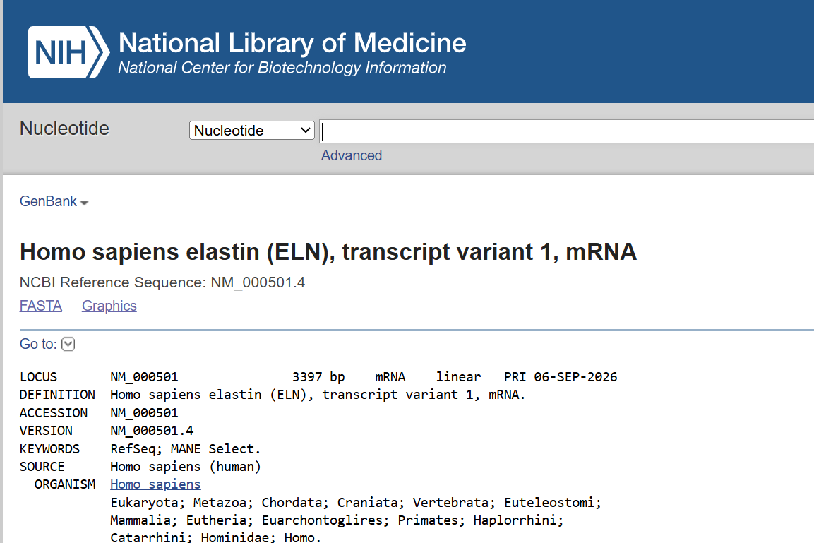
Figure 2. NCBI RefSeq record for human ELN transcript variant 1 (NM_000501.4), used as the reference sequence for this project.
The intended expression system is mammalian, specifically human bladder cells in the context of functional bladder tissue.
For this reason, the expression design uses a mammalian promoter and a 5′ UTR containing a Kozak sequence, rather than a bacterial ribosome-binding site.
The coding-sequence optimization tool in Kernel was also run using Homo sapiens as the host.
The intended destination of the protein is the extracellular environment.
Human elastin is synthesized as a secretory-pathway protein, and the human ELN protein sequence contains an N-terminal signal peptide in the reference protein sequence (UniProt, n.d.; Wise & Weiss, 2009).
In the Kernel design, I did not add a separate artificial secretion signal. Instead, I retained the N-terminal region of the ELN coding sequence supplied by Kernel and treated the targeting information as part of the native ELN sequence.
This is a design assumption rather than experimental evidence of secretion. The actual efficiency of secretion would need to be tested experimentally in the intended cell type.
The reason for this design is that tropoelastin needs to reach the extracellular environment in order to participate in extracellular-matrix assembly.
I created a simple mammalian expression cassette in Kernel.
The functional elements of the expression cassette are:
| Layer | Part | Type | Length | Source | Reason for selection |
|---|---|---|---|---|---|
| Transcription start | Short HsEef1a1 promoter | Promoter | 233 bp | Kernel | Provides a mammalian promoter for transcription of the ELN expression cassette. |
| Translation start | Unspecified 5′ UTR with Kozak | 5′ UTR with Kozak | 30 bp | Kernel | Provides a mammalian translation-initiation context. |
| Coding sequence | Human ELN CDS | CDS (+start +stop) | 2,178 bp | Human ELN sequence in Kernel | Encodes the ELN protein used as the target of the design. |
| Targeting | Native ELN N-terminal sequence | Included within CDS | — | Human ELN | No separate artificial secretion signal was added. |
| Transcription termination | BBa_K2217005 | Terminator | 122 bp | Kernel / existing part | Provides a downstream transcription termination element. |
| Polyadenylation | Unspecified Poly(A) signal | Poly(A) signal | 30 bp | Kernel | Provides the 3′ polyadenylation signal. |
The short HsEef1a1 promoter is annotated in Kernel with functional features including a G-rich element, an Sp1/GC box, a TATA box, and a transcription start site. These annotations are features within the 233-bp promoter and are not additional parts that were separately added to the cassette.
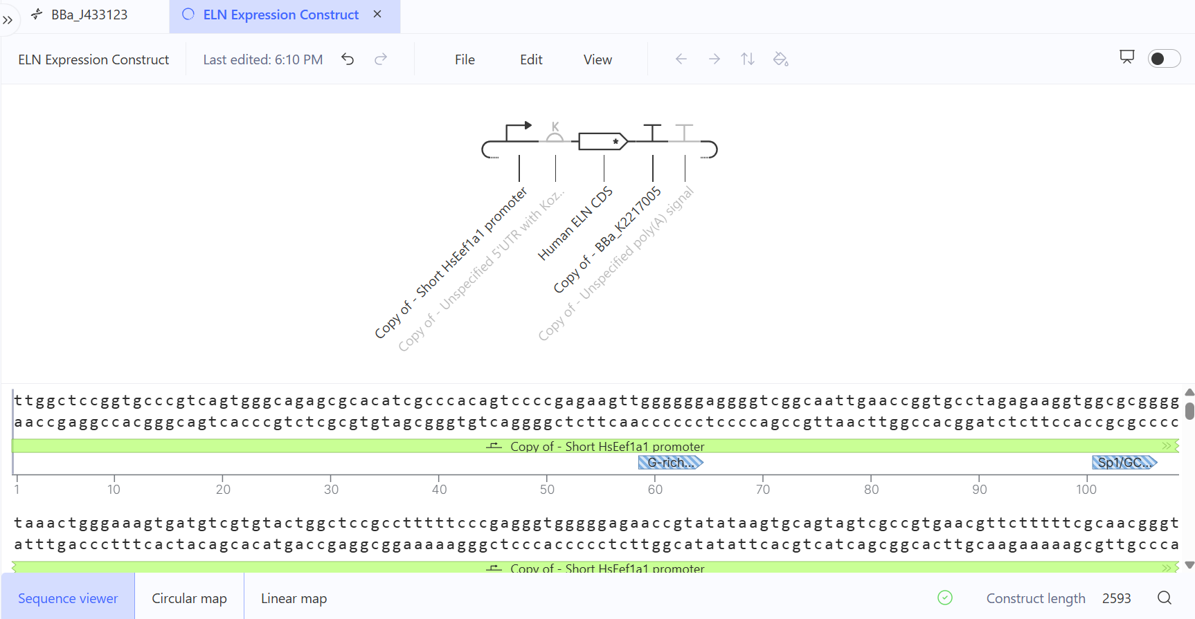
Figure 3. Kernel view showing the genetic parts selected for the ELN expression cassette.
The design uses the BBa_J433123 / AmpR-ColE1 backbone as the vector context in Kernel.
The backbone is considered separately from the expression cassette. The expression cassette contains the components directly involved in transcription, translation, ELN coding, termination, and polyadenylation. The BBa_J433123 / AmpR-ColE1 backbone provides the plasmid context, including the elements associated with plasmid propagation and selection.
Therefore, the overall design can be represented as:
BBa_J433123 / AmpR-ColE1 vector backbone + ELN expression cassette
The backbone is not an additional element inserted between the promoter, 5′ UTR/Kozak sequence, CDS, terminator, and poly(A) signal. It is therefore not included in the calculated length of the expression cassette.
I assembled the ELN expression design in Kernel using the BBa_J433123 / AmpR-ColE1 backbone as the vector context.
Within the vector, the expression cassette follows the intended 5′→3′ organization:
Short HsEef1a1 promoter
↓
5′ UTR with Kozak
↓
Human ELN CDS (+start +stop)
↓
BBa_K2217005 terminator
↓
Poly(A) signal
The expression-cassette components have the following lengths:
The total expression-cassette length is therefore:
233 + 30 + 2,178 + 122 + 30 = 2,593 bp
The value of 2,593 bp refers only to the expression cassette and does not include the BBa_J433123 / AmpR-ColE1 backbone.
The final Kernel construct map should show both the vector context and the expression cassette in its intended orientation.
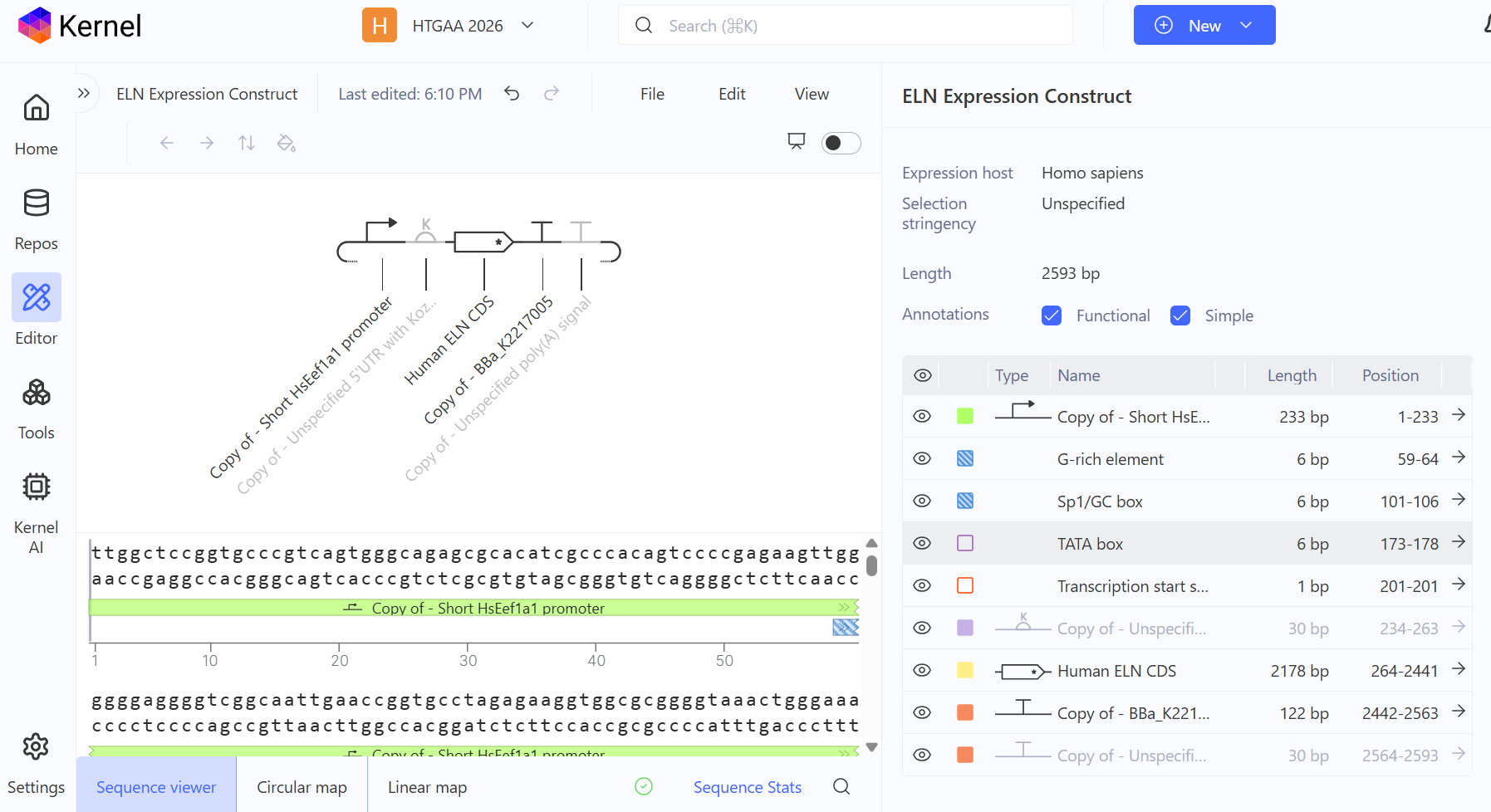
Figure 4. Final ELN Expression Construct assembled in Kernel. The map shows the BBa_J433123 / AmpR-ColE1 backbone together with the mammalian ELN expression cassette.
Kernel lists the Human ELN sequence as a CDS with both a start and a stop. The coding sequence begins with an ATG start codon and terminates with a stop codon.
The CDS length is 2,178 bp, which is divisible by three:
2,178 / 3 = 726 codons
Because the CDS is annotated as including both the start and stop codons, this value should be interpreted as 726 codons including the terminal stop codon, rather than 726 amino acids.
I therefore do not use this calculation to claim that the Kernel sequence corresponds exactly to the canonical human elastin protein sequence. The external UniProt reference contains multiple ELN isoforms and reports a canonical human elastin sequence of 786 amino acids (UniProt, n.d.). The exact relationship between the Kernel CDS and a particular reference isoform should therefore be established by direct sequence alignment rather than inferred from CDS length alone.
The Kernel sequence also contains the N-terminal region of the ELN coding sequence, so no separate artificial secretion signal was added.
I applied Kernel’s Coding Sequence Optimization tool to the Human ELN CDS.
The final optimization run used:
The initial Human ELN CDS had a length of 2,178 bp, and the sequence returned by the optimization tool was also 2,178 bp.
I compared the nucleotide sequence entered into the optimization run with the sequence returned under optimized sequences. In this final run, the two sequences were identical.
Therefore, I cannot claim that Kernel introduced specific nucleotide substitutions during this final optimization run. The appropriate conclusion is that the optimization workflow was completed using the selected mammalian host and settings, but the final run produced no nucleotide-level change in the CDS.
Codon optimization can be useful because synonymous codons are not used at identical frequencies by all organisms, and sequence composition can influence aspects of heterologous expression. However, in this particular run there was no before-and-after nucleotide difference that could be attributed to codon optimization.
The ELN coding sequence also contains substantial repetitive sequence character. Tropoelastin has a compositionally distinctive sequence containing repeated regions enriched in residues such as glycine, proline, alanine, and valine (Wise & Weiss, 2009; Keeley, 2025). This repetitive character is relevant when considering subsequent DNA synthesis and sequence verification.
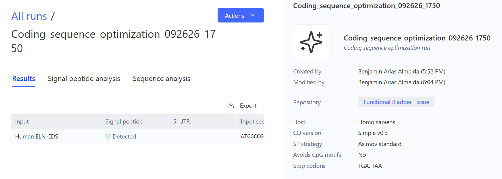
Figure 5. Kernel Coding Sequence Optimization settings used for the final ELN optimization run.
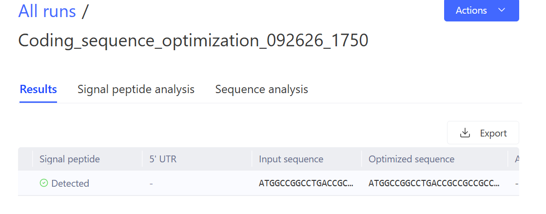
Figure 6. Kernel optimized-sequence output showing the final optimization result. The input and optimized sequences were identical in this final run.
I carried out a nucleotide sequence search using NCBI BLASTN against the core_nt database.
The query sequence used in the BLAST run, identified by RID BFXSCTE2014, had a length of 2,178 bp.
The reported result was:
No significant similarity found.
The correct interpretation of this result is that no significant nucleotide similarity was detected under the conditions of this particular search. It does not by itself demonstrate that the sequence is unrelated to human ELN.
This result differs from the earlier BLAST analysis, in which human ELN reference sequences including NM_000501.4 were identified.
One possible reason for the difference is that the sequences submitted in the two searches were not identical. A nucleotide sequence can also encode the same protein while differing at synonymous positions. Therefore, protein identity and nucleotide-level identity should not be treated as equivalent.
For this reason, I report the two BLAST observations separately rather than choosing one result as the definitive description of the sequence.
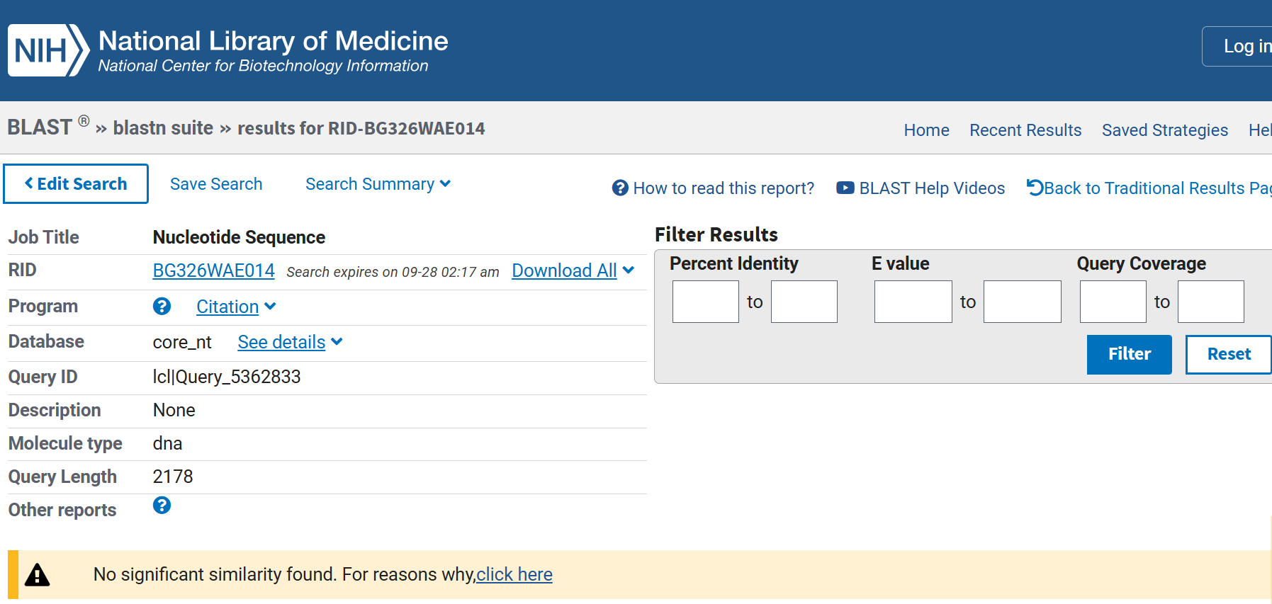
Figure 7. NCBI BLASTN result for the 2,178-bp sequence used in the later analysis, showing “No significant similarity found.”
I also analysed the sequence using the Biosecurity Sequence Scanner.
The reported result was:
No flagged sequences detected.
Thus, the submitted sequence did not produce a flagged result in this screening step.
This should be interpreted specifically as a screening result. It is not a general biological-safety certification and does not establish that a construct is universally safe. It only records the result produced by the screening system for the sequence that was submitted.
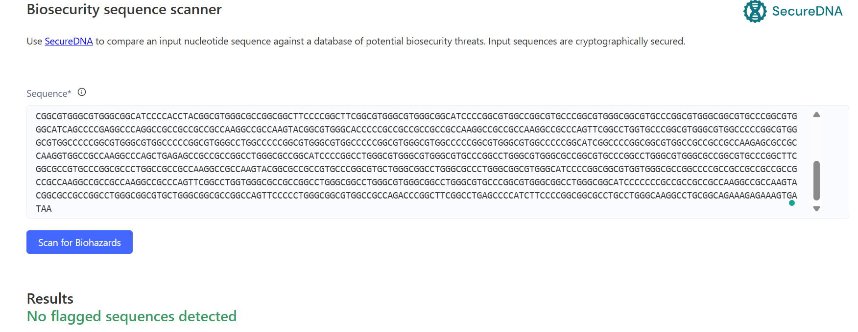
Figure 8. Biosecurity Sequence Scanner result for the submitted sequence, showing “No flagged sequences detected.”
The expression cassette is approximately 2.59 kb, with most of its length corresponding to the ELN CDS.
For an initial prototype, the designed DNA could be obtained as synthetic DNA and incorporated into the intended vector context. Because the ELN coding sequence has a repetitive sequence composition, synthesis fidelity and sequence verification would be important considerations.
Rather than assuming that one uninterrupted synthetic fragment is always optimal, the design could also be considered as a set of DNA fragments that can subsequently be assembled into the desired construct.
The HTBAA DNA-design workflow similarly distinguishes between a DNA insert/expression cassette and its plasmid backbone, emphasizing that the cassette can be designed separately before being placed into a vector context (HTGAA, 2026).
The main sequence-level consideration for this design is the repetitive character of the ELN coding sequence.
Errors introduced during DNA synthesis are particularly important for a coding sequence because nucleotide changes can alter the encoded protein or disrupt the reading frame.
Carr et al. (2004) demonstrated a protein-mediated error-correction approach for synthetic DNA and showed that error correction can improve the fidelity of de novo DNA synthesis, including for constructs on the scale of a few kilobases.
The repetitive nature of the ELN sequence therefore makes synthesis fidelity and subsequent sequence verification important considerations for this design.
If synthesis of a single continuous fragment proved problematic, dividing the design into smaller fragments would provide an alternative assembly strategy. Gibson assembly, for example, was developed to join overlapping DNA fragments into larger constructs (Gibson et al., 2009).
For the first prototype, the main objective would be to determine whether the designed expression cassette produces human tropoelastin and whether the protein reaches the extracellular environment.
At this stage, the objective is to evaluate the biological design rather than to optimize large-scale production.
For future scale-up, DNA manufacturing, assembly fidelity, sequence verification, reproducibility, and production throughput would become additional considerations. Kosuri et al. (2010) described a scalable gene-synthesis approach combining DNA microchips, selective amplification, gene assembly, and enzymatic error correction.
Thus, the priorities differ between the prototype and a potential scale-up stage: the prototype focuses on validating the biological concept, whereas later development would also require optimization of manufacturing and reproducibility.
Several important questions remain unresolved by the sequence design alone.
The presence of a promoter, Kozak sequence, and ELN CDS does not establish the expression level that will actually be obtained in the intended bladder-cell context.
How I would test it:
I would evaluate ELN expression at the RNA level and measure the resulting tropoelastin protein.
This would distinguish successful transcription from successful protein production.
The design does not contain an additional artificial secretion signal. The ELN coding sequence retains its native N-terminal region, and reference human elastin sequences contain an N-terminal signal peptide.
However, identifying a signal peptide from a sequence does not demonstrate how efficiently the resulting protein will be secreted in the intended cellular context.
How I would test it:
I would compare the amount of tropoelastin associated with the cells with the amount detected in the extracellular medium and verify the identity of the detected protein.
Expression and secretion of tropoelastin do not by themselves demonstrate the formation of a functional elastic matrix.
Extracellular assembly, crosslinking, organization, and tissue-level properties are additional biological questions.
How I would test it:
I would characterize extracellular-matrix formation and evaluate structural and functional properties of the resulting tissue construct.
These experiments would distinguish between a construct that simply expresses ELN and one that contributes to the intended functional bladder-tissue phenotype.
I used ChatGPT as an AI-assisted learning and writing tool to help organize the report according to the HTBAA Homework #2 requirements, review the logic of the genetic design, identify inconsistencies in the written interpretation, and improve the presentation of sequence-analysis results.
Example prompt:
“Can you help me organize the report according to the HTBAA Homework #2 protocol?”
The final construct design, sequence information, Kernel settings, BLAST result, and Biosecurity Sequence Scanner result were checked against my actual Kernel outputs and sequence-analysis results.
Carr, P. A., Park, J. S., Lee, Y.-J., Yu, T., Zhang, S., & Jacobson, J. M. (2004). Protein-mediated error correction for de novo DNA synthesis. Nucleic Acids Research, 32(20), e162.
Gibson, D. G., Young, L., Chuang, R.-Y., Venter, J. C., Hutchison, C. A., III, & Smith, H. O. (2009). Enzymatic assembly of DNA molecules up to several hundred kilobases. Nature Methods, 6, 343–345.
Keeley, F. W. (2025). Evolutionary constraints on positional sequence, collective properties and sequence style of tropoelastin dictated by fundamental requirements for formation and function of the extracellular elastic matrix. Matrix Biology Plus, 28, 100183.
National Center for Biotechnology Information. (n.d.). Homo sapiens elastin (ELN), transcript variant 1, mRNA (NCBI Reference Sequence: NM_000501.4). NCBI Nucleotide.
Ozsvar, J., Yang, C., Cain, S. A., Baldock, C., Tarakanova, A., & Weiss, A. S. (2021). Tropoelastin and elastin assembly. Frontiers in Bioengineering and Biotechnology, 9, 643110.
UniProt Consortium. (n.d.). Elastin (ELN_HUMAN), UniProtKB P15502. UniProt.
Wise, S. G., & Weiss, A. S. (2009). Tropoelastin. International Journal of Biochemistry & Cell Biology, 41(3), 494–497.
Cell-free protein synthesis (CFPS) lets us make proteins outside of living cells by using cell lysate, DNA templates, energy sources, amino acids, nucleotides, salts, and other ingredients.
For this assignment, I designed reaction mixes for three proteins, each with different requirements for expression and folding:
The main goal is to find reaction conditions that produce functional protein while keeping the cost per gram as low as possible.
Potassium glutamate and magnesium glutamate are important because they help control the ionic environment of the CFPS reaction.
Potassium glutamate mainly affects ionic strength and helps create suitable conditions for transcription and translation.
Magnesium glutamate supplies Mg²⁺, which is required by many enzymes and is especially important for ribosome function, nucleotide interactions, and ATP-dependent reactions.
The optimal concentrations of potassium and magnesium are different because they have different roles in the reaction.
Magnesium is generally more sensitive. Too little Mg²⁺ can reduce translation efficiency, while too much can also interfere with the reaction. Therefore, relatively small changes in magnesium concentration can have a strong effect on protein production.
A PEP + NTP formulation uses phosphoenolpyruvate (PEP) as an energy substrate and ATP, CTP, GTP, and UTP as nucleotide triphosphates.
PEP supports ATP regeneration, while the NTPs provide the substrates needed for RNA synthesis.
A Ribose + NMP formulation uses ribose as an alternative metabolic substrate and nucleotide monophosphates such as AMP, CMP, GMP, and UMP instead of nucleotide triphosphates.
The main difference is therefore the way energy and nucleotide substrates are supplied.
The Ribose + NMP approach is interesting for cost optimization because NMPs can be less expensive than NTPs. However, the CFPS system still needs to convert these substrates into the appropriate forms for transcription and energy metabolism.
I compared the sfGFP PEP and sfGFP Ribose reference formulations available in the Coopetition reaction designer.
The main difference is the energy and nucleotide system:
| Feature | sfGFP PEP | sfGFP Ribose |
|---|---|---|
| Main energy strategy | PEP-based | Ribose-based |
| Nucleotide strategy | NTPs | NMPs |
| ATP | Included | Included |
| CTP/GTP/UTP | Included as NTPs | Supplied through the NMP strategy |
| Main purpose | Efficient conventional CFPS | Potential cost reduction |
| Main optimization idea | Strong energy regeneration and transcription | Reduce the cost of nucleotide inputs |
The reference formulations also differ in their salt, amino acid, buffer, and other reagent concentrations.
This comparison shows that CFPS does not require one fixed recipe. Different combinations of energy sources, nucleotides, salts, and cofactors can be used to obtain functional protein.
For my designs, I used the reference compositions as starting points and adjusted the formulation according to the requirements of each target protein.
Each reaction has a final volume of 20 µL (20,000 nL).
The following components are fixed in all three reactions:
The remaining volume was used for the variable components.
sfGFP is the simplest of the three proteins to work with. It has no disulfide bonds, although it contains two free cysteines.
Therefore, I chose a standard reducing CFPS setup rather than an oxidizing one.
My main considerations were:
| Reagent | Volume (nL) | Final concentration |
|---|---|---|
| Base buffer | 2000 | Fixed |
| Lysate | 5000 | Fixed |
| sfGFP DNA template | 2000 | Fixed |
| Potassium glutamate | 3500 | 153.1 mM |
| Magnesium glutamate | 300 | 7.5 mM |
| HEPES-KOH | 1000 | 50 mM |
| 17 Amino Acid Mix | 1500 | 3.75 mM |
| PEP mono | 4000 | 20 mM |
| ATP | 250 | 1.25 mM |
| CTP | 125 | 0.625 mM |
| GTP | 125 | 0.625 mM |
| UTP | 125 | 0.625 mM |
| Spermidine | 75 | 0.94 mM |
| Nuclease-free water | 0 | — |
| Total | 20,000 | 20 µL |
The design uses a PEP-based energy system and standard NTPs for transcription. Since sfGFP does not require disulfide bonds, I did not add DsbC, GSSG, or GSH.
sfGFP activity is measured directly through fluorescence. Correctly folded sfGFP produces the fluorescent signal, so this assay provides a direct measurement of functional folded protein.
PETase is more challenging than sfGFP because it contains one disulfide bond.
However, the assignment indicates that PETase can be produced under standard CFPS conditions. Therefore, I kept a mainly reducing formulation instead of using the fully oxidizing formulation required for Reteplase.
I also included DsbC as a folding support.
My main considerations were:
| Reagent | Volume (nL) | Final concentration |
|---|---|---|
| Base buffer | 2000 | Fixed |
| Lysate | 5000 | Fixed |
| PETase DNA template | 2000 | Fixed |
| Potassium glutamate | 3500 | 153.1 mM |
| Magnesium glutamate | 300 | 7.5 mM |
| HEPES-KOH | 1000 | 50 mM |
| 17 Amino Acid Mix | 1500 | 3.75 mM |
| PEP mono | 3500 | 17.5 mM |
| ATP | 250 | 1.25 mM |
| CTP | 125 | 0.625 mM |
| GTP | 125 | 0.625 mM |
| UTP | 125 | 0.625 mM |
| Spermidine | 75 | 0.94 mM |
| DsbC | 1000 | 5 µM |
| Nuclease-free water | 0 | — |
| Total | 20,000 | 20 µL |
Compared with my sfGFP formulation, the PETase formulation uses slightly less PEP and adds DsbC.
The idea is that DsbC could help PETase achieve its correct folded state while avoiding the more complex oxidizing environment required by Reteplase.
PETase activity is measured by tracking the release of p-nitrophenol at 405 nm. Therefore, the assay measures enzymatic activity rather than simply the total amount of protein produced.
Reteplase is the most difficult of the three proteins because it contains nine disulfide bonds.
Unlike sfGFP and PETase, Reteplase requires an oxidizing environment so that its disulfide bonds can form correctly.
Therefore, my design includes:
The goal is not just to produce the Reteplase polypeptide, but to produce correctly folded and enzymatically active protein.
| Reagent | Volume (nL) | Final concentration |
|---|---|---|
| Base buffer | 2000 | Fixed |
| Lysate | 5000 | Fixed |
| Reteplase DNA template | 2000 | Fixed |
| Potassium glutamate | 3000 | 131.25 mM |
| Magnesium glutamate | 300 | 7.5 mM |
| HEPES-KOH | 1000 | 50 mM |
| 17 Amino Acid Mix | 1500 | 3.75 mM |
| PEP mono | 2825 | 14.125 mM |
| ATP | 250 | 1.25 mM |
| CTP | 125 | 0.625 mM |
| GTP | 125 | 0.625 mM |
| UTP | 125 | 0.625 mM |
| Spermidine | 75 | 0.94 mM |
| GSSG | 475 | 4.04 mM |
| GSH | 200 | 1.00 mM |
| DsbC | 1000 | 5 µM |
| Nuclease-free water | 0 | — |
| Total | 20,000 | 20 µL |
The GSSG/GSH pair establishes an oxidizing redox environment, while DsbC can help rearrange incorrectly paired disulfide bonds.
Reteplase activity is measured using the fluorogenic substrate IPR-AMC. When active Reteplase cleaves the substrate, fluorescent AMC is released.
Therefore, this assay checks for functional activity, not just total protein production.
For the Community Reagents section, I selected FkpA, an E. coli chaperone with peptidyl-prolyl isomerase activity.
I chose FkpA because one of the biggest challenges in producing Reteplase is correct protein folding. Reteplase contains nine disulfide bonds, so producing the polypeptide chain alone is not enough.
FkpA could provide additional folding assistance and potentially reduce aggregation of difficult proteins.
This is particularly interesting because the Reteplase formulation already uses DsbC. DsbC mainly helps rearrange incorrectly formed disulfide bonds, while FkpA could provide additional folding support.
My hypothesis is that combining different folding-support mechanisms could increase the amount of functional Reteplase.
I would test this by comparing Reteplase activity with and without FkpA and by considering the additional cost of the reagent.
The FkpA suggestion in the Community Reagents section was supported by previous work showing that FkpA can reduce aggregation and support folding in cell-free systems, including in combination with DsbC.
I also checked the CFPS Coopetition forum to follow discussions about CFPS optimization.
One optimization idea that I found interesting is the use of alternative energy sources such as maltodextrin.
Maltodextrin is relatively inexpensive and could provide a slower and more controlled energy supply compared with PEP.
This could potentially improve protein production while reducing the cost of the reaction.
I used ChatGPT (GPT-5.6 Luna) as an AI tool during the design process.
I used AI to:
Examples of prompts used included:
I selected the final reaction designs based on the assignment information, the reference formulations available in the Coopetition website, and the biological requirements of each protein.
The AI was used as a design and reasoning aid, while the final choices were made based on the experimental constraints and biological requirements described in the assignment.
Jewett, M. C., & Swartz, J. R. (2004). Mimicking the Escherichia coli cytoplasmic environment activates long-lived and efficient cell-free protein synthesis. Biotechnology and Bioengineering, 86(1), 19–26. DOI: 10.1002/bit.20026.
Silverman, A. D., Karim, A. S., & Jewett, M. C. (2020). Cell-free gene expression: an expanded repertoire of applications. Nature Reviews Genetics, 21, 151–170. DOI: 10.1038/s41576-019-0186-3.
Rasor, B. J., Vögeli, B., Landwehr, G. M., Bogart, J. W., Karim, A. S., & Jewett, M. C. (2021). Toward sustainable, cell-free biomanufacturing. Current Opinion in Biotechnology, 69, 136–144. DOI: 10.1016/j.copbio.2020.12.012.
Smith, A. A., et al. (2026). Using a GPT-5-driven autonomous lab to optimize the cost and titer of cell-free protein synthesis. bioRxiv. DOI: 10.64898/2026.02.05.703998.
Olsen, M. L., Copeland, C. E., Sundberg, C. A., Aw, R., Shaver, Z. M., Rao, G., Swartz, J. R., Karim, A. S., & Jewett, M. C. (2026). Design-driven optimization of low-cost reagent formulations for reproducible and high-yielding cell-free gene expression. Nature Communications. DOI: 10.1038/s41467-026-69605-8.