Week 1 HW: Final Project Ideas
21st September 2026
Week 1 Homework - Biomanufactured Phage Particles With Enhanced Receptor and Host Specificity for Antibiotic Resistance
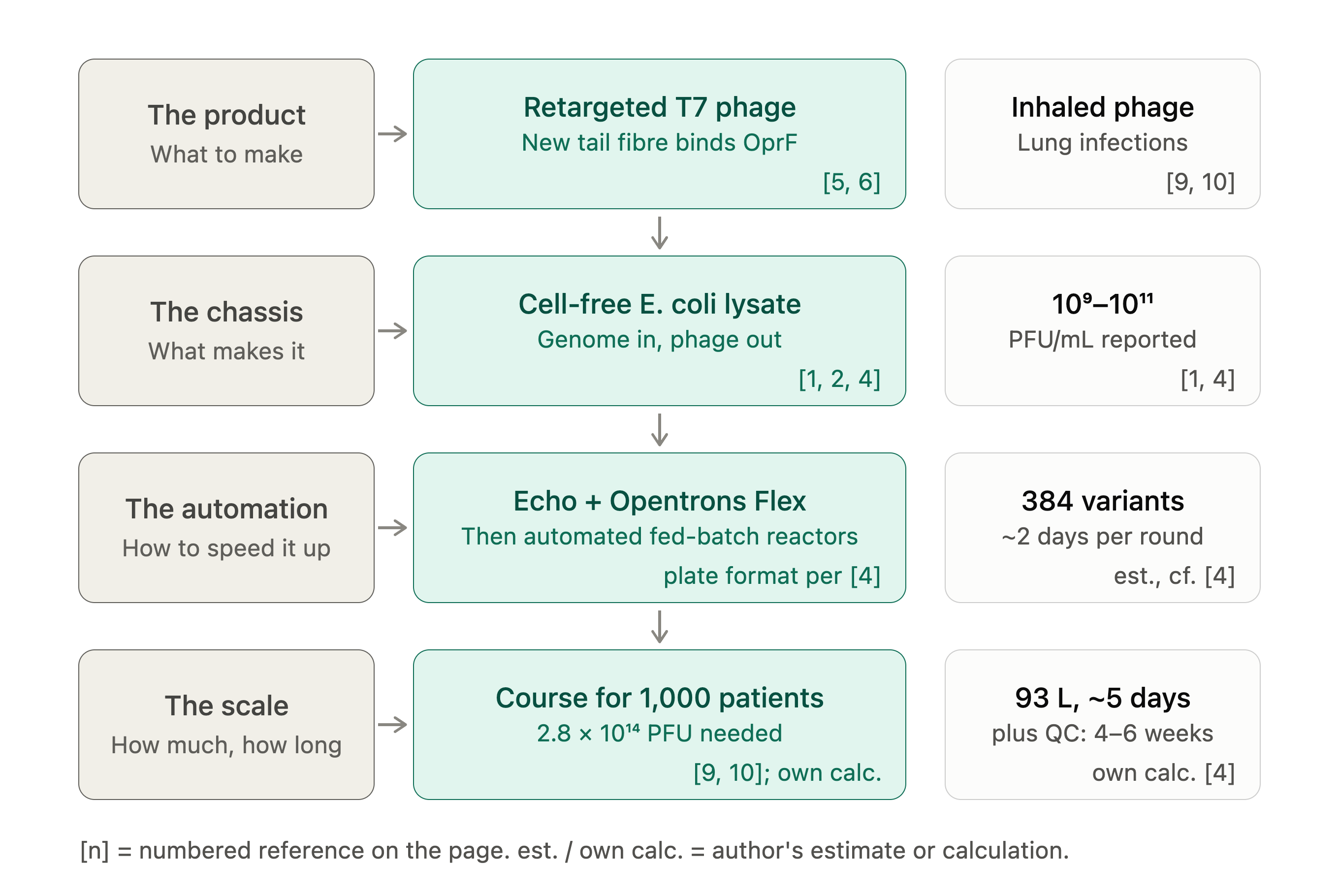
Figure 1. Overview of the proposed cell-free biomanufacturing route for retargeted T7 phage. Bracketed numbers refer to the References below; “est.” and “own calc.” mark the author’s estimates and calculations.
1. What I would like to biomanufacture, and why?
I would like to manufacture engineered bacteriophage particles, specifically T7 phage carrying a redesigned gp17 receptor-binding domain (RBD) that targets the outer-membrane protein OprF of Pseudomonas aeruginosa — as a therapeutic product.
The product is a purified, endotoxin-reduced suspension of infectious phage particles, formulated at a defined titre (plaque-forming units per millilitre) for nebulised or topical delivery. It is intended for people with chronic P. aeruginosa infection: cystic fibrosis and bronchiectasis patients with established airway colonisation, and burns or chronic-wound patients where the organism is multidrug-resistant.
My motivation is partly utilitarian and partly personal to the work I have already done. P. aeruginosa is on the WHO priority pathogens list, where carbapenem-resistant isolates are classed as high priority (BPPL 2024) [12], and these isolates leave very little left to prescribe. Phage therapy works, but almost every successful case to date has been artisanal: a clinician finds a matching phage in a collection, one patient at a time, over weeks (for example, the personalised aerosolised treatment reported in [9]). The bottleneck is not biology, it is manufacturing — we cannot yet make a specified phage quickly, safely, and reproducibly.
This connects directly to my existing computational work, where I redesigned the gp17 RBD of T7 to bind OprF using ProteinMPNN [13], ESM3 [14], and AlphaFold-Multimer [15], with a lead candidate (LC-0.36) predicted to improve interface confidence substantially over wild type. That work stops at a sequence on a screen. This proposal is the natural continuation: how do you actually turn a designed RBD sequence into doses? What makes it interesting to me is that the design step is now fast and the manufacturing step is not, so the manufacturing side is where the real leverage is.
A design note worth stating up front: T7 is a lytic phage with a short, well-characterised genome (~40 kb) [4], and its host range is dominated by the tail-fibre/RBD [4–6], which makes it unusually amenable to retargeting. I would also delete or inactivate any genes not required for lysis and assembly (a 35.8 kb T7 with three non-essential regions deleted has been made in cell-free reactions [4]), and would not carry antibiotic-resistance markers in the therapeutic construct.
2. What living system would I use?
Primary chassis: a cell-free protein synthesis (TXTL) system — E. coli lysate-based used to bootstrap whole infectious phage from purified genomic DNA.
This is the part of the proposal I think is genuinely interesting, and it is not hypothetical: T7, T4, and several other phages have been shown to self-assemble into infectious particles in E. coli cell extracts supplied only with their genome, an energy regeneration system, amino acids, and NTPs (the Noireaux group’s TXTL work is the key reference here [1–4]).
Inputs: (i) the engineered T7 genome, assembled from synthetic or PCR-amplified fragments with the redesigned gp17 RBD swapped in — by Gibson or yeast assembly, or by exonuclease III–based annealing, which unlike Gibson can be added directly to the cell-free reaction without clean-up [4]; (ii) E. coli S30-type lysate supplying ribosomes, tRNAs, and translation factors; (iii) an energy/substrate buffer — amino acids, NTPs, Mg²⁺/K⁺, and a phosphate-based energy regeneration system such as 3-PGA or creatine phosphate.
Process: the lysate’s endogenous RNA polymerase transcribes the T7 early genes from the phage’s own promoters; T7 RNAP is then produced and drives the middle and late genes, including capsid, scaffolding, tail, and tail-fibre proteins. Capsid assembly, genome packaging by the terminase, and tail attachment all occur spontaneously in the tube, because phage morphogenesis is self-assembling and requires no host membrane.
Output: infectious phage particles in a cell-free reaction, at reported titres on the order of 10⁹–10¹¹ PFU/mL [1, 4].
Why cell-free rather than infecting live E. coli? Three reasons, and they all bear on manufacturing. First, there is no living host, so there is no endotoxin-laden bacterial culture to remove and no risk of lysogeny or host-genome carryover — a major regulatory advantage for an injectable or inhaled product. Second, retargeted phage often cannot be propagated on its intended host (you do not want to culture P. aeruginosa at manufacturing scale) nor on E. coli (the redesigned RBD no longer binds E. coli LPS) — cell-free sidesteps this host-range trap entirely [4, 7], which is the single biggest practical obstacle to retargeted phage production. Third, the reaction is linear and additive: one genome sequence in, particles out, with no strain-construction step.
Assumptions I am making: that the redesigned RBD folds and trimerises correctly in a cell-free context; that cell-free titres reported for wild-type T7 hold approximately for the retargeted variant (they may not — see §5); and that a scaled reaction behaves like a scaled-up batch rather than requiring a fundamentally different format.
Secondary/backup chassis: a surrogate E. coli strain engineered to surface-display OprF, allowing conventional high-titre lytic propagation. Cheaper per particle, but reintroduces endotoxin and host-DNA removal burden.
3. How would I use automation?
I would use automation at two distinct stages, and they call for different technology.
(a) Prototyping — acoustic dispensing plus an Opentrons Flex for design-build-test cycling.
The computational pipeline gives me not one RBD but a ranked panel of perhaps 96–384 designs. The step that actually limits me is not designing them, it is testing them. I would automate:
Reaction assembly: nanolitre acoustic dispensing (Echo-style) to set up cell-free reactions in 384-well plates — each well a different RBD variant genome — at 5–10 µL per reaction [4], conserving lysate, which is the expensive reagent. Screening readout: plate-reader kinetics on a fluorescent reporter encoded in the phage genome to confirm expression, followed by automated spotting of each well onto P. aeruginosa lawns (Opentrons Flex with a pipetting head) to score infectivity and host range. Optimisation: the cell-free reaction composition itself (Mg²⁺, K⁺, energy source, DNA concentration) has a strong, non-obvious optimum per construct. This is a classic design-of-experiments problem, and I would run active-learning-driven DoE across the plate rather than one-factor-at-a-time. This turns a design-build-test cycle from a week or more for in vivo phage engineering methods [4] to roughly two days per round, which is where most of the value sits.
(b) Manufacturing — automated fed-batch cell-free reactors with in-line monitoring.
At scale the format changes. Rather than thousands of wells, I would run litre-scale stirred cell-free reactions with automated feeding of energy substrates (cell-free reactions stall on phosphate depletion and pH drift, not on ribosome availability), automated sampling into an at-line plaque or qPCR titre assay, and automated tangential-flow filtration and chromatography for particle purification. This is also where a Cloud Lab or a CDMO-style automated suite would be used, because the process must be reproducible under a documented protocol, not just successful once.
The important point is that the prototyping automation and the manufacturing automation share almost nothing except the biology — and I think that gap is itself one of the main themes of this course.
4. How much, and how long?
(a) How much? Scale I am defining: a full therapeutic course for 1,000 patients with chronic P. aeruginosa airway infection.
Assumptions (with sources noted):
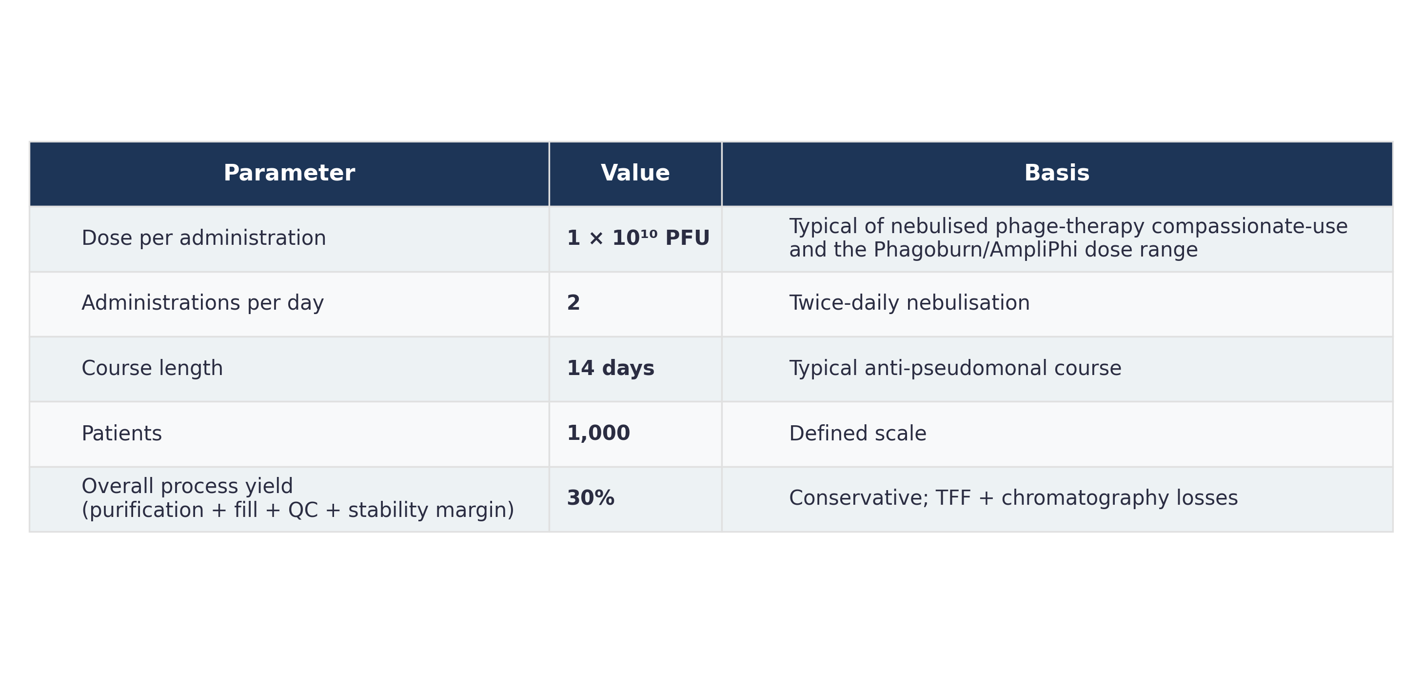
Sources for the assumptions: the dose per patient sits above the 5 × 10⁹ PFU/day (five days) used in a published aerosol case [9] and follows a twice-daily nebulised schedule like that in a phase 1b/2a study [10]; the titre range comes from [1, 4]; the 30% overall yield and the patient number are my own assumptions.
Phage required per patient:
1 × 10¹⁰ PFU × 2 /day × 14 days = 2.8 × 10¹¹ PFU per course
For 1,000 patients:
2.8 × 10¹¹ × 1,000 = 2.8 × 10¹⁴ PFU
Correcting for 30% overall yield:
2.8 × 10¹⁴ / 0.30 ≈ 9.3 × 10¹⁴ PFU must be produced in the reactor
Converting to reaction volume, assuming a cell-free titre of 1 × 10¹⁰ PFU/mL (a deliberately conservative figure from the reported 10⁹–10¹¹ range [1, 4]):
9.3 × 10¹⁴ PFU ÷ 1 × 10¹⁰ PFU/mL = 9.3 × 10⁴ mL ≈ 93 litres of cell-free reaction
If the higher end of the reported range (10¹¹ PFU/mL) is achievable, this drops to ~9.3 L — which is the difference between a benchtop process and a plant, and is why titre is the number I care most about (§5).
(b) How long?
Per-batch time: a T7 cell-free reaction reaches plateau in roughly 6–8 hours at 29–30 °C (T7 reached its reported titre after about 3 hours at 29 °C in [4], so this is conservative). Adding reactor turnaround, clarification, TFF, and polishing chromatography, I will assume 24 hours per batch end-to-end, with purification of batch n overlapping the reaction of batch n+1.
Configuration: 4 × 5 L reactors run in parallel = 20 L of reaction per batch cycle.
93 L ÷ 20 L per cycle = ~4.7 → 5 batch cycles
5 cycles × 24 h = 120 hours ≈ 5 days of production
Adding genome assembly and QC release (sterility, endotoxin, titre, sequence identity), realistically 4–6 weeks from sequence to released doses, with the QC and release steps — not the biology — dominating the timeline.
Prototyping timeline, for contrast: 384 variants × 10 µL = 3.84 mL of total reaction volume (lysate is about a third of this [2]), one 8-hour reaction, one overnight plaque readout. One design-build-test round per ~48 hours. Roughly four orders of magnitude less material to test 384 designs than to treat 1,000 patients (3.84 mL versus 93 L) — which I think is the clearest illustration of why prototyping and manufacturing need different automation.
Sanity check: 93 L of cell-free reaction is large but not absurd — it is comparable to a mid-size pilot bioreactor batch, and it treats 1,000 patients for two weeks. Scaling to all ~100,000 people with CF worldwide (an estimated 105,000 are diagnosed [11]) would need ~9,300 L, at which point the lysate supply chain, not the phage biology, becomes the binding constraint.
5. What I don't know yet
Does the retargeted phage actually assemble and remain infectious — and at what titre? Every number in §4 scales inversely with titre, and the 10⁹–10¹¹ PFU/mL spread [1, 4] is two orders of magnitude, i.e. the difference between 9 L and 930 L. In the PHEIGES study, libraries of tail-fibre mutants gave lower titres than wild-type T7, and titre fell as mutation load rose [4], which is a first sign that retargeting may cost yield. The redesigned RBD must fold, trimerise, and be incorporated into the tail at wild-type-like efficiency; a design that binds OprF beautifully in an AlphaFold-Multimer model may still assemble poorly, or may bind so tightly that it fails to release and eject its genome. To determine: measure cell-free titre for the top designs directly, and separately measure adsorption rate constant and burst behaviour on P. aeruginosa — binding affinity and infectivity are not the same variable, and I have so far only optimised the first.
Cost and scalability of the lysate itself. Cell-free is elegant at 10 µL and potentially prohibitive at 93 L. The reactions in [4] were run at 1–10 µL, and I have not found litre-scale cell-free phage data. Lysate is currently the dominant cost and is produced by, ironically, growing and lysing large volumes of E. coli — so the endotoxin problem I claimed to avoid may simply move upstream into the lysate. To determine: cost per PFU in cell-free versus surrogate-host propagation, and residual endotoxin in the final product from the lysate route.
Resistance and regulatory identity. P. aeruginosa can downregulate or modify OprF, and a single-receptor phage invites escape mutants [8] — a cocktail of several RBD designs against different receptors is almost certainly the real product. But a cocktail multiplies the manufacturing and regulatory burden by its number of components, and current frameworks do not cleanly accommodate a product whose composition is meant to change per patient. To determine: escape frequency against single designs versus a cocktail in vitro, and which parts of the process must be re-validated when one component is swapped.
References
- Shin, J., Jardine, P., & Noireaux, V. (2012). Genome replication, synthesis, and assembly of the bacteriophage T7 in a single cell-free reaction. ACS Synthetic Biology, 1(9), 408–413. https://doi.org/10.1021/sb300049p
- Rustad, M., Eastlund, A., Marshall, R., Jardine, P., & Noireaux, V. (2017). Synthesis of infectious bacteriophages in an E. coli-based cell-free expression system. Journal of Visualized Experiments, 56144. https://doi.org/10.3791/56144
- Rustad, M., Eastlund, A., Jardine, P., & Noireaux, V. (2018). Cell-free TXTL synthesis of infectious bacteriophage T4 in a single test tube reaction. Synthetic Biology, 3(1), ysy002. https://doi.org/10.1093/synbio/ysy002
- Levrier, A., Karpathakis, I., Nash, B., Bowden, S. D., Lindner, A. B., & Noireaux, V. (2024). PHEIGES: all-cell-free phage synthesis and selection from engineered genomes. Nature Communications, 15, 2223. https://doi.org/10.1038/s41467-024-46585-1
- Huss, P., Meger, A., Leander, M., Nishikawa, K., & Raman, S. (2021). Mapping the functional landscape of the receptor binding domain of T7 bacteriophage by deep mutational scanning. eLife, 10, e63775.
- Garcia-Doval, C., & van Raaij, M. J. (2012). Structure of the receptor-binding carboxy-terminal domain of bacteriophage T7 tail fibers. Proceedings of the National Academy of Sciences, 109, 9390–9395.
- Emslander, Q., Vogele, K., Braun, P., Stender, J., Willy, C., Joppich, M., Hammerl, J. A., Abele, M., Meng, C., Pichlmair, A., Ludwig, C., Bugert, J. J., Simmel, F. C., & Westmeyer, G. G. (2022). Cell-free production of personalized therapeutic phages targeting multidrug-resistant bacteria. Cell Chemical Biology, 29(9), 1434–1445.e7. https://doi.org/10.1016/j.chembiol.2022.06.003
- Yehl, K., Lemire, S., Yang, A. C., Ando, H., Mimee, M., Torres, M. D. T., de la Fuente-Nunez, C., & Lu, T. K. (2019). Engineering phage host-range and suppressing bacterial resistance through phage tail fiber mutagenesis. Cell, 179, 459–469.e9.
- Personalized aerosolised bacteriophage treatment of a chronic lung infection due to multidrug-resistant Pseudomonas aeruginosa [Case report]. https://pmc.ncbi.nlm.nih.gov/articles/PMC10300124/
- Nebulized phage therapy for patients with cystic fibrosis with chronic Pseudomonas aeruginosa pulmonary infection: a phase 1b/2a randomized, double-blind, placebo-controlled, multicenter study [Conference abstract 2891]. https://www.ncbi.nlm.nih.gov/pmc/articles/PMC10678098/
- Guo, J., Garratt, A., & Hill, A. (2022). Worldwide rates of diagnosis and effective treatment for cystic fibrosis. Journal of Cystic Fibrosis, 21, 456–462. https://pubmed.ncbi.nlm.nih.gov/35125294/
- World Health Organization. (2024). WHO bacterial priority pathogens list, 2024: Bacterial pathogens of public health importance to guide research, development and strategies to prevent and control antimicrobial resistance. Geneva: WHO. https://www.who.int/publications/i/item/9789240093461
- Dauparas, J., et al. (2022). Robust deep learning-based protein sequence design using ProteinMPNN. Science, 378, 49–56.
- Hayes, T., et al. (2025). Simulating 500 million years of evolution with a language model. Science, 387, 850–858.
- Evans, R., et al. (2021). Protein complex prediction with AlphaFold-Multimer. bioRxiv. https://doi.org/10.1101/2021.10.04.463034
Course readings
- Karim, A. S., & Jewett, M. C. (2021). Toward sustainable, cell-free biomanufacturing. Current Opinion in Biotechnology, 69, 136–144. https://doi.org/10.1016/j.copbio.2020.12.012
- Petzold, C. J., & Mukhopadhyay, A. (2025). From bench to biofactory: High-throughput technologies and automated workflows to accelerate biomanufacturing. Current Opinion in Biotechnology, 94, 103320. https://doi.org/10.1016/j.copbio.2025.103320
AI use
Claude (Anthropic, Claude Sonnet 5) was used to check this page against the assignment, to find and verify sources, to build the overview figure, and to insert in-text citations.