I'm a bioengineer from UTEC (Lima, Peru) with a background that bridges wet lab research and regulatory affairs. During a research internship at the University of Alberta, I worked on cell culture, PCR, and fluorescence microscopy, and I led a fermentation kinetics project on chicha de jora funded by the Harvard-UTEC Seed Fund. I currently work at a pathology laboratory in Peru, verifying compliance of immunohistochemistry results for oncology partnerships, an experience that sparked my interest in biomanufacturing, particularly cell-free systems and automation applied to diagnostics. I'm excited to join HTBAA as a Committed Listener and explore how to translate lab-based discoveries into real, scalable products.
See the nav bar on the left to look through my work for How To
Biomanufacture Almost Anything (Fall 2026)!
Subsections of Tania Barreda — HTBAA Fall 2026
Week 1 HW: Final Project Ideas
HTBAA Homework #1
1. What you would like to biomanufacture and why?
Target product: a single-domain antibody fragment (nanobody, VHH) directed against PD-L1, intended for use as an immunohistochemistry (IHC) diagnostic reagent, designed to integrate with an automated digital quantification workflow.
Why: In triple-negative breast cancer (TNBC), eligibility for atezolizumab depends on a specific assay, SP142, which evaluates PD-L1 expression in tumor-infiltrating immune cells using a threshold of just 1%. This type of scoring presents challenges related to the pathologist’s visual assessment and inter-pathologist agreement when evaluating low thresholds in tissue. Without prior training, visual interpretation tends to be more subjective, and while training improves outcomes, it does not eliminate reliance on individual visual interpretation (Kim et al., 2021). This substantial variability among pathologists when evaluating immunological biomarkers has direct implications for therapeutic decisions (Van Bockstal et al., 2023). I have encountered this type of problem firsthand in my current position at a pathology laboratory in Peru. The accuracy of a reagent is not merely a technical issue; rather, it determines whether a patient has access to treatment. A nanobody produced through a standardized cell-free process, combined with an automated digital quantification system rather than a pathologist’s visual reading, could offer a more objective alternative to the current manual scoring. As far as I can tell from the literature, this combination has not been specifically explored for this problem.
Who it is for and why it matters: this reagent would be intended for patients with advanced or metastatic TNBC being evaluated for atezolizumab eligibility, and for pathology laboratories that currently rely on manual immune-cell scoring to make that determination. By combining a standardized biomanufactured reagent with an automated reading workflow, this approach could offer a more objective and reproducible alternative to current visual interpretation, particularly relevant in metastatic sites, where documented variability is greatest.
2. What living system would you use?
Proposed chassis: a cell-free system based on E. coli lysate, following the commercial NEBExpress-type approach, well documented for producing single-chain antibody fragments quickly, including nanobodies (Haueis & Kubick, 2022).
How it would work: The system needs a DNA template (linear or plasmid-based) encoding each candidate anti-PD-L1 nanobody sequence, fused to a histidine tag for later purification. It also needs E. coli lysate, which supplies the transcription and translation machinery, plus an energy mix of amino acids, NTPs, and an energy-regeneration system. The reaction runs through coupled in vitro transcription-translation (IVTT). A nanobody is a single domain with no light chain and no strict glycosylation requirement for binding activity, so a simple bacterial chassis works fine here. A full IgG antibody would need a mammalian system instead. The reaction finishes within a few hours. What comes out is a soluble nanobody, purified by affinity chromatography using Ni-NTA magnetic beads that capture the histidine tag.
Assumptions I’m making:
Preliminary research, such as a phage display library derived from immunized camelids, has already yielded a small set of candidate VHH sequences against PD-L1. Not a single final sequence, and not an unfiltered full library. This project focuses on manufacturing and selecting the best option from that small pool, not on the full initial discovery of the binder.
The E. coli lysate, whether produced in-house or purchased commercially, maintains consistent activity across batches.
3. How would you use automation?
Proposed technology: a desktop liquid-handling robot, Opentrons OT-2 type, organized around the biofoundry Design-Build-Test-Learn (DBTL) cycle.
What I would automate, step by step:
Build: automated assembly of the small pool of nanobody candidates from Question 2, using a standard method like Golden Gate, dispensed in parallel across 96-well plates.
Cell-free reaction: automated dispensing of the IVTT reaction mix into each well, then controlled incubation.
Purification: automated magnetic separation of Ni-NTA beads to isolate the nanobody from each candidate.
Test: each purified nanobody gets coupled to a staining assay on tissue samples (or a simulated model). Then comes image capture and automated digital quantification of the staining pattern on immune cells, replacing the pathologist’s visual reading identified as a source of subjectivity in Question 1.
Why automate these specific steps: it lets me compare candidates from the small pool in parallel to find the best one for affinity and specificity, instead of testing them one at a time. That matters, but what matters more for this proposal is that it builds a quantitative, reproducible reading step into the process from the start. No falling back on a pathologist’s visual read again.
4. How much would you need to make, and how long would it take?
(a) How much?
Peru records roughly 20,759 new breast cancer cases every year (Johnston, 2008). A cross-sectional study of 1,176 patients at the Regional Institute of Neoplastic Diseases in northern Peru found that 18.2% of immunohistochemical profiles were triple-negative (Gómez-Rázuri et al., 2024). That works out to about 3,777 patients per year in Peru who would need PD-L1 evaluation via SP142 to determine eligibility for atezolizumab.
I’m assuming each test requires on the order of 1 µg of reagent, based on typical primary antibody volumes in IHC (roughly 100-200 µL applied at a working concentration of 1-5 µg/mL). Adding a ×3 factor for controls and possible repeat testing gives 3,777 × 3 µg, or about 11.3 mg of nanobody per year to cover this need in Peru.
(b) How long would it take?
New England Biolabs has published real yield data for their NEBExpress system showing nanobody (VHH) production in the range of 50-250 ng/µL, or 0.05-0.25 mg/mL, in E. coli cell-free reactions completed within 2-4 hours (NEB, 2026; Buss, n.d.). Taking a conservative 0.1 mg/mL and a 96-well plate at 200 µL per well, each well yields roughly 20 µg. A full plate run produces about 1.92 mg in a single 2-4 hour run. To cover Peru’s 11.3 mg annual need, that comes out to roughly 6 plate runs per year.
5. What don’t you know yet?
Real specificity and affinity: it’s unclear whether any candidate from the reduced pool of VHH sequences would reach sufficient affinity and specificity against the relevant PD-L1 epitope on TNBC immune cells. Class 7 (Measurement & Assays) should give me the conceptual grounding I’m missing here, general principles of quantitative measurement of biomolecular interactions, so I know what kind of affinity assay would actually make sense to design.
Compatibility with existing staining protocols: a nanobody, typically detected through its tag rather than a traditional secondary-antibody system, would likely require reworking the current visualization protocol. What I don’t understand yet, at a general level, is what alternative diagnostic detection formats exist beyond conventional IHC. Class 5 (Digital Microfluidics) seems like the right place to build that understanding, and Lab #2 should help me get a better feel for the proposed chassis hands-on. Neither would hand me a ready-made solution for this specific reagent, but together they’d give me a reference framework for thinking about different detection platforms.
Real yield and reproducibility of this specific VHH: the NEB data (0.05-0.25 mg/mL) comes from generic nanobodies, not the anti-PD-L1 VHH in this project. I still need the general principles behind what drives yield and reproducibility in cell-free systems, lysate quality, reaction conditions, scale-up, and Class 3 (Cell-Free Systems, Cloud Labs) plus Lab #1 are where I’d expect to build that conceptual base.
Gómez-Rázuri, K., Abad-Licham, M., Astigueta, J., & Moreno, J. (2024). Cáncer de mama en el norte del Perú: subtipos moleculares y HER2 low. Revista Peruana de Medicina Experimental y Salud Pública, 41(1), 62–68. https://doi.org/10.17843/rpmesp.2024.411.13424
Haueis, S., & Kubick, S. (2022). A cell-free expression pipeline for the generation and functional characterization of nanobodies. Frontiers in Bioengineering and Biotechnology, 10, 896763. https://doi.org/10.3389/fbioe.2022.896763
Kim, S. (2021). Programmed death ligand 1 immunohistochemistry in triple-negative breast cancer: Evaluation of inter-pathologist concordance and inter-assay variability. Journal of Breast Cancer, 24(3), 266–279. https://doi.org/10.4048/jbc.2021.24.e29
Van Bockstal, M. R., Cooks, M., Nederlof, I., Brinkhuis, M., Dutman, A., Koopmans, M., Kooreman, L., van der Vegt, B., Verhoog, L., Vreuls, C., Westenend, P., Kok, M., van Diest, P. J., Nauwelaers, I., Laudus, N., Denkert, C., Rimm, D., Siziopikou, K. P., Ely, S., Zardavas, D., … van Deurzen, C. H. M. (2021). Interobserver agreement of PD-L1/SP142 immunohistochemistry and tumor-infiltrating lymphocytes (TILs) in distant metastases of triple-negative breast cancer: A proof-of-concept study. Cancers, 15(6), 1690.
AI disclosure: Claude (Anthropic) was used throughout the development of this assignment to verify internal consistency across answers, refine technical wording, translate content between Spanish and English, and format this document in Markdown. All scientific claims were reviewed against cited primary sources.
Week 2 HW: Genetic Design in Kernel
HTBAA Homework #2
0. Set up in Kernel
My repository is tania-barreda-PDL1
1. Define your expression target
Molecule: a protein, the anti-PD-L1 nanobody (VHH) KN035.
Sequence and source: the primary sequence comes from PDB entry 5JDS (chain B, “Nanobody,” Camelidae), also indexed in NCBI Structure under MMDB ID 149666, from Zhang et al. (2017). Because KN035 is an engineered antibody rather than a natural gene product, it has no dedicated UniProt entry; the target antigen, human PD-L1, does (UniProt Q9NZQ7). As a backup source for the same molecule, KN035 is also disclosed as SEQ ID NO. 1 in patent US10556954B2. The final CDS I designed adds an initiator Met before this sequence and, separately, a double stop codon (TGA-TAA) at the end, since the deposited sequence has neither.
Host or chassis: cell-free system based on E. coli lysate (NEBExpress-type), as proposed in Homework #1.
Where the product goes: the nanobody stays soluble in the cell-free reaction; no compartments exist in this system, so no signal peptide is used.
2. Design your genetic parts
Layer
Part (type and short name)
Sequence and source
Justification
Transcription start
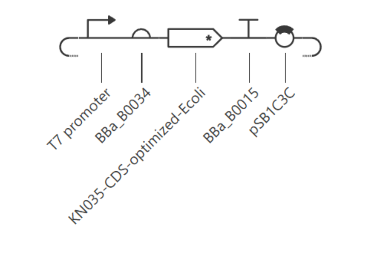
Promoter: T7 promoter
TAATACGACTCACTATAGGG (Studier & Moffatt, 1986); found in Kernel’s repository
NEBExpress transcribes only from a T7 promoter, since T7 RNA polymerase is supplied in the kit.
Translation start
RBS: BBa_B0034
Kernel/iGEM Registry public part
A well-characterized strong ribosome binding site, functional with mRNA regardless of which RNA polymerase transcribed it.
Coding sequence
CDS: KN035, +start +stop
Based on Kernel part BBa_K4266004 (KN035 DNA sequence).
Encodes the product defined in Part 1; the added start/stop codons were required for Kernel’s Construct Checker to recognize it as a complete CDS, and for correct translation in the simulator
Targeting
Tag: 6xHis (already included at the C-terminus of the deposited KN035 sequence)
Part of the same CDS part above
Enables Ni-NTA magnetic bead purification, matching the automation workflow from Homework #1. No separate signal peptide part is used, consistent with a soluble, compartment-free cell-free reaction.
Transcription stop
Terminator: BBa_B0015
Kernel/iGEM Registry public part (double terminator)
Standard, well-characterized terminator compatible with either T7 or native E. coli RNA polymerase.
Vector context
Backbone: pSB1C3C (Kernel’s internal copy of the standard pSB1C3 backbone)
Kernel public repository; confirmed to carry chloramphenicol resistance, a high-copy origin, and an empty size close to the standard ~2,070 bp
Provides origin of replication and selection marker to propagate the construct in E. coli; not required by the cell-free reaction itself.
3. Assemble your construct
The construct, named PD-L1-nanobody-construct, was assembled in Kernel with parts in this order, 5’ to 3’: backbone (pSB1C3C) → T7 promoter → RBS (BBa_B0034) → CDS (KN035, +start +stop) → terminator (BBa_B0015).
Kernel’s Construct Checker initially flagged two real issues and three expected, non-applicable warnings:
Real issues (fixed): the CDS did not start with ATG and did not end with a double stop codon, because the source part (BBa_K4266004) held only the coding sequence without these elements. I resolved this by creating a new CDS part with ATG and TGA-TAA added, and by correcting that new part’s type from “(-start -stop)” to “(+start +stop)” after noticing the simulator (Part 4) was not translating it correctly despite the sequence being correct.
Expected, non-applicable warnings: Kernel also flagged missing Kozak sequences, 5’ and 3’ UTRs, and poly(A) signals. These apply to eukaryotic expression architecture, not to a prokaryotic, RBS-based system, so I did not add them.
4. Optimize and quality-check
Codon optimization. I ran Kernel’s codon optimizer on the amino acid sequence, with preset “None” (the CHO/LV/AAV presets do not apply to a bacterial host), expression organism E. coli, no signal peptide algorithm, and stop codons set to TGA then TAA (double stop, per Kernel’s own recommendation). The tool returned a new, optimized DNA sequence as a separate part, which I then used to replace the manually-built CDS in the construct.
Sequence search (BLASTx). I ran BLASTx. The best hit was “Chain B, Nanobody [Camelidae]”, confirming that the CDS encodes the intended KN035 nanobody and not an unrelated sequence.
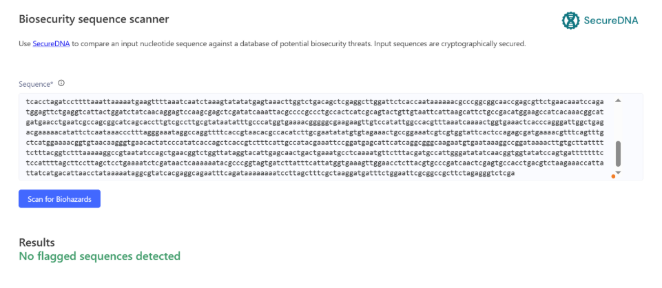
Safety screen. I ran Kernel’s SecureDNA biosecurity scanner on the full assembled construct. It reported “No flagged sequences detected.”
5. From design to build
How I would build it. My assembled expression cassette (promoter through terminator) is under 1 kb, and the full construct with backbone is a few kb, well within the range for a single synthetic gene order, sequence-verified by the vendor in their standard cloning vector. Gibson assembly (Gibson et al., 2009) would only be worth using if I needed to combine this cassette with other fragments I already had, joining overlapping pieces in one isothermal reaction with a 5’ exonuclease, a polymerase, and a ligase. Chip-derived oligo pool synthesis, as in Kosuri et al. (2010), who assembled 47 genes (42 of them therapeutic antibody sequences) from a 13,000-oligo pool using selective amplification and enzymatic error correction, becomes worthwhile mainly when many sequence variants must be built and screened in parallel, closer to the discovery step assumed in Homework #1 than to this single, defined construct.
What synthesis constrains. Carr et al. (2004) report a typical error rate near one error per 600 bp for conventional gene synthesis, reduced more than 15-fold, to about one per 10,000 bp, using a MutS mismatch-binding protein to remove failure products. Applying those rates to my construct (assuming independent, Poisson-distributed errors), a several-kb construct would have a substantially lower error-free fraction at the uncorrected rate than at the corrected one, which is why I would rely on vendor sequence verification of the final clone rather than skip verification. Nothing in my current design (his-tag repeats aside) is an unusually difficult synthesis target, but the deposited KN035 sequence does contain a short repetitive stretch (the poly-His tag) that vendors sometimes flag for extra QC, since homopolymer-like repeats are harder to synthesize accurately.
Prototype vs. scale. For the prototype, I would order one sequence-verified plasmid and test both circular and linearized template in the NEBExpress reaction, since linear DNA is compatible with the system but more exposed to nuclease degradation in lysate; NEB offers a nuclease inhibitor (GamS) for this case. For scale, DNA demand stays small: using the Homework #1 estimate of about 6 plate runs of 96 wells at 200 µL per year, and NEB’s suggested starting point of 250 ng of template per 50 µL reaction, the annual template requirement is under 1 mg. I would meet this by growing and purifying the same sequence-verified plasmid in E. coli in batches, verified by sequencing, rather than repeating de novo gene synthesis for each batch.
6. What don’t you know yet?
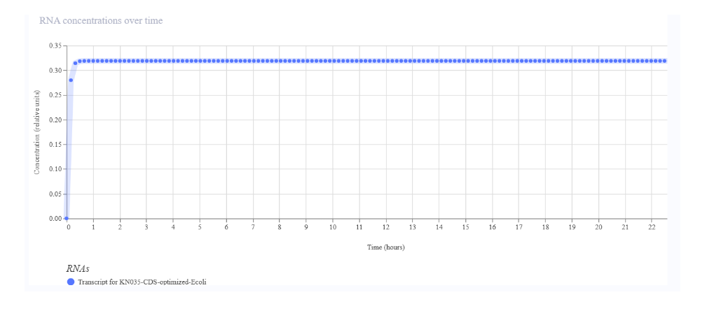
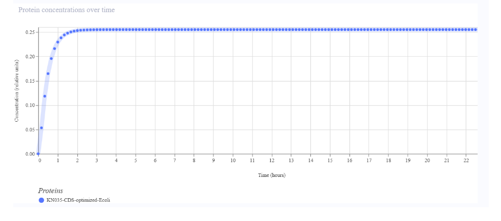
Whether the metadata correction I made (part type “+start +stop”) reflects reality, not just the simulator’s expectations. My CDS part initially simulated zero protein despite RNA accumulating; the fix was changing the part’s start/stop flags, not the sequence itself. This raises a real unknown: whether Kernel’s simulated protein output (0.25, stabilizing around 2 hours) reflects a biologically meaningful expression level, or only that the simulator can now correctly locate the reading frame. How I would find out: run the actual cell-free reaction and measure protein by SDS-PAGE and His-tag capture, rather than relying on the simulation’s absolute values.
Disulfide bond formation in a reducing, cell-free E. coli environment. KN035 carries two disulfide bonds (Zhang et al., 2017), and E. coli lysates are normally reducing, so the protein may misfold or lose PD-L1 binding activity. How I would find out: run the reaction with and without NEB’s PURExpress Disulfide Bond Enhancer, and compare soluble yield and binding activity (ELISA or biolayer interferometry) between conditions.
Whether KN035, developed and structurally characterized as a PD-1/PD-L1 blocker, retains its epitope on formalin-fixed, paraffin-embedded (FFPE) tissue, as required for IHC use. How I would find out: stain FFPE cell pellets with known PD-L1 expression levels and serial TNBC tissue sections, and compare staining pattern and intensity against an approved clinical PD-L1 clone.
References
Carr, P. A., Park, J. S., Lee, Y.-J., Yu, T., Zhang, S., & Jacobson, J. M. (2004). Protein-mediated error correction for de novo DNA synthesis. Nucleic Acids Research, 32(20), e162. https://doi.org/10.1093/nar/gnh160
Gibson, D. G., Young, L., Chuang, R.-Y., Venter, J. C., Hutchison, C. A., III, & Smith, H. O. (2009). Enzymatic assembly of DNA molecules up to several hundred kilobases. Nature Methods, 6(5), 343–345. https://doi.org/10.1038/nmeth.1318
Kosuri, S., Eroshenko, N., LeProust, E. M., Super, M., Way, J., Li, J. B., & Church, G. M. (2010). Scalable gene synthesis by selective amplification of DNA pools from high-fidelity microchips. Nature Biotechnology, 28(12), 1295–1299. https://doi.org/10.1038/nbt.1716
Studier, F. W., & Moffatt, B. A. (1986). Use of bacteriophage T7 RNA polymerase to direct selective high-level expression of cloned genes. Journal of Molecular Biology, 189(1), 113–130. https://doi.org/10.1016/0022-2836(86)90385-2
Zhang, F., Wei, H., Wang, X., Bai, Y., Wang, P., Wu, J., Jiang, X., Wang, Y., Cai, H., Xu, T., & Zhou, A. (2017). Structural basis of a novel PD-L1 nanobody for immune checkpoint blockade. Cell Discovery, 3, 17004. https://doi.org/10.1038/celldisc.2017.4
Zhou, A., & Wei, H. (2017). Crystal structure of PD-L1 complexed with a nanobody at 1.7 Angstrom resolution (PDB ID: 5JDS) [Data set]. RCSB Protein Data Bank. https://doi.org/10.2210/pdb5JDS/pdb
AI disclosure: Claude (Anthropic) was used throughout this assignment to check NEBExpress template requirements, diagnose the Construct Checker warnings and improve the wording of this document. All Kernel outputs (part creation, construct assembly, codon optimization, BLAST, biosecurity screen, and simulation) were run directly by the author inside Kernel.
Week 3 HW: Cell-Free Protocols
A. Compare cell-free reaction compositions
1. Why does the concentration of potassium glutamate and magnesium glutamate differ between compositions? Which is more sensitive?
The concentrations of potassium glutamate and magnesium glutamate differ between formulations because cell-free protein synthesis (CFPS) performance depends on the overall chemical environment, so the reagent concentrations have to be optimized together rather than one at a time. The glutamate is only the counter-ion; what matters is the K⁺ and Mg²⁺ it delivers. K⁺ mainly sets the ionic strength of the reaction, while Mg²⁺ stabilizes the ribosome and is required for ATP and other nucleotide triphosphates to be usable by the enzymes. Because NTPs, ATP and the inorganic phosphate released from PEP all bind Mg²⁺, a formulation with a different energy and nucleotide system needs a different total amount of Mg²⁺ to keep the right amount free. The lysate and the fixed base buffer also contribute their own ions.
Magnesium is the more sensitive of the two. Its concentration may need to be re-optimized for different lysate batches, and protein synthesis depends nonlinearly on free Mg²⁺ and nucleotide concentrations, so a small deviation can reduce yield sharply. Potassium tolerates a much wider range. This is also visible in the reference formulations on the coopetition site: K-glutamate ranges from 200 to 345 mM (about 1.7-fold), whereas Mg-glutamate stays within roughly 8 to 10 mM (about 1.2-fold), which points to a narrow optimum for Mg²⁺.
2. Explain how a PEP+NTP reaction formulation is different from a Ribose+NMP reaction formulation.
Both formulations must supply nucleotides to build mRNA and energy to regenerate ATP, but they do it in different ways.
A PEP+NTP formulation supplies the nucleotides already phosphorylated (ATP, CTP, GTP and UTP, each with three phosphate groups), which makes them ready to use. PEP is added as the secondary energy substrate: it donates its phosphate to ADP to regenerate ATP. This works well, but NTPs are among the most expensive reagents in the menu, and PEP releases inorganic phosphate that binds Mg²⁺ and can acidify the reaction. These formulations also usually carry extra cofactors such as NAD, CoA, folinic acid, polyamines and oxalate. PEP is also needed at high concentration (about 30 mM), so it takes up more than half of the 11 µL of variable volume.
A Ribose+NMP formulation supplies the nucleotides as monophosphates (AMP, CMP, GMP and UMP, with only one phosphate group), which are cheaper but not ready to use. Ribose is a simple sugar that serves as the starting material for energy and carbon. Because the lysate still contains the metabolic machinery of the original cells, its enzymes can convert these simpler starting materials into the forms needed for protein synthesis, phosphorylating the NMPs up to NTPs. Olsen et al. (2026) report that their optimized formulation can produce NTPs from nucleotides and ribose. This removes NTPs, PEP, NAD, CoA and folinic acid from the recipe, and the formulation relies instead on nicotinamide and phosphate buffering.
In terms of cost, using the reagent prices given in the assignment, CTP, GTP and UTP at about 175 nL each of a 100 mM stock cost roughly $0.015 to $0.020 per 20 µL reaction, and CoA (about 100 nL at about $104/mL) costs roughly $0.010. Together they already match or exceed the lysate cost (5 µL at $4/mL, or $0.020). The trade-off is that Ribose+NMP formulations depend more on the activity of the lysate and on good pH buffering, while PEP+NTP formulations depend more on directly supplied components.