Week 2 HW: Genetic Design in Kernel
HTBAA Homework #2
0. Set up in Kernel
My repository is tania-barreda-PDL1
1. Define your expression target
Molecule: a protein, the anti-PD-L1 nanobody (VHH) KN035.
Sequence and source: the primary sequence comes from PDB entry 5JDS (chain B, “Nanobody,” Camelidae), also indexed in NCBI Structure under MMDB ID 149666, from Zhang et al. (2017). Because KN035 is an engineered antibody rather than a natural gene product, it has no dedicated UniProt entry; the target antigen, human PD-L1, does (UniProt Q9NZQ7). As a backup source for the same molecule, KN035 is also disclosed as SEQ ID NO. 1 in patent US10556954B2. The final CDS I designed adds an initiator Met before this sequence and, separately, a double stop codon (TGA-TAA) at the end, since the deposited sequence has neither.
Host or chassis: cell-free system based on E. coli lysate (NEBExpress-type), as proposed in Homework #1.
Where the product goes: the nanobody stays soluble in the cell-free reaction; no compartments exist in this system, so no signal peptide is used.
2. Design your genetic parts
| Layer | Part (type and short name) | Sequence and source | Justification |
|---|---|---|---|
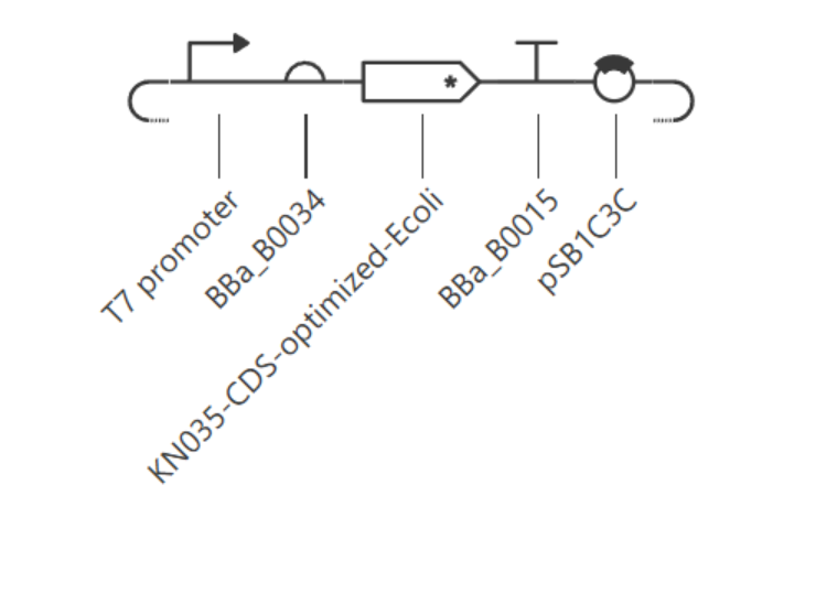
| Transcription start | Promoter: T7 promoter | TAATACGACTCACTATAGGG (Studier & Moffatt, 1986); found in Kernel’s repository | NEBExpress transcribes only from a T7 promoter, since T7 RNA polymerase is supplied in the kit. |
| Translation start | RBS: BBa_B0034 | Kernel/iGEM Registry public part | A well-characterized strong ribosome binding site, functional with mRNA regardless of which RNA polymerase transcribed it. |
| Coding sequence | CDS: KN035, +start +stop | Based on Kernel part BBa_K4266004 (KN035 DNA sequence). | Encodes the product defined in Part 1; the added start/stop codons were required for Kernel’s Construct Checker to recognize it as a complete CDS, and for correct translation in the simulator |
| Targeting | Tag: 6xHis (already included at the C-terminus of the deposited KN035 sequence) | Part of the same CDS part above | Enables Ni-NTA magnetic bead purification, matching the automation workflow from Homework #1. No separate signal peptide part is used, consistent with a soluble, compartment-free cell-free reaction. |
| Transcription stop | Terminator: BBa_B0015 | Kernel/iGEM Registry public part (double terminator) | Standard, well-characterized terminator compatible with either T7 or native E. coli RNA polymerase. |
| Vector context | Backbone: pSB1C3C (Kernel’s internal copy of the standard pSB1C3 backbone) | Kernel public repository; confirmed to carry chloramphenicol resistance, a high-copy origin, and an empty size close to the standard ~2,070 bp | Provides origin of replication and selection marker to propagate the construct in E. coli; not required by the cell-free reaction itself. |
3. Assemble your construct
The construct, named PD-L1-nanobody-construct, was assembled in Kernel with parts in this order, 5’ to 3’: backbone (pSB1C3C) → T7 promoter → RBS (BBa_B0034) → CDS (KN035, +start +stop) → terminator (BBa_B0015).
Kernel’s Construct Checker initially flagged two real issues and three expected, non-applicable warnings:
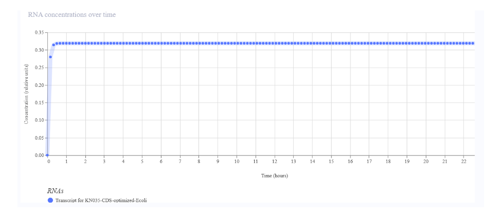
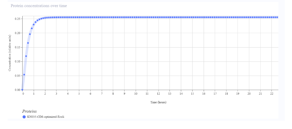
- Real issues (fixed): the CDS did not start with ATG and did not end with a double stop codon, because the source part (BBa_K4266004) held only the coding sequence without these elements. I resolved this by creating a new CDS part with ATG and TGA-TAA added, and by correcting that new part’s type from “(-start -stop)” to “(+start +stop)” after noticing the simulator (Part 4) was not translating it correctly despite the sequence being correct.
- Expected, non-applicable warnings: Kernel also flagged missing Kozak sequences, 5’ and 3’ UTRs, and poly(A) signals. These apply to eukaryotic expression architecture, not to a prokaryotic, RBS-based system, so I did not add them.
4. Optimize and quality-check
Codon optimization. I ran Kernel’s codon optimizer on the amino acid sequence, with preset “None” (the CHO/LV/AAV presets do not apply to a bacterial host), expression organism E. coli, no signal peptide algorithm, and stop codons set to TGA then TAA (double stop, per Kernel’s own recommendation). The tool returned a new, optimized DNA sequence as a separate part, which I then used to replace the manually-built CDS in the construct.
Sequence search (BLASTx). I ran BLASTx. The best hit was “Chain B, Nanobody [Camelidae]”, confirming that the CDS encodes the intended KN035 nanobody and not an unrelated sequence.
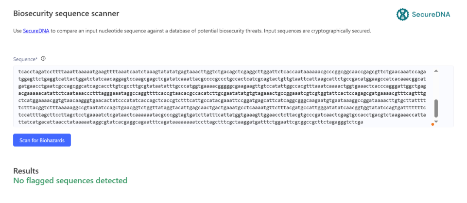
Safety screen. I ran Kernel’s SecureDNA biosecurity scanner on the full assembled construct. It reported “No flagged sequences detected.”
5. From design to build
How I would build it. My assembled expression cassette (promoter through terminator) is under 1 kb, and the full construct with backbone is a few kb, well within the range for a single synthetic gene order, sequence-verified by the vendor in their standard cloning vector. Gibson assembly (Gibson et al., 2009) would only be worth using if I needed to combine this cassette with other fragments I already had, joining overlapping pieces in one isothermal reaction with a 5’ exonuclease, a polymerase, and a ligase. Chip-derived oligo pool synthesis, as in Kosuri et al. (2010), who assembled 47 genes (42 of them therapeutic antibody sequences) from a 13,000-oligo pool using selective amplification and enzymatic error correction, becomes worthwhile mainly when many sequence variants must be built and screened in parallel, closer to the discovery step assumed in Homework #1 than to this single, defined construct.
What synthesis constrains. Carr et al. (2004) report a typical error rate near one error per 600 bp for conventional gene synthesis, reduced more than 15-fold, to about one per 10,000 bp, using a MutS mismatch-binding protein to remove failure products. Applying those rates to my construct (assuming independent, Poisson-distributed errors), a several-kb construct would have a substantially lower error-free fraction at the uncorrected rate than at the corrected one, which is why I would rely on vendor sequence verification of the final clone rather than skip verification. Nothing in my current design (his-tag repeats aside) is an unusually difficult synthesis target, but the deposited KN035 sequence does contain a short repetitive stretch (the poly-His tag) that vendors sometimes flag for extra QC, since homopolymer-like repeats are harder to synthesize accurately.
Prototype vs. scale. For the prototype, I would order one sequence-verified plasmid and test both circular and linearized template in the NEBExpress reaction, since linear DNA is compatible with the system but more exposed to nuclease degradation in lysate; NEB offers a nuclease inhibitor (GamS) for this case. For scale, DNA demand stays small: using the Homework #1 estimate of about 6 plate runs of 96 wells at 200 µL per year, and NEB’s suggested starting point of 250 ng of template per 50 µL reaction, the annual template requirement is under 1 mg. I would meet this by growing and purifying the same sequence-verified plasmid in E. coli in batches, verified by sequencing, rather than repeating de novo gene synthesis for each batch.
6. What don’t you know yet?
Whether the metadata correction I made (part type “+start +stop”) reflects reality, not just the simulator’s expectations. My CDS part initially simulated zero protein despite RNA accumulating; the fix was changing the part’s start/stop flags, not the sequence itself. This raises a real unknown: whether Kernel’s simulated protein output (0.25, stabilizing around 2 hours) reflects a biologically meaningful expression level, or only that the simulator can now correctly locate the reading frame. How I would find out: run the actual cell-free reaction and measure protein by SDS-PAGE and His-tag capture, rather than relying on the simulation’s absolute values.
Disulfide bond formation in a reducing, cell-free E. coli environment. KN035 carries two disulfide bonds (Zhang et al., 2017), and E. coli lysates are normally reducing, so the protein may misfold or lose PD-L1 binding activity. How I would find out: run the reaction with and without NEB’s PURExpress Disulfide Bond Enhancer, and compare soluble yield and binding activity (ELISA or biolayer interferometry) between conditions.
Whether KN035, developed and structurally characterized as a PD-1/PD-L1 blocker, retains its epitope on formalin-fixed, paraffin-embedded (FFPE) tissue, as required for IHC use. How I would find out: stain FFPE cell pellets with known PD-L1 expression levels and serial TNBC tissue sections, and compare staining pattern and intensity against an approved clinical PD-L1 clone.
References
Carr, P. A., Park, J. S., Lee, Y.-J., Yu, T., Zhang, S., & Jacobson, J. M. (2004). Protein-mediated error correction for de novo DNA synthesis. Nucleic Acids Research, 32(20), e162. https://doi.org/10.1093/nar/gnh160
Gibson, D. G., Young, L., Chuang, R.-Y., Venter, J. C., Hutchison, C. A., III, & Smith, H. O. (2009). Enzymatic assembly of DNA molecules up to several hundred kilobases. Nature Methods, 6(5), 343–345. https://doi.org/10.1038/nmeth.1318
Kosuri, S., Eroshenko, N., LeProust, E. M., Super, M., Way, J., Li, J. B., & Church, G. M. (2010). Scalable gene synthesis by selective amplification of DNA pools from high-fidelity microchips. Nature Biotechnology, 28(12), 1295–1299. https://doi.org/10.1038/nbt.1716
New England Biolabs. (n.d.). NEBExpress® cell-free E. coli protein synthesis system [Instruction manual, E5360]. https://www.neb.com/-/media/nebus/files/manuals/manuale5360.pdf
Studier, F. W., & Moffatt, B. A. (1986). Use of bacteriophage T7 RNA polymerase to direct selective high-level expression of cloned genes. Journal of Molecular Biology, 189(1), 113–130. https://doi.org/10.1016/0022-2836(86)90385-2
Zhang, F., Wei, H., Wang, X., Bai, Y., Wang, P., Wu, J., Jiang, X., Wang, Y., Cai, H., Xu, T., & Zhou, A. (2017). Structural basis of a novel PD-L1 nanobody for immune checkpoint blockade. Cell Discovery, 3, 17004. https://doi.org/10.1038/celldisc.2017.4
Zhou, A., & Wei, H. (2017). Crystal structure of PD-L1 complexed with a nanobody at 1.7 Angstrom resolution (PDB ID: 5JDS) [Data set]. RCSB Protein Data Bank. https://doi.org/10.2210/pdb5JDS/pdb
AI disclosure: Claude (Anthropic) was used throughout this assignment to check NEBExpress template requirements, diagnose the Construct Checker warnings and improve the wording of this document. All Kernel outputs (part creation, construct assembly, codon optimization, BLAST, biosecurity screen, and simulation) were run directly by the author inside Kernel.