Designer and researcher exploring the relationship between living matter,
spatial design, and immersive experiences.
BIO ATMA is a research framework investigating biomaterials as a medium
for imagining new forms of temporary, experimental, and sensory spatial experiences.
Research
Exploring how living materials can become spatial systems rather than simply objects or surfaces.
Experimentation
Material tests, cultivation, transformation, observation, and prototyping.
Living Sculpture
Ephemeral artifacts shaped by living matter.
The project investigates temporary structures that can change, grow,
transform, and develop through time.
Process
Research · Material experiments · Prototypes · Observations · Documentation
Living systems · Temporary structures · Spatial experiences
Subsections of Vitalia Espinoza
Final Project
Week 01 — Grown Artifact
HTBAA Homework #1
Grown Artifact Biological Fabrication through Designed Growth
1. What would I like to biomanufacture and why?
I propose to biomanufacture an ephemeral three-dimensional sculptural artifact combining a lightweight digitally designed scaffold with a biological membrane grown from bacterial cellulose.
The proposed artifact would be approximately 1 meter tall and 50–60 cm wide. Its geometry would be vertical, volumetric, abstract, and organic, positioned somewhere between an architectural structure and a biological form.
The scaffold would establish an initial geometric framework, but the final appearance of the artifact would not be completely predetermined. Instead, bacterial cellulose would develop in relation to the scaffold over time, potentially producing variations in thickness, density, transparency, texture, and spatial distribution.
The artifact would therefore be experienced as a spatial object rather than a frontal image. Its surfaces, openings, layers, and translucency could change depending on the viewer’s position and the way light passes through the material. The relationship between the scaffold and the biological material would remain partially visible, making the fabrication process part of the final aesthetic.
Why this is interesting to me
My interest in this project comes from the intersection of design, spatial experience, digital fabrication, and biological processes.
I became particularly interested in bacterial cellulose through a previous experience growing a kombucha SCOBY. Observing how its texture, thickness, and appearance changed over time suggested the possibility of treating biological growth itself as a design and fabrication process.
This led me to question a conventional assumption in design: that the designer should determine the final form before fabrication begins.
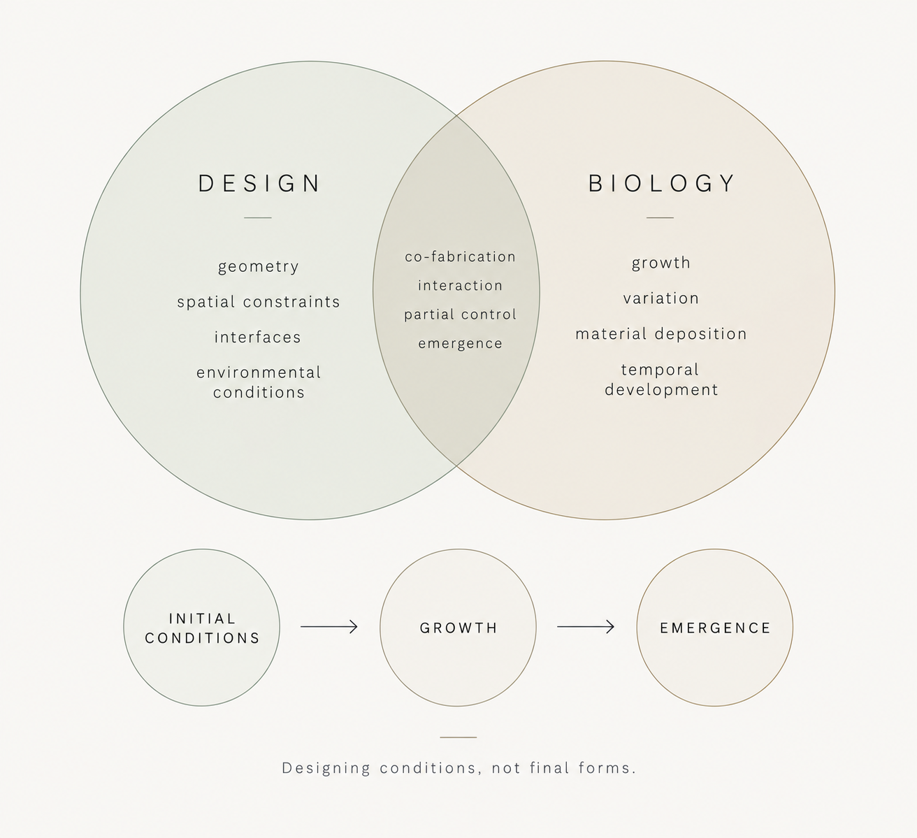
Instead, I am interested in a process where the designer establishes an initial geometry and a set of conditions, while the biological system participates in determining the final morphology.
Rather than designing the final object directly, I am interested in designing the conditions under which the object can emerge.
This approach is informed by Neri Oxman’s work on Material Ecology and, more specifically, by her concept of templating, which explores how top-down design frameworks can guide or influence bottom-up biological and material processes. In Templating Design for Biology and Biology for Design, Oxman describes the possibility of combining top-down design procedures with bottom-up biological or material growth (Oxman, 2015).
Grown Artifact adapts this principle to bacterial cellulose: the scaffold establishes a spatial framework and environmental interface, while biological growth contributes to the formation of the final morphology.
The project can therefore be understood as a form of co-fabrication between a designer and a biological system.
The designer establishes:
geometry;
spatial constraints;
interfaces;
environmental conditions.
The biological system contributes:
growth;
material deposition;
variation;
temporal development;
emergent morphology.
The final artifact would consequently be neither completely designed nor completely spontaneous.
It would emerge from the interaction between both systems.
2. What living system would I use?
The proposed biological manufacturing system is bacterial cellulose produced by a microbial culture associated with kombucha, particularly cellulose-producing bacteria from the Komagataeibacter genus.
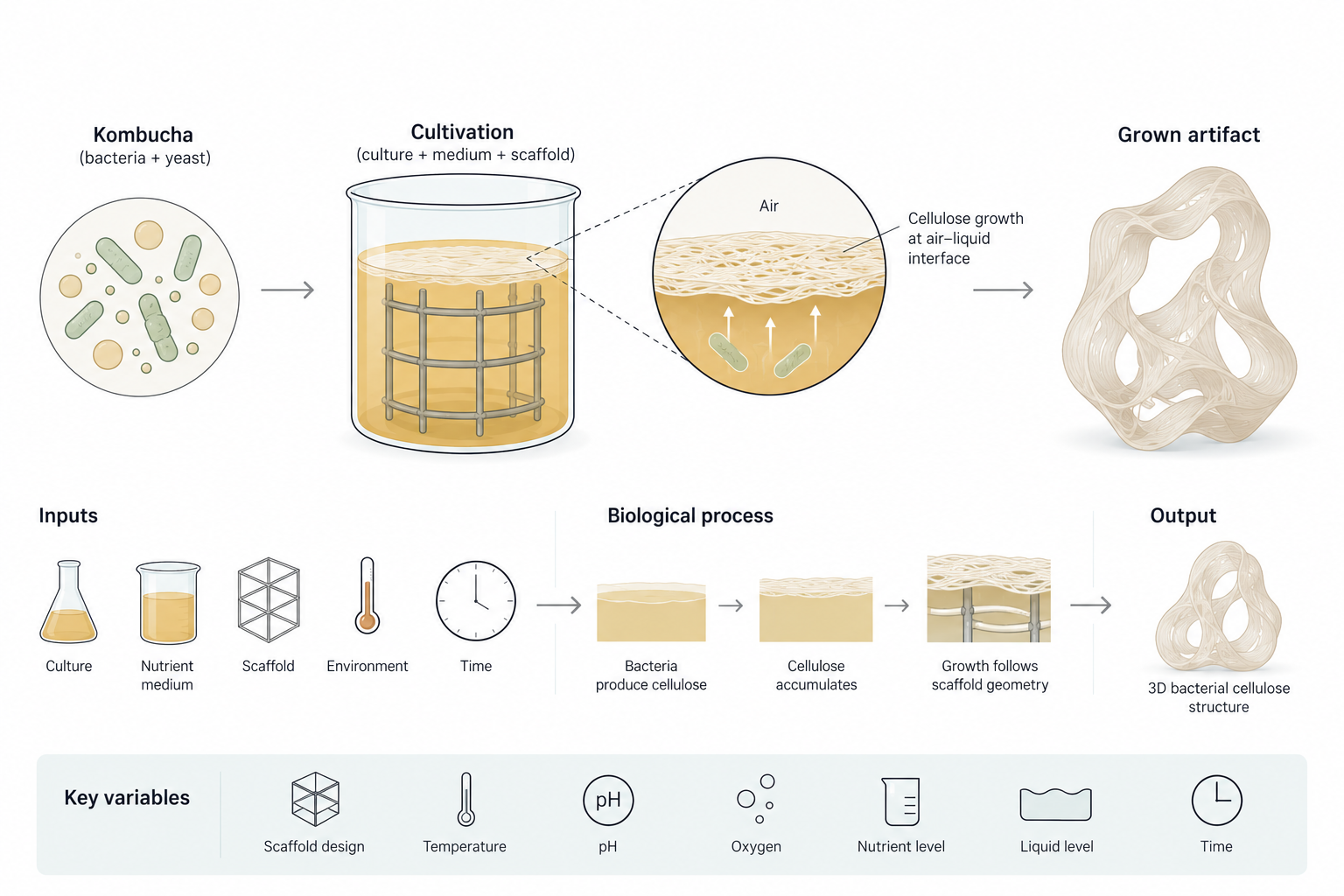
Kombucha is a symbiotic culture of bacteria and yeasts. Within this system, cellulose-producing bacteria generate cellulose extracellularly, forming a cellulose-rich membrane, particularly at the air–liquid interface.
This behavior is important because it establishes both an opportunity and a constraint for the project.
The opportunity is that the microorganisms naturally produce a visible, macroscopic material layer.
The constraint is that bacterial cellulose does not simply grow uniformly around an arbitrary three-dimensional object. Its formation is strongly related to cultivation conditions and the air–liquid interface.
Recent research provides two particularly relevant precedents.
Loh et al. (2025) developed a bacterial cellulose bioreactor combining a supportive mesh scaffold with intermittent nutrient feeding. The mesh anchored the growing pellicle at the air–liquid interface, allowing continuous growth and greater control over thickness. Their system was tested from 250 mL to 10 L and produced pellicles exceeding 80 mm in thickness.
Caro-Astorga et al. (2021) demonstrated another approach to three-dimensional bacterial cellulose fabrication. They produced millimetre-scale bacterial cellulose spheroids from Komagataeibacter rhaeticus and used them as building blocks to grow continuous 3D shapes. Their experimental structures grew and fused over approximately 4–10 days.
These studies demonstrate that bacterial cellulose can be produced in thicker and three-dimensional configurations. However, they use different fabrication strategies from the one proposed here.
Grown Artifact therefore asks a more specific question:
Can a deliberately designed scaffold and interface guide bacterial cellulose growth into a larger, continuous spatial artifact while retaining biological variation?
This becomes the central biological question of the project.
What goes into the system?
At a high level, the biological system would require:
an established bacterial-cellulose-producing culture;
a nutrient-containing cultivation medium;
a suitable cultivation environment;
a designed scaffold or support structure;
time for biological growth.
What happens biologically?
The cellulose-producing bacteria synthesize cellulose extracellularly.
The cellulose accumulates as a network at the growth interface, progressively forming a material layer.
The resulting material would then interact with the scaffold and its geometry.
What comes out?
The desired output is not simply a quantity of bacterial cellulose.
The desired output is a spatially organized biological material system in which the morphology of cellulose is influenced by the relationship between:
scaffold geometry + biological growth + environmental conditions + time
This is an important distinction.
The project is not assuming that the final one-meter artifact can already be produced reliably.
Instead, the ability to guide bacterial cellulose toward the proposed spatial configuration is itself part of the experimental question.
Proposed hypothesis
I hypothesize that a scaffold designed around the spatial and environmental requirements of bacterial cellulose growth can influence the distribution, thickness, continuity, and morphology of cellulose deposition, enabling the formation of a coherent three-dimensional artifact while retaining biological variation.
This hypothesis does not assume complete control over biological growth.
Instead, the goal is to investigate whether biological variability can be constrained sufficiently to produce a coherent spatial artifact.
Design and biological variables
The experiment would distinguish between design variables and biological/environmental variables.
Design and scaffold variables
Possible scaffold variables include:
geometry;
curvature;
surface area;
aperture size;
density;
orientation;
porosity;
surface characteristics;
distance from the air–liquid interface;
relationship between scaffold and growing cellulose.
Biological and environmental variables
Relevant variables could include:
temperature;
pH;
oxygen availability;
nutrient concentration;
cultivation time;
liquid level;
culture volume;
surface-to-volume relationship.
The intention would not be to change every variable simultaneously.
Instead, experiments would progressively investigate relationships between variables to determine which conditions produce observable changes in cellulose morphology.
How would I know if it worked?
The experiment would be considered successful if changes in scaffold geometry or cultivation conditions produced measurable differences in the resulting bacterial cellulose.
The main observations would include:
surface coverage;
thickness;
continuity;
spatial distribution;
attachment to the scaffold;
morphology;
variation between experimental conditions.
The objective would not be to obtain an identical result in every experiment.
Instead, the objective would be to determine whether changes in the designed conditions produce repeatable and observable changes in biological morphology.
This would allow the project to evaluate the relationship between:
designed conditions → biological response → resulting morphology
rather than evaluating the artifact only through its visual appearance.
3. How would I use automation?
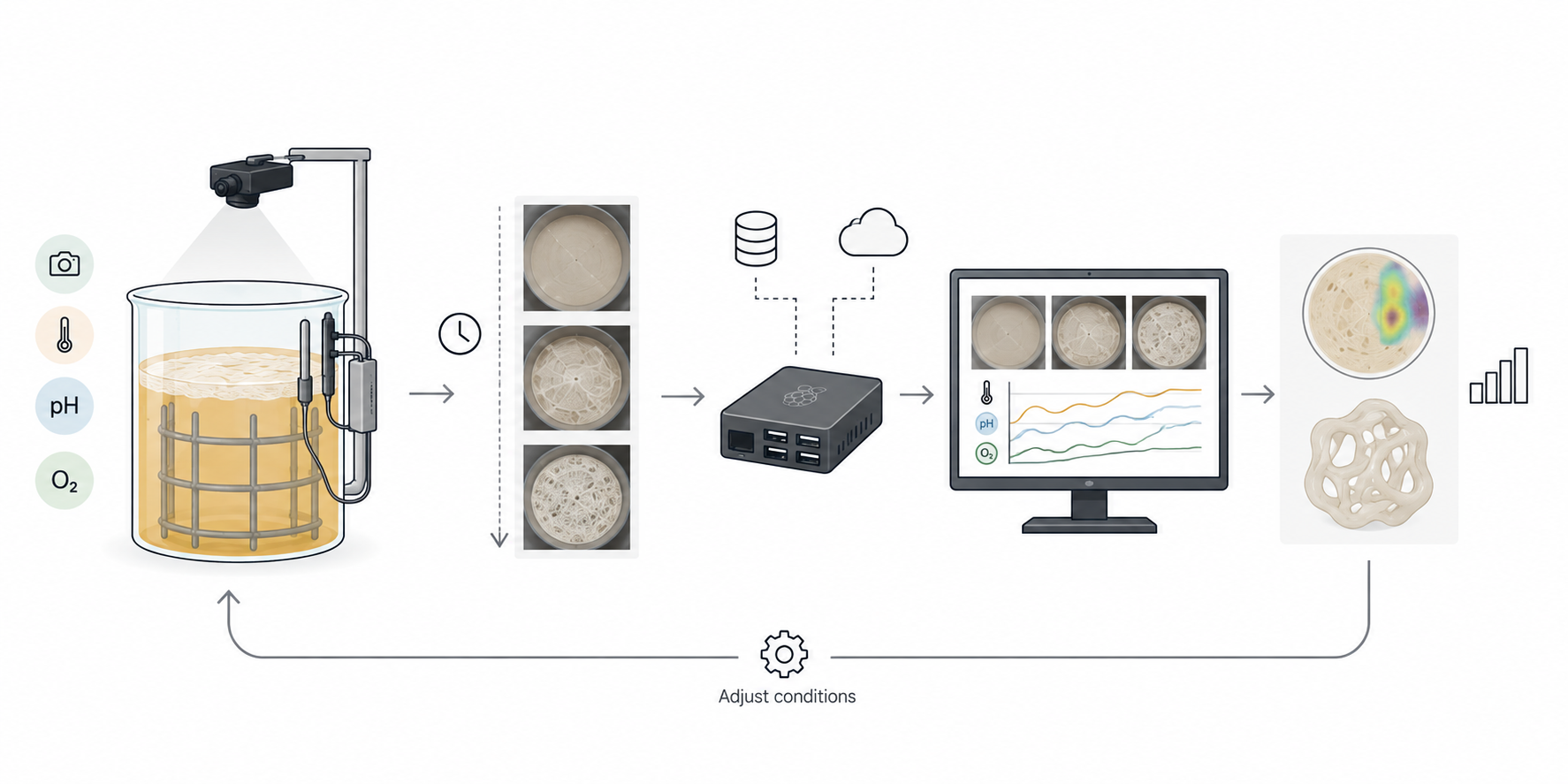
Automation would primarily be used to monitor, document, and compare biological growth over time.
Rather than initially trying to completely automate fabrication, I would use automation as a research instrument.
A camera-based system could periodically capture images of the developing bacterial cellulose.
These images could be used to document changes in:
surface coverage;
morphology;
thickness;
transparency;
density;
spatial distribution.
Environmental sensors could simultaneously record relevant cultivation conditions.
A programmable controller could collect and associate these measurements with the corresponding stages of biological growth.
The resulting dataset could connect:
time + environmental conditions + scaffold configuration + biological morphology
This would make it possible to move beyond simple visual observation toward a more measurable and repeatable experimental process.
Existing bacterial cellulose research demonstrates that automation can also be integrated directly into the cultivation process. Loh et al. (2025), for example, used automated peristaltic pumps to introduce fresh growth medium into their mesh-supported bioreactor, allowing continued growth at the air–liquid interface.
Grown Artifact would extend this logic toward monitoring and adaptive experimentation, rather than only automating nutrient delivery.
Feedback loop
The automation system could eventually support a feedback loop:
biological growth → observation → data → adjustment → new growth condition
For example, if a particular scaffold geometry consistently produces greater cellulose accumulation in a specific region, that information could inform the next scaffold iteration.
The purpose of automation is therefore not to make biology behave like a conventional manufacturing machine.
Instead, it is to make biological variability observable, measurable, and progressively understandable.
This approach is consistent with broader biomanufacturing research emphasizing automation, data collection, process monitoring, and enabling infrastructure as important components of biological production systems.
Prototype versus scale
The automation used during experimentation would likely differ from the system used for larger-scale production.
For the prototype stage, a relatively simple system could be sufficient:
camera + environmental sensors + programmable controller + data logging
The purpose would be to understand the biological process.
At a larger manufacturing scale, automation would likely need to address:
environmental control;
monitoring of multiple cultivation systems;
nutrient delivery;
sampling;
data management;
process consistency;
parallel production.
This distinction is important because the automation required to learn the process is not necessarily the same as the automation required to manufacture at scale.
4. How much would I need to make, and how long would it take?
(a) How much?
For this project, I define a meaningful scale as one exhibition-scale artifact approximately 1 meter tall and 50–60 cm wide.
The purpose of this calculation is not to determine a final manufacturing specification, but to estimate the approximate quantity of bacterial cellulose involved.
Assume that the biological membrane covers approximately:
1.5 m²
and has an average dry thickness of:
0.12 mm
First, estimate the volume of dry bacterial cellulose:
1.5 m² × 0.12 mm ≈ 180 cm³
For a preliminary mass estimate, I use the dry-density range reported by Loh et al. (2025) for their mesh-supported system:
91.5–156 kg/m³
which corresponds to:
0.0915–0.156 g/cm³
Using this range:
180 cm³ × 0.0915–0.156 g/cm³ ≈ 16.5–28.1 g
Therefore, under this literature-based assumption, the target artifact would contain approximately:
17–28 g of dry bacterial cellulose
This estimate should be treated cautiously because bacterial cellulose density depends on cultivation method, thickness, drying method, and material structure. The values reported by Loh et al. correspond to their specific bioreactor and should not be assumed to represent a universal density for bacterial cellulose.
Culture volume estimate
Avcioglu et al. (2021) reported a bacterial cellulose production value of 18.68 g/L using an optimized kombucha consortium under specific experimental conditions: static cultivation at 30 °C for 10 days with an optimized medium.
Using this value only as a theoretical reference:
17–28 g ÷ 18.68 g/L ≈ 0.9–1.5 L
This suggests that approximately 0.9–1.5 L of culture medium could theoretically produce the estimated dry mass under comparable production conditions.
However, this should not be interpreted as the actual culture volume required for the sculpture.
The proposed artifact is three-dimensional, the biological material would not necessarily grow uniformly, and the relationship between culture volume, scaffold geometry, and usable three-dimensional material has not yet been established.
The calculation is therefore a back-of-the-envelope mass-to-yield comparison, rather than a validated production specification.
The first experimental prototypes would be used to determine the actual relationship between:
The production time of the final artifact cannot currently be established with sufficient confidence.
Published bacterial cellulose studies demonstrate that production rate depends strongly on cultivation configuration and environmental conditions.
Loh et al. (2025) demonstrated continuous growth at approximately 1 mm of thickness per day in their mesh-supported bioreactor, with the system tested over extended cultivation periods. Their 10 L system produced pellicles up to approximately 82 mm thick.
Caro-Astorga et al. (2021), using a different fabrication strategy based on bacterial cellulose spheroids, observed growth and fusion into continuous 3D shapes over approximately 4–10 days.
These results demonstrate that bacterial cellulose can grow at useful rates, but they cannot be directly transferred to the proposed artifact because the geometry, interface configuration, scale, culture conditions, and fabrication strategy are different.
Therefore, the project would first establish an empirical relationship between:
time → cellulose accumulation → thickness → spatial coverage
A hypothetical growth rate can illustrate the calculation.
For example, if a particular region accumulated:
1 mm/day
then producing:
10 mm
in that region would theoretically require:
10 mm ÷ 1 mm/day = 10 days
However, this does not mean that the complete one-meter artifact would require ten days.
Different regions may grow at different rates, and increasing scale may introduce environmental gradients, oxygen limitations, structural issues, and other constraints.
For overall project planning, I would initially allocate approximately:
10–14 weeks
for the complete development process, including:
scaffold design;
biological experiments;
growth;
monitoring;
iteration;
final cultivation;
drying;
stabilization;
documentation.
This is a project-planning estimate, not a demonstrated manufacturing time.
The project would therefore progress from small experimental prototypes toward the final artifact rather than assuming that the final object can simply be scaled proportionally from a small sample.
5. What don’t I know yet?
After considering the proposed biological system, automation, and scale, several major technical uncertainties remain.
1. Can a designed scaffold guide bacterial cellulose into a larger continuous spatial form?
Existing research demonstrates that bacterial cellulose can be produced in three-dimensional configurations, including both mesh-supported pellicles and modular spheroid-based structures.
The remaining question for Grown Artifact is whether a deliberately designed scaffold and interface can guide the growth of a continuous spatial artifact at a larger scale.
The first experiments would therefore need to determine whether scaffold geometry produces measurable differences in cellulose distribution and attachment.
2. Which scaffold properties most strongly influence growth?
The project does not yet establish which properties of the scaffold will have the greatest effect on bacterial cellulose formation.
Potential factors include:
geometry;
porosity;
surface area;
surface characteristics;
curvature;
aperture size;
orientation;
distance from the air–liquid interface.
Determining which of these variables produce observable changes would help identify which aspects of the scaffold should be actively designed and which can remain uncontrolled.
3. How predictable can the final morphology become?
The project intentionally allows biological growth to influence the final form.
This introduces variability.
I do not yet know how consistently factors such as:
temperature;
pH;
oxygen availability;
nutrient concentration;
scaffold geometry;
surface area;
cultivation time
will influence thickness and spatial distribution.
The key challenge is therefore not complete control.
It is finding a useful balance between:
design control ↔ biological variability
The research would need to determine whether this balance is sufficient to produce a coherent artifact while preserving the biological character of the growth process.
4. Can the system be scaled from prototype to a one-meter artifact?
A small prototype and a one-meter artifact cannot necessarily be treated as the same system at different scales.
Increasing scale may introduce challenges related to:
oxygen availability;
environmental gradients;
surface-to-volume ratio;
structural support;
contamination;
uniformity of growth;
drying;
cultivation volume.
The transition from experimental biological fabrication toward reproducible and scalable production remains a significant challenge in biomanufacturing.
The prototype would therefore function not only as a visual test, but as a calibration stage for determining whether the biological fabrication process can realistically be scaled.
Expected Contribution
The expected contribution of Grown Artifact is not the invention of bacterial cellulose as a material, nor the claim that biological growth can be completely controlled.
Instead, the project investigates a specific design question:
How can a designer construct the spatial and environmental conditions that allow biological material to participate in the formation of an artifact?
The project proposes a shift from material selection toward material cultivation, and from designing a finished geometry toward designing a system capable of generating geometry.
The research therefore has two interconnected outcomes:
a biological fabrication process that can be observed, measured, and iterated;
a final spatial artifact produced through that process.
The final artifact would consequently function simultaneously as:
object + experiment + evidence
The research could contribute to future explorations of:
biological fabrication;
computationally guided growth;
ephemeral spatial artifacts;
material ecology;
biofabrication;
responsive design systems.
Ultimately, Grown Artifact explores a different role for the designer: not solely as the author of a predetermined form, but as the designer of the conditions, interfaces, and feedback systems through which form can emerge.
References
Bacterial Cellulose
Avcioglu, N. H., Birben, M., & Seyis Bilkay, I. (2021). Optimization and physicochemical characterization of enhanced microbial cellulose production with a new Kombucha consortium. Process Biochemistry, 108, 60–68. https://doi.org/10.1016/j.procbio.2021.06.005
Caro-Astorga, J., Walker, K. T., Herrera, N., Lee, K.-Y., & Ellis, T. (2021). Bacterial cellulose spheroids as building blocks for 3D and patterned living materials and for regeneration. Nature Communications, 12, 5027. https://doi.org/10.1038/s41467-021-25350-8
Loh, J., Arnardottir, T., Gilmour, K., Zhang, M., & Dade-Robertson, M. (2025). Enhanced production of bacterial cellulose with a mesh dispenser vessel-based bioreactor. Cellulose, 32, 2209–2226. https://doi.org/10.1007/s10570-024-06367-w
Wang, J., Tavakoli, J., & Tang, Y. (2019). Bacterial cellulose production, properties and applications with different culture methods – A review. Carbohydrate Polymers, 219, 63–76. https://doi.org/10.1016/j.carbpol.2019.05.008
Lee, K.-Y., Buldum, G., Mantalaris, A., & Bismarck, A. (2014). More than meets the eye in bacterial cellulose: Biosynthesis, bioprocessing, and applications in advanced fiber composites. Macromolecular Bioscience, 14(1), 10–32. https://doi.org/10.1002/mabi.201300298
Czaja, W., Krystynowicz, A., Bielecki, S., & Brown, R. M. Jr. (2006). Microbial cellulose—the natural power to heal wounds. Biomaterials, 27(2), 145–151. https://doi.org/10.1016/j.biomaterials.2005.07.035
Biological and Computational Design
Oxman, N. (2015). Templating Design for Biology and Biology for Design. Architectural Design, 85(5), 100–107. https://doi.org/10.1002/ad.1961
Oxman, N., Keel, M., Tsai, L., et al. (2014). Silk Pavilion: A Case Study in Fiber-based Digital Fabrication. Fabricate: Negotiating Design & Making.
Oxman, N. (2010). Material-Based Design Computation. Massachusetts Institute of Technology.
Biomanufacturing and Automation
Asin-Garcia, E., Fawcett, J. D., Batianis, C., & Martins dos Santos, V. A. P. (2025). A snapshot of biomanufacturing and the need for enabling research infrastructure. Trends in Biotechnology, 43(5), 1000–1014. https://doi.org/10.1016/j.tibtech.2024.10.014
Petzold, C. J., & Mukhopadhyay, A. (2025). From bench to biofactory: High-throughput technologies and automated workflows to accelerate biomanufacturing. Current Opinion in Biotechnology, 94, 103320. https://doi.org/10.1016/j.copbio.2025.103320
Zhang, Y.-H. P., Sun, J., & Ma, Y. (2017). Biomanufacturing: History and perspective. Journal of Industrial Microbiology & Biotechnology, 44, 773–784. https://doi.org/10.1007/s10295-016-1863-2
Averesch, N. J. H., Berliner, A. J., Nangle, S. N., et al. (2023). Microbial biomanufacturing for space-exploration—what to take and when to make. Nature Communications, 14, 2311. https://doi.org/10.1038/s41467-023-37910-1
AI Assistance
I used ChatGPT (OpenAI) as a brainstorming and research-support tool during the development of this project.
Selected prompts included:
“How could bacterial cellulose be used as a biological fabrication system for an ephemeral sculpture?”
“How can scaffold geometry and controlled growth conditions influence bacterial cellulose morphology?”
“How can bacterial cellulose be grown using a mesh scaffold at the air–liquid interface?”
“What would be a realistic back-of-the-envelope calculation for scaling a bacterial cellulose artifact to approximately 1 meter?”
“How could automation be used to monitor bacterial cellulose growth?”
“How can this project be framed as a biomanufacturing research question rather than only an art or design project?”
AI-generated information was treated as preliminary and used to identify concepts, technical questions, quantitative assumptions, and potential references. Scientific and quantitative claims were checked against external sources where possible.
Week 02 — DNA Design
Part 0 — Set up in Kernel
I created my project in the HTBAA 2026 workspace using the vitalia-espinoza-bacterial-cellulose repository and the vitalia-espinoza-bcsd folder.
Part 1 — Define your expression target
What molecule are you expressing?
I am designing the expression of BcsD, also referred to as AcsD, a component of the bacterial cellulose synthase system. I selected BcsD because it is associated with the organization and crystallization of cellulose during bacterial cellulose biosynthesis, connecting the genetic design to my goal of studying the organization of a grown cellulose-based artifact.
What is the sequence?
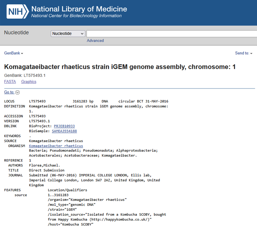
The reference genome for this project is the Komagataeibacter rhaeticus iGEM genome available in NCBI GenBank (accession LT575493.1).
For the expression design, I am using the candidate coding sequence acsD_KRIGEM_00627, a 471 nt DNA sequence associated with Komagataeibacter rhaeticus. This sequence was imported into VectorBee and will be used as the candidate BcsD/AcsD coding sequence in Kernel.
Figure 1. NCBI GenBank record for the Komagataeibacter rhaeticus iGEM genome (LT575493.1), used as the reference genome for the target sequence in this project.
What host or chassis?
The host/chassis is Komagataeibacter rhaeticus, a bacterial cellulose-producing bacterium. This keeps the DNA design connected to the biological system proposed in Homework #1, where bacterial cellulose is the living material used to grow the artifact.
Where does the product need to go?
BcsD needs to function as part of the bacterial cellulose synthase system rather than as an independently secreted protein. Therefore, no additional secretion signal or targeting tag is planned at this stage.
Part 2 — Design the Genetic Parts
For this project, I designed a genetic construct in Kernel to express the candidate BcsD/AcsD protein in Komagataeibacter rhaeticus.
The construct includes the following functional genetic elements:
Layer
Part
Type
Length
Source
Reason for selection
Transcription start
PJ23104
Promoter
35 bp
Kernel
Provides transcriptional initiation for the BcsD expression cassette.
Translation start
BBa_B0034
Ribosome binding site (RBS)
12 bp
iGEM Part Registry / Kernel
Provides a bacterial translation-initiation site upstream of the BcsD CDS.
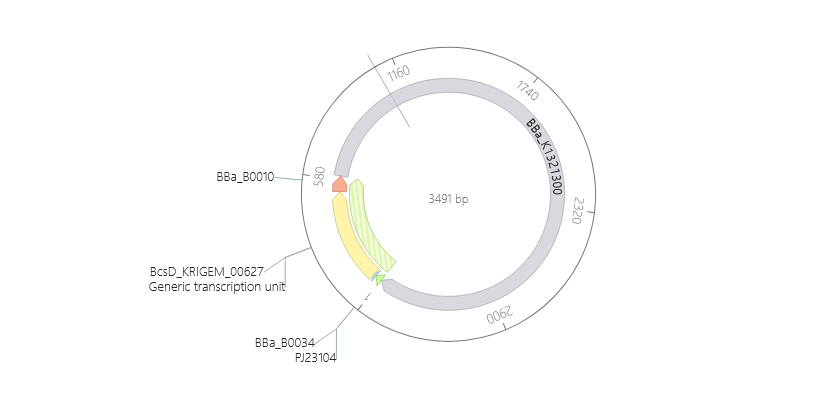
The expression cassette is contained within the BBa_K1321300 backbone.
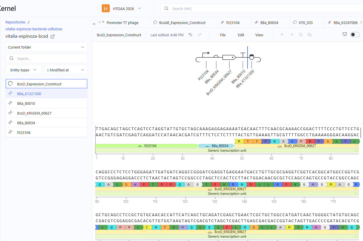
Figure 2. Linear map of the BcsD expression construct assembled in Kernel, showing the promoter, RBS, BcsD coding sequence, and terminator in their 5′→3′ order.
Figure 3. Circular map of the complete construct, showing the BBa_K1321300 backbone and the BcsD expression cassette.
Part 3 — Assemble your construct
I assembled the genetic parts selected in Part 2 in Kernel to create a
single expression construct for Komagataeibacter rhaeticus. The purpose of
this step was to bring the regulatory elements, BcsD coding sequence,
transcriptional terminator, and backbone together into one organized genetic
design.
Construct organization
The expression cassette follows the expected 5′→3′ order for a prokaryotic
expression system:
PJ23104 provides the promoter for transcription initiation, followed by
BBa_B0034 as the ribosome binding site. The BcsD_KRIGEM_00627 coding sequence
is positioned downstream of the RBS and is followed by the BBa_B0010
terminator.
The expression cassette is contained within the BBa_K1321300 backbone, which
provides the vector context for the construct.
Construct check
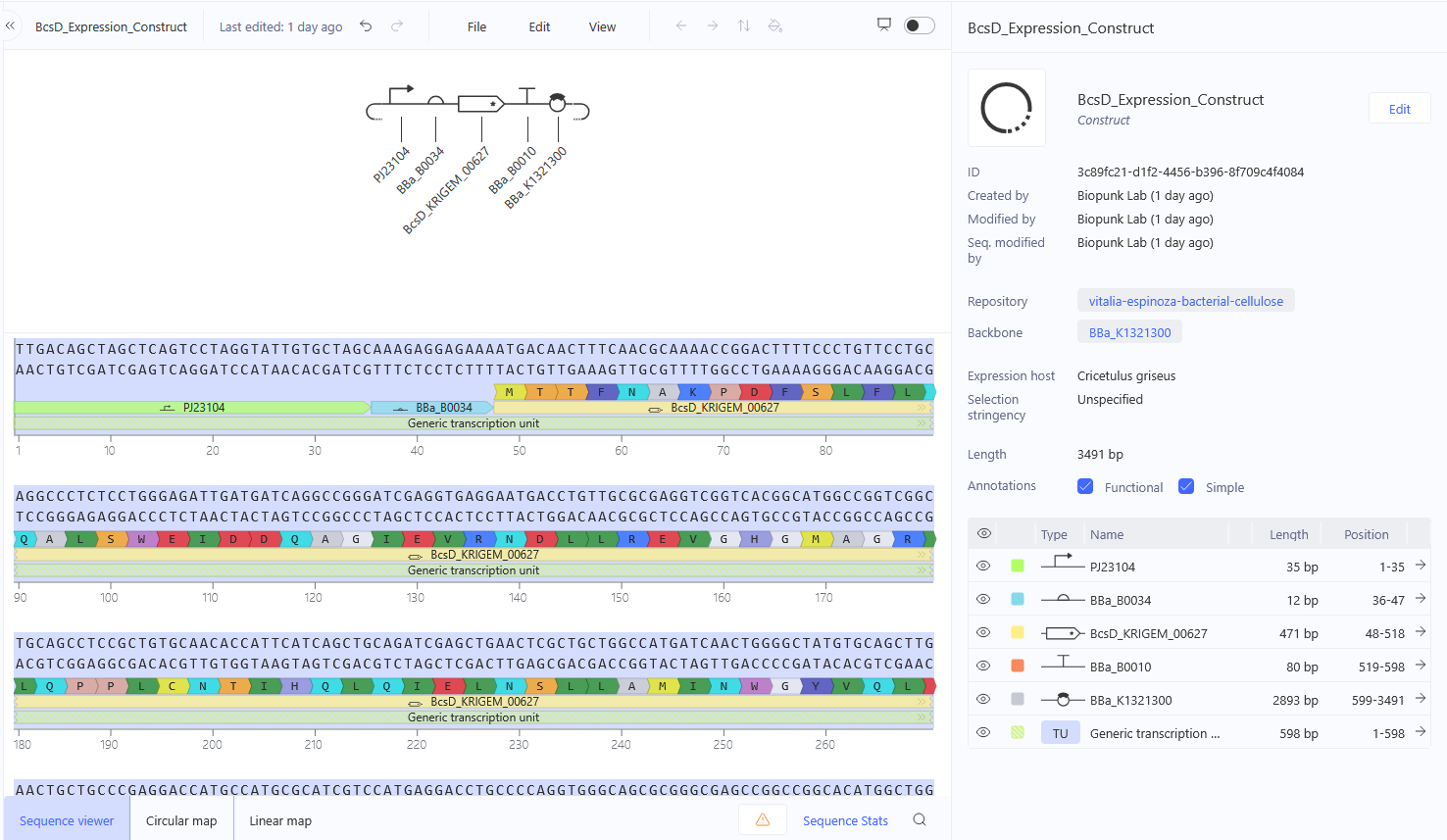
I checked the assembled construct in Kernel to confirm that the selected
parts are present in the intended order and orientation. The sequence viewer
also shows the boundaries and lengths of the individual parts.
The final construct has a total length of 3,491 bp.
Figure 4. Final BcsD expression construct assembled in Kernel. The
sequence viewer shows the PJ23104 promoter, BBa_B0034 ribosome binding site,
BcsD_KRIGEM_00627 coding sequence, BBa_B0010 terminator, and BBa_K1321300
backbone, together with their positions and lengths within the 3,491 bp
construct.
Final design
The resulting construct provides a complete genetic design for expressing
the candidate BcsD/AcsD coding sequence in the selected bacterial host. This
assembled construct will serve as the DNA design basis for the next stages
of the project.
Part 4 — Optimize and quality-check
The BcsD coding sequence was analyzed using Kernel’s sequence tools to evaluate the design before considering further experimental work. The first analysis was codon optimization, which allowed me to compare the original coding sequence with an optimized version while checking whether the encoded protein remained unchanged.
Codon optimization
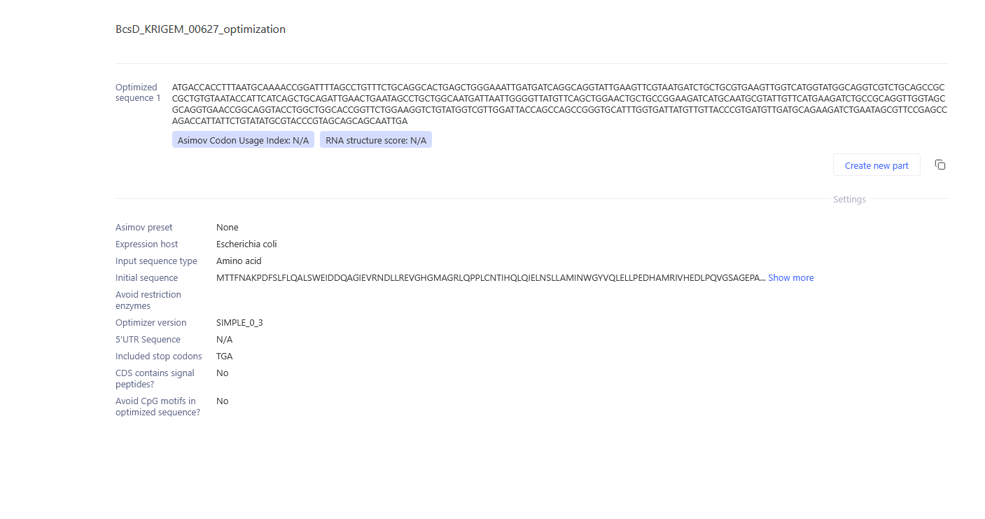
I ran the BcsD coding sequence through Kernel’s Codon Optimizer to compare the original sequence with a codon-optimized version.
The original coding sequence is 471 bp long, and the optimized sequence is also 471 bp long. The optimization changed 94 of the 471 nucleotide positions, while preserving the encoded BcsD amino acid sequence, including the terminal stop codon.
Kernel’s Codon Optimizer does not provide Komagataeibacter rhaeticus as an available expression organism. Therefore, I selected Escherichia coli, the available bacterial option, for this comparison. This means that the resulting sequence should not be considered a codon optimization specifically for K. rhaeticus. Instead, the result demonstrates how synonymous codon changes can modify the DNA sequence while preserving the same protein sequence.
Codon usage is relevant because different organisms can have different preferences for synonymous codons, which can influence how efficiently a coding sequence is translated. For this project, the comparison is therefore useful for understanding the effect of codon usage, while the original BcsD sequence remains the reference sequence for the K. rhaeticus design.
Figure 5. Kernel Codon Optimizer result for the BcsD coding sequence. The tool generated an alternative DNA sequence while preserving the amino acid sequence of the target protein.
Sequence search (BLAST)
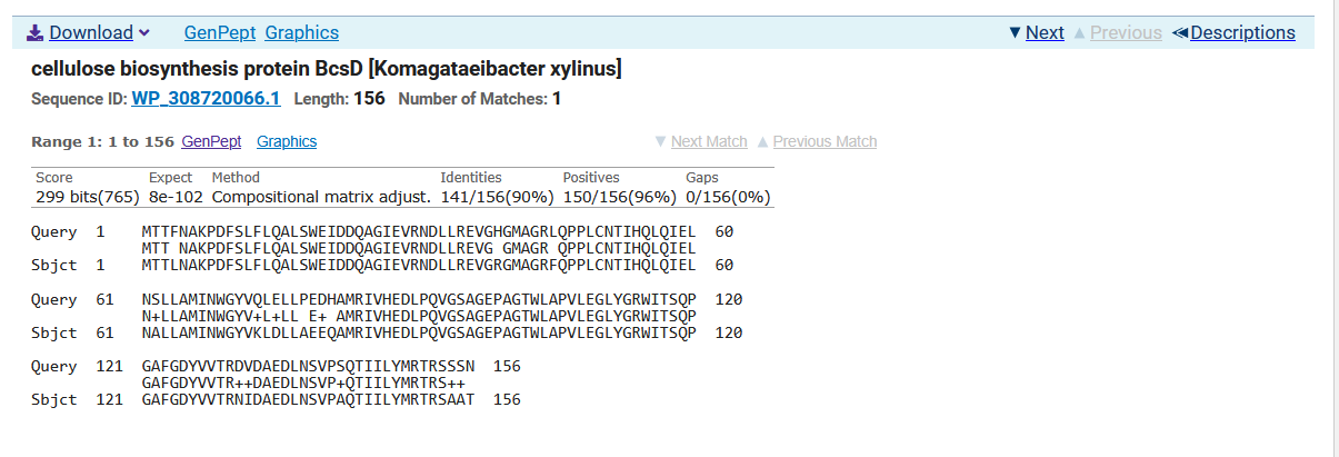
I searched the BcsD amino acid sequence using NCBI Protein BLAST (BLASTP) against the ClusteredNR protein database. The query sequence was 156 amino acids long. The closest significant match was identified as cellulose biosynthesis protein BcsD from Komagataeibacter xylinus (NCBI Reference Sequence WP_308720066.1).
The alignment covered the full query sequence (156/156 amino acids) and showed 141 identical amino acids, corresponding to 90.38% sequence identity. The alignment also showed 150/156 positive matches (96%), no gaps, and an E-value of 8e-102. These results indicate a strong sequence similarity between my candidate protein and a naturally occurring BcsD protein.
The NCBI record for WP_308720066.1 identifies the protein as the natural gene bcsD, encoding cellulose biosynthesis protein BcsD. Therefore, the BLAST result supports the identification of BcsD_KRIGEM_00627 as a BcsD homolog. The candidate sequence is not identical to the K. xylinus reference sequence, but its full-length alignment and high sequence identity indicate that it is closely related to a natural BcsD protein rather than a novel unrelated sequence.
Figure 6. NCBI BLASTP alignment of the candidate BcsD protein with a cellulose biosynthesis protein BcsD from Komagataeibacter xylinus (WP_308720066.1). The alignment shows 100% query coverage, 90.38% sequence identity, 96% positive matches, and no gaps.
Biosecurity screen
I screened the complete BcsD_Expression_Construct sequence using Kernel’s Biosecurity Sequence Scanner, which uses SecureDNA to compare an input nucleotide sequence against a database of potential biosecurity threats. The complete 3,491 bp construct was used for the screening, including the promoter, RBS, BcsD coding sequence, terminator, and backbone.
The scan returned “No flagged sequences detected.” This means that the screening tool did not identify any sequence in the construct that matched a potential biosecurity threat in the database used for the analysis. No sequence was therefore flagged for further review by this screen. This result provides a biosecurity check of the designed construct, while recognizing that it represents the result of this specific screening tool and database rather than a general guarantee of biological safety.
Figure 7. Kernel Biosecurity Sequence Scanner result for the complete BcsD_Expression_Construct. The SecureDNA-based screen returned “No flagged sequences detected.”
Part 5 — From Design to Build
How would you build it?
For the first prototype, I would not start by producing a large library of BcsD variants. I would first build a small number of variants and keep the rest of the construct constant. The main variable would be the BcsD coding sequence, while the promoter, RBS, terminator, and backbone would remain the same.
For these first versions, I would use synthetic DNA or DNA fragments and assemble them with the same backbone. A fragment-based assembly approach such as Gibson assembly could be useful because it allows DNA fragments to be combined into a larger construct. The assigned Gibson et al. reading describes enzymatic assembly of multiple DNA molecules into larger DNA constructs. :contentReference[oaicite:1]{index=1}
My goal would be to test whether different BcsD variants could lead to measurable differences in the physical properties of bacterial cellulose, such as thickness or density. I would also be interested in whether these differences could eventually be used to create regions with different material characteristics within a grown 3D artifact.
What does synthesis constrain?
DNA synthesis is not error-free, so I would not assume that every synthesized sequence is identical to the sequence I designed. This becomes especially important when generating many variants.
Kosuri et al. showed that DNA synthesis from oligonucleotide pools can be affected by synthesis errors and that assembly becomes more difficult as the diversity of the oligonucleotide mixture increases. They also describe challenges associated with sequence characteristics such as high GC content, repetitive sequences, and sequence similarity. :contentReference[oaicite:2]{index=2} :contentReference[oaicite:3]{index=3}
For my construct, I would therefore verify the sequences before using the variants for comparison. If I used an oligo pool to generate many variants, error correction and sequence verification would become particularly important. Kosuri et al. used enzymatic error correction and sequence verification as part of their workflow for improving the quality of assembled genes. :contentReference[oaicite:4]{index=4}
Carr et al. also studied protein-mediated error correction for de novo DNA synthesis, supporting the idea that synthesized DNA can require an error-correction step before being used as a final construct. :contentReference[oaicite:5]{index=5}
At this stage, I do not identify a specific sequence feature in my BcsD design as a known synthesis problem. However, the synthesis process itself is a constraint because errors can be introduced during oligonucleotide synthesis and assembly.
Prototype vs. scale
For the prototype stage, I would work with a small number of BcsD variants. This would allow me to test the main idea without generating a large DNA library. I would keep the overall construct architecture constant and compare the resulting bacterial cellulose to see whether the BcsD variants are associated with differences in properties such as thickness or density.
If the prototype showed an interesting relationship between the BcsD sequence and the physical properties of the cellulose, I would then scale the design to a larger number of variants. At that stage, a DNA pool synthesized on a microchip could become useful because it allows many different oligonucleotide sequences to be generated in parallel. Kosuri et al. demonstrated highly parallel gene synthesis from oligonucleotide pools and showed how this approach can increase the number of sequences that can be explored. :contentReference[oaicite:6]{index=6}
However, scaling would also introduce additional challenges. Kosuri et al. found that increasing sequence diversity can make assembly more difficult and that error correction and sequence verification remain important parts of the process. :contentReference[oaicite:7]{index=7}
Overall, my build strategy would therefore change with scale: a small number of individually designed variants for the first prototype, followed by a more parallel synthesis strategy if the initial results justify exploring a larger number of BcsD variants.
Part 5 — From Design to Build
How would you build it?
For the first prototype, I would not start by producing a large library of BcsD variants. I would first build a small number of variants and keep the rest of the construct constant. The main variable would be the BcsD coding sequence, while the promoter, RBS, terminator, and backbone would remain the same.
For these first versions, I would use synthetic DNA or DNA fragments and assemble them with the same backbone. A fragment-based assembly approach such as Gibson assembly could be useful because it allows DNA fragments to be combined into a larger construct (Gibson et al., 2009).
My goal would be to test whether different BcsD variants could lead to measurable differences in the physical properties of bacterial cellulose, such as thickness or density. I would also be interested in whether these differences could eventually be used to create regions with different material characteristics within a grown 3D artifact.
What does synthesis constrain?
DNA synthesis is not error-free, so I would not assume that every synthesized sequence is identical to the sequence I designed. This becomes especially important when generating many variants.
Kosuri et al. showed that DNA synthesis from oligonucleotide pools can be affected by synthesis errors and that assembly becomes more difficult as the diversity of the oligonucleotide mixture increases. They also describe challenges associated with sequence characteristics such as high GC content, repetitive sequences, and sequence similarity (Kosuri et al., 2010).
For my construct, I would therefore verify the sequences before using the variants for comparison. If I used an oligo pool to generate many variants, error correction and sequence verification would become particularly important. Kosuri et al. used enzymatic error correction and sequence verification as part of their workflow for improving the quality of assembled genes (Kosuri et al., 2010).
Carr et al. also studied protein-mediated error correction for de novo DNA synthesis, supporting the use of error-correction strategies to improve the quality of synthesized DNA (Carr et al., 2004).
At this stage, I do not identify a specific sequence feature in my BcsD design as a known synthesis problem. However, the synthesis process itself is a constraint because errors can be introduced during oligonucleotide synthesis and assembly.
Prototype vs. scale
For the prototype stage, I would work with a small number of BcsD variants. This would allow me to test the main idea without generating a large DNA library. I would keep the overall construct architecture constant and compare the resulting bacterial cellulose to see whether the BcsD variants are associated with differences in properties such as thickness or density.
If the prototype showed an interesting relationship between the BcsD sequence and the physical properties of the cellulose, I would then scale the design to a larger number of variants. At that stage, a DNA pool synthesized on a microchip could become useful because it allows many different oligonucleotide sequences to be generated in parallel. Kosuri et al. demonstrated highly parallel gene synthesis from oligonucleotide pools and showed how this approach can increase the number of sequences that can be explored (Kosuri et al., 2010).
However, scaling would also introduce additional challenges. Kosuri et al. found that increasing sequence diversity can make assembly more difficult and that error correction and sequence verification remain important parts of the process (Kosuri et al., 2010).
Overall, my build strategy would therefore change with scale: a small number of individually designed variants for the first prototype, followed by a more parallel synthesis strategy if the initial results justify exploring a larger number of BcsD variants.
Part 6 — What Don’t You Know Yet?
The main uncertainties in my design are related to expression, the effect of BcsD on the material, and whether the overall genetic design works as expected in Komagataeibacter rhaeticus.
Will BcsD be expressed and produce an observable effect?
One uncertainty is whether the BcsD construct will actually be expressed in Komagataeibacter rhaeticus and whether this expression will have an observable effect on bacterial cellulose.
I would first check whether BcsD is expressed and then compare the resulting cellulose with an appropriate reference condition. This would help determine whether the genetic design is producing an observable biological effect.
Will different BcsD variants affect the physical properties and organization of bacterial cellulose?
Another uncertainty is whether changing the BcsD sequence will actually affect the material. I am interested in whether different variants could produce differences in properties such as thickness or density, as well as differences in how the cellulose is organized.
I would first compare the resulting cellulose samples visually to identify differences in morphology or distribution. I would then use physical measurements, such as thickness or density, together with observations of the cellulose structure, to characterize these differences more systematically.
Will the promoter, RBS, and complete construct work as expected?
A third uncertainty is whether the promoter and RBS selected for the construct will provide an appropriate level of expression, and whether the complete genetic design will function as expected in Komagataeibacter rhaeticus.
I would compare the expression and cellulose production associated with the construct against an appropriate reference condition. If the construct does not behave as expected, I would test design variants, such as changing the promoter or RBS, rather than assuming that the original design is optimal.
These uncertainties define the next stage of the project: moving from a sequence that is computationally designed and checked to a biological prototype that can provide evidence about how BcsD affects bacterial cellulose as a material.
AI
I used ChatGPT as a support tool throughout this project. I used it to help me understand the assignment requirements, organize my ideas, review my genetic construct, and clarify concepts related to DNA design, synthesis, and bacterial cellulose.
I also used ChatGPT to question my assumptions and improve the clarity of my explanations. The final design choices, sequence selection, and project decisions were reviewed and made by me.
AI tool: ChatGPT (OpenAI)
Example prompts used:
“I am designing a BcsD expression construct for Komagataeibacter rhaeticus. Help me identify the main assumptions in my promoter, RBS, CDS, and terminator choices.”
“I want to compare several BcsD variants in the future. How could I approach the first prototype differently from a larger-scale variant library?”
“What are the main limitations of DNA synthesis and oligonucleotide pools that could affect my build strategy?”
“What are the biggest unknowns in my current BcsD design, and how could I test each one experimentally?”
References
Carr, P. A., et al. (2004). Protein-mediated error correction for de novo DNA synthesis. Nucleic Acids Research, 32(20), e162. https://doi.org/10.1093/nar/gnh160
Gibson, D. G., et al. (2009). Enzymatic assembly of DNA molecules up to several hundred kilobases. Nature Methods, 6, 343–345. https://doi.org/10.1038/nmeth.1318
Kondo, T., Nakamura, Y., Nojima, S., Yao, M., & Imai, T. (2022). The BcsD subunit of type I bacterial cellulose synthase interacts dynamically with the BcsAB catalytic core complex. FEBS Letters. https://doi.org/10.1002/1873-3468.14495
Kosuri, S., et al. (2010). Scalable gene synthesis by selective amplification of DNA pools from high-fidelity microchips. Nature Biotechnology, 28, 1295–1299. https://doi.org/10.1038/nbt.1716
Römling, U., & Galperin, M. Y. (2015). Bacterial cellulose biosynthesis: diversity of operons, subunits, products, and functions. Trends in Microbiology, 23(9), 545–557.
This homework explores the design and optimization of cell-free protein synthesis (CFPS) reactions for three target proteins: superfolder green fluorescent protein (sfGFP), PETase (TfCut2), and Reteplase.
The objective is to reduce the cost per gram of functional protein, while accounting for differences in expression, folding, and activity requirements. A low-cost reaction is not necessarily the best formulation if it produces less active protein.
The designs below combine literature-based reasoning with initial hypotheses developed using the CFPS Reaction Designer. The proposed modifications have not yet been experimentally validated.
A. Compare Cell-Free Reaction Compositions
A1. Why do potassium glutamate and magnesium glutamate concentrations differ between compositions? Which is more sensitive?
Potassium glutamate and magnesium glutamate influence different aspects of the chemical environment required for cell-free protein synthesis. Potassium helps establish the ionic conditions needed for transcription and translation, while magnesium is directly involved in ribosome function and interacts with RNA, nucleotides, and phosphate.
Magnesium availability is particularly sensitive to changes in reaction composition. ATP, other nucleotides, and phosphate can bind or interact with magnesium, so changing the energy system or nucleotide concentrations can alter the amount of magnesium available for biochemical processes. Potassium glutamate also requires optimization, but its effects depend on the ionic environment and the other components in the reaction.
Jewett and Swartz (2004) showed that both potassium glutamate and magnesium glutamate affect protein production in CFPS. They also found that the optimal magnesium concentration differed between the PANOx-SP and Cytomim systems, illustrating that the appropriate concentration depends on the energy-regeneration strategy.
Conclusion: Magnesium glutamate is often a particularly sensitive variable because its effective availability depends on interactions with other reaction components. However, neither salt has a universal optimum; both must be optimized in the context of the complete formulation and the lysate used.
A2. How does a PEP + NTP formulation differ from a ribose + NMP formulation?
PEP + NTP and ribose + NMP formulations use different strategies to supply energy and nucleotide precursors for cell-free protein synthesis.
PEP + NTP
Traditional formulations such as PANOx-SP supply nucleotide triphosphates directly:
ATP
CTP
GTP
UTP
They also use phosphoenolpyruvate (PEP) as a high-energy substrate that supports ATP regeneration.
A commonly reported PANOx-SP formulation contains 1.2 mM ATP, 0.85 mM each of GTP, UTP, and CTP, and 33 mM PEP. The same formulation includes 170 mM potassium glutamate and 16 mM magnesium glutamate, among other components (Knapp et al., 2007; Jewett & Swartz, 2004).
This approach provides nucleotides in a form that can be used directly for transcription. However, PEP and several nucleotide triphosphates can contribute substantially to reagent costs. PEP metabolism also generates inorganic phosphate, which can affect magnesium availability and the chemical environment over the course of a reaction.
Ribose + NMP
Ribose/NMP-based strategies use nucleotide monophosphates as precursors:
AMP
CMP
GMP
UMP
The cell lysate contains metabolic pathways that can convert these precursors into the nucleotide triphosphates required for transcription. Ribose can support endogenous metabolism, depending on the formulation and metabolic capacity of the lysate.
Calhoun and Swartz (2005) demonstrated that a glucose/NMP-based CFPS system could maintain protein production while reducing reagent costs by more than 75% under the conditions they studied. This result supports the general principle of using less expensive nucleotide precursors, although the exact performance depends on the formulation.
Olsen et al. (2026) further demonstrated that low-cost CFPS formulations can generate NTPs from less expensive precursors and sustain protein production without relying on a conventional phosphorylated energy substrate such as PEP.
Comparison
Feature
PEP + NTP
Ribose/NMP-based strategy
Nucleotide input
NTPs supplied directly
NMPs supplied as precursors
Energy strategy
PEP supports ATP regeneration
Depends more on endogenous metabolism and the selected substrates
Reagent cost
Can be high because of PEP and NTPs
Potentially lower, depending on the formulation
Metabolic dependence
Supplies more nucleotide resources directly
Requires the lysate to convert precursors into usable nucleotides
Phosphate balance
PEP metabolism can generate substantial phosphate
Depends on the selected substrates and metabolic pathways
Main advantage
Established, direct nucleotide supply
Potential reduction in reagent cost
Main challenge
Cost and changing phosphate/Mg²⁺ balance
Performance depends on lysate metabolism and substrate balance
For this project, I consider a lower-cost nucleotide strategy a useful starting point. However, the objective is not to minimize reagent cost alone. It is to maximize functional protein produced per unit cost.
A3. Compare two cell-free reaction formulations from the literature
I chose PANOx-SP, described by Jewett and Swartz (2004), and RFopt, the optimized low-cost formulation developed by Olsen et al. (2026). These formulations illustrate two different approaches to cell-free protein synthesis: supplying energy and nucleotide resources directly, or relying more heavily on metabolic conversion of less expensive precursors.
PANOx-SP
PANOx-SP is a conventional cell-free protein synthesis (CFPS) formulation that uses phosphoenolpyruvate (PEP) as an energy substrate and supplies nucleotide triphosphates (NTPs) directly. A commonly reported composition includes the following components:
Component
Reported concentration
ATP
1.2 mM
GTP
0.85 mM
UTP
0.85 mM
CTP
0.85 mM
PEP
33 mM
Potassium glutamate
175 mM
Magnesium glutamate
20 mM
Ammonium glutamate
10 mM
Spermidine
1.5 mM
Putrescine
1.0 mM
NAD
0.33 mM
Coenzyme A
0.26 mM
Sodium oxalate
2.7 mM
Amino acids
2 mM each
These values correspond to the PANOx-SP formulation reported by Jewett and Swartz (2004). The complete reaction also includes components such as folinic acid, tRNA, DNA template, T7 RNA polymerase, and cell extract. Exact concentrations can vary between protocols and modified versions of the system.
The main advantage of PANOx-SP is that it supplies important substrates directly to the transcription and translation machinery. However, PEP and several nucleotide triphosphates can contribute substantially to reagent costs. PEP consumption and degradation can also lead to phosphate accumulation, affecting the chemical environment during the reaction (Jewett & Swartz, 2004).
RFopt
Olsen et al. (2026) developed RFopt through a systematic optimization campaign involving 1,231 reaction formulations. Their objective was to reduce reagent costs while maintaining high protein production. The optimized formulation uses a smaller set of added components and supports the production of nucleotide triphosphates from nitrogenous bases and ribose
B. Design One Reaction Composition for Each of the Three Proteins
The three reaction designs use the fixed-volume framework provided by the CFPS Reaction Designer. My objective is to reduce the cost per gram of functional protein rather than simply minimize the cost per reaction or maximize total protein expression.
The submitted formulations are initial hypotheses informed by the literature and the reference compositions available in the designer. Their performance has not yet been experimentally validated.
B1. sfGFP: Cost-efficient protein synthesis
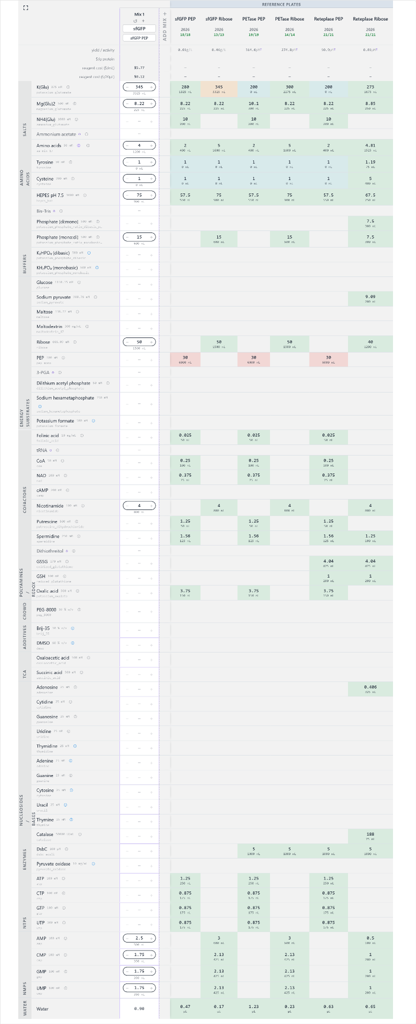
For sfGFP, I selected a nucleotide-focused modification within a ribose/NMP-based reference formulation. sfGFP is a useful reporter because its fluorescence provides a practical readout of correctly folded protein.
My submitted composition reduces AMP from 3 to 2.5 mM and reduces CMP, GMP, and UMP from 2.13 to 1.75 mM each. All other reaction settings remain unchanged from the starting composition.
The hypothesis is that reducing the supplied NMP concentrations may lower reagent cost while retaining sufficient nucleotide availability for transcription and translation. However, lowering nucleotide precursor concentrations could also limit protein synthesis. The direction and magnitude of the effect cannot be established without experimental measurements.
Hypothesis: Reducing NMP concentrations may lower reagent cost without substantially reducing sfGFP fluorescence.
Evaluation: Compare fluorescence and reagent cost against the reference formulation. A lower reaction cost alone would not establish an improvement if functional protein output also decreased.
B2. TfCut2/PETase: Functional enzyme production
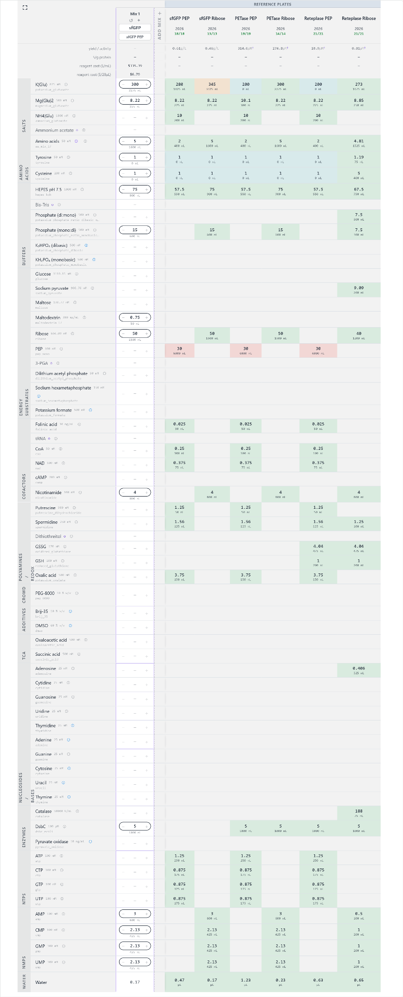
For TfCut2, I used the ribose/NMP-based PETase reference formulation and increased maltodextrin to 0.75 mg/mL. The other reaction components remain unchanged from the reference formulation.
This modification tests whether additional maltodextrin can support the metabolic processes required for protein synthesis and maintain or improve the production of functional enzyme. The rationale is exploratory: the effect depends on the metabolic capacity of the lysate, and adding a carbon source does not necessarily increase protein yield.
Olsen et al. (2026) found that individual additions of several alternative energy substrates, including maltodextrin and ribose, did not improve yield in their initial tests. Therefore, my proposed increase should be treated as a specific hypothesis for this formulation rather than a result established by the literature.
TfCut2 is a cutinase-family enzyme whose function depends on correct folding and catalytic activity. Total protein production alone would not be sufficient to evaluate this design.
Hypothesis: Increasing maltodextrin to 0.75 mg/mL may maintain or improve TfCut2 activity without a disproportionate increase in reagent cost.
Evaluation: I would measure enzyme activity using an appropriate p-nitrophenyl ester substrate and monitor the initial increase in absorbance at 405 nm. The assay should include suitable controls and use the same conditions for the reference and modified reactions. I would compare initial enzyme activity and reagent cost against the reference formulation. The modification would be useful only if the measured functional output justifies its cost.
B3. Reteplase: Functional protein production and oxidative folding
Reteplase presents a folding challenge because its biological activity depends on the formation of the correct disulfide bonds. Producing the polypeptide alone does not guarantee that the resulting protein will be functional.
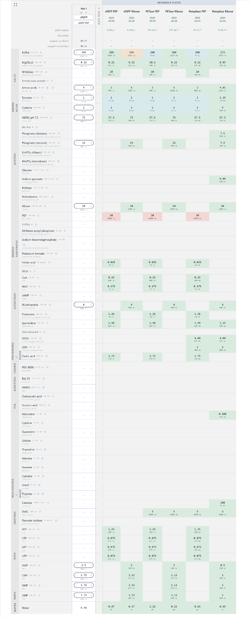
For this target, I selected the Reteplase Ribose reference formulation and reduced the amino acid concentration from 4.81 to 4 mM. All other reaction settings remain unchanged. The purpose is to investigate whether a moderate reduction in amino acid input can lower reagent costs while maintaining sufficient substrate availability for functional protein production.
I retained the folding-related components included in the reference formulation: DsbC, GSSG, GSH, and catalase. These components serve different roles in the reaction, and retaining them allows me to focus this initial modification on amino acid concentration rather than changing several variables simultaneously.
Previous research on cell-free synthesis of disulfide-bonded proteins highlights the importance of controlling the redox environment and providing suitable folding conditions (Yin & Swartz, 2004). Groff et al. (2014) investigated folding helpers, including DsbC and FkpA, in a cell-free antibody-expression system. However, those findings do not establish that the same strategy will improve Reteplase production. FkpA is therefore a possible direction for future investigation, not an additional component in my current formulation.
Hypothesis: Reducing the amino acid concentration from 4.81 to 4 mM may lower reagent costs while preserving functional Reteplase production.
Evaluation: I would compare the modified reaction with the reference formulation by measuring Reteplase activity using a validated functional assay. One possible approach is to measure the initial rate of substrate cleavage using IPR-AMC, provided the assay conditions are appropriate for Reteplase and include suitable controls. I would compare functional activity and reagent cost to determine whether the reduction offers a meaningful benefit. If activity decreases substantially, the lower reagent cost may not justify the modification.
Since these reactions have not yet been experimentally evaluated, the effect of reducing amino acid concentration on Reteplase activity remains unknown.
Design limitations and evaluation
These three formulations are initial experimental hypotheses, not validated improvements.
Each reaction must total 20,000 nL, with fixed volumes of lysate, DNA, and base buffer, variable reagent volumes in multiples of 25 nL, and the remaining variable volume filled with nuclease-free water.
The proposed modifications target different potential constraints:
sfGFP: nucleotide precursor availability and reagent cost.
TfCut2: the possible contribution of additional maltodextrin to metabolic support and enzyme activity.
Reteplase: amino acid input while retaining the selected folding-related components.
A successful formulation must balance reagent cost with functional output. The relevant readouts are sfGFP fluorescence, TfCut2 enzymatic activity, and Reteplase enzymatic activity. Until those measurements are available, I cannot conclude that any of the proposed modifications improves protein production or reduces cost per gram of functional protein.
B4. Community Reagent: FkpA
I upvoted FkpA in the Community Reagents section.
FkpA is an Escherichia coli periplasmic protein with chaperone and peptidyl-prolyl isomerase activity. It is a candidate folding helper because it can support protein folding and reduce aggregation in certain contexts.
Groff et al. (2014) identified FkpA and the disulfide isomerase DsbC as positive effectors of immunoglobulin folding and assembly in an open cell-free synthesis system. Their results suggest that these proteins can act synergistically in that particular system.
I consider FkpA a candidate for future investigation in reactions that require substantial folding support. However, FkpA is not a substitute for DsbC’s disulfide-isomerase function, and the published antibody results do not demonstrate that FkpA improves Reteplase activity.
My rationale for upvoting FkpA is that it may expand the available set of folding helpers for future CFPS optimization. Its contribution would need to be evaluated experimentally by comparing functional protein activity and reagent cost with and without the additional component.
Relevant research:
Groff et al. (2014), Engineering toward a bacterial “endoplasmic reticulum” for the rapid expression of immunoglobulin proteins.
I used ChatGPT (GPT-6) throughout this assignment as a tool to help me understand and compare different cell-free protein synthesis strategies. I focused on how energy substrates, nucleotide precursors, salt concentrations, and folding helpers influence reaction design, and how these choices relate to the cost of producing functional protein.
I also used it to explore possible modifications to the reference formulations for sfGFP, TfCut2, and Reteplase. Rather than changing several components at once, I focused on specific adjustments that could be evaluated individually: reducing nucleotide monophosphate concentrations for sfGFP, increasing maltodextrin for TfCut2, and reducing amino acid concentration for Reteplase while retaining the selected folding-related components.
The main prompts I used during this process included:
Compare PEP/NTP-based and ribose/NMP-based cell-free protein synthesis formulations. Explain their differences in energy regeneration, nucleotide availability, and reagent cost, and identify the main trade-offs when choosing between them.
Explain how potassium glutamate and magnesium glutamate affect cell-free protein synthesis, and why changing their concentrations can influence reaction performance.
Help me compare the PANOx-SP formulation with the RFopt formulation reported in the literature. Separate published experimental results from possible explanations and identify what can reasonably be concluded about protein yield and production cost.
Based on the reference formulations available in the CFPS Reaction Designer, help me identify one specific component to modify for sfGFP, TfCut2, and Reteplase. For each target, explain the rationale, the potential trade-offs, and how I could evaluate the effect without assuming the modification will improve the outcome.
Explain the folding requirements of Reteplase, including the role of disulfide bonds, DsbC, and the redox environment. Could FkpA be a useful additional folding helper, and what are the limitations of applying findings from antibody expression to Reteplase?
Help me define meaningful evaluation criteria for the three reaction designs, considering functional protein activity and reagent cost rather than total protein yield alone.
An important part of this process was distinguishing between what has been demonstrated in published research and what remains an assumption in my own designs. The literature helped inform the rationale behind the proposed changes, but it does not establish that these modifications will improve my reactions. I therefore treated each composition as a hypothesis to be tested, with functional protein activity and reagent cost as the main criteria for evaluating the results.
ChatGPT also helped me examine the potential role of FkpA as a folding helper. This was useful for identifying a possible direction for future investigation, while keeping in mind that results obtained with antibodies cannot be directly assumed to apply to Reteplase.
The final compositions reflect my design decisions informed by the reference formulations, published research, and this AI-assisted analysis. Since the reactions have not yet been experimentally evaluated, their effects on protein activity and cost remain unknown.
The prompts listed above are representative examples of the questions explored during the design process, rather than verbatim transcripts of every interaction.
B6. CFPS Coopetition Forum
I checked access to the CFPS Coopetition forum as part of the assignment. The forum provides a space to discuss reaction design, compare ideas, and exchange feedback related to cell-free protein synthesis.
References
Borkowski, O., et al. (2020). Large scale active-learning-guided exploration for in vitro protein production optimization. Nature Communications, 11, Article 1872. https://doi.org/10.1038/s41467-020-15798-5
Calhoun, K. A., & Swartz, J. R. (2005). An economical method for cell-free protein synthesis using glucose and nucleoside monophosphates. Biotechnology Progress, 21(4), 1146–1153. https://doi.org/10.1021/bp050052y
Groff, D., Armstrong, S., Rivers, P. J., Zhang, J., Yang, J., Green, E., Rozzelle, J., Liang, S., Kittle, J. D., Jr., Steiner, A. R., Baliga, R., Thanos, C. D., Hallam, T. J., Sato, A. K., & Yam, A. Y. (2014). Engineering toward a bacterial “endoplasmic reticulum” for the rapid expression of immunoglobulin proteins. mAbs, 6(3), 671–678. https://doi.org/10.4161/mabs.28172
Jewett, M. C., & Swartz, J. R. (2004). Mimicking the Escherichia coli cytoplasmic environment activates long-lived and efficient cell-free protein synthesis. Biotechnology and Bioengineering, 86(1), 19–26. https://doi.org/10.1002/bit.20026
Knapp, K. G., Goerke, A. R., & Swartz, J. R. (2007). Cell-free synthesis of proteins that require disulfide bonds using glucose as an energy source. Biotechnology and Bioengineering, 97, 901–908. https://doi.org/10.1002/bit.21296
Olsen, M. L., Copeland, C. E., Sundberg, C. A., Aw, R., Shaver, Z. M., Rao, G., Swartz, J. R., Karim, A. S., & Jewett, M. C. (2026). Design-driven optimization of low-cost reagent formulations for reproducible and high-yielding cell-free gene expression. Nature Communications, 17, Article 3478. https://doi.org/10.1038/s41467-026-69605-8
Silverman, A. D., Karim, A. S., & Jewett, M. C. (2020). Cell-free gene expression: An expanded repertoire of applications. Nature Reviews Genetics, 21, 151–170. https://doi.org/10.1038/s41576-019-0186-3
Smith, A. A., et al. (2026). Using a GPT-5-driven autonomous lab to optimize the cost and titer of cell-free protein synthesis. bioRxiv. https://doi.org/10.64898/2026.02.05.703998
Yin, G., & Swartz, J. R. (2004). Enhancing multiple disulfide bonded protein folding in a cell-free system. Biotechnology and Bioengineering, 86(2), 188–195. https://doi.org/10.1002/bit.10827
Silverman, A. D., Karim, A. S., & Jewett, M. C. (2020). Cell-free gene expression: An expanded repertoire of applications. Nature Reviews Genetics, 21, 151–170. https://doi.org/10.1038/s41576-019-0186-3
Smith, A. A., et al. (2026). Using a GPT-5-driven autonomous lab to optimize the cost and titer of cell-free protein synthesis. bioRxiv. https://doi.org/10.64898/2026.02.05.703998
Yin, G., & Swartz, J. R. (2004). Enhancing multiple disulfide bonded protein folding in a cell-free system. Biotechnology and Bioengineering, 86(2), 188–195. https://doi.org/10.1002/bit.10827