Hi, I’m Wissal, a molecular biology engineer fascinated by synthetic biology, mammalian cell engineering, and anything related to using biology to solve real-world problems 💫
I love learning new things, meeting new people, and discovering new cultures. I enjoy discussions about philosophy, history, and psychology.
I believe interdisciplinarity holds great potential for addressing some of the most challenging issues of our century, and I particularly value HTB(G)AA’s constructionist approach to learning and its vision of radical inclusion.
learning should be accessible to everyone, no matter where they are or what their background is.
I’m eager to dive into this learning journey, discover more about myself and the world, and gain a deeper understanding of how we can harness incredibly complex biological systems to design advanced therapies that can translate into meaningful change. 🧬🌸
Contact info
If you enjoy anything related to biology and want to discuss feel free to reach out !
A Programmable SynNotch CAR-T Platform for Neuroblastoma
1.🪄 What I would like to biomanufacture, and Why ?
Neuroblastoma is a cancer of the developing nervous system that primarily affects children. Although treatment outcomes have improved, patients with high-risk, relapsed, or refractory disease continue to have limited effective therapeutic options.
CAR-T-cell therapy has shown encouraging activity in neuroblastoma, but its effectiveness is limited immune escape, limited or variable T-cell persistence, functional exhaustion within the tumor microenvironment, and the potential for on-target, off-tumor toxicity.
To address some of these limitations and to improve the specificity and functionality of CAR-T-cell therapy, I propose to biomanufacture a programmable, dual-antigen, logic-gated theranostic CAR-T-cell system that restricts full therapeutic activation to the tumor environment.
The engineered T cells would use a synthetic IF–THEN recognition circuit to increase tumor specificity while reducing constitutive CAR signaling providing autonomous control over T-cell activity.
The core of my proposed system would therefore use dual-antigen recognition rather than constitutive recognition of a single tumor antigen.
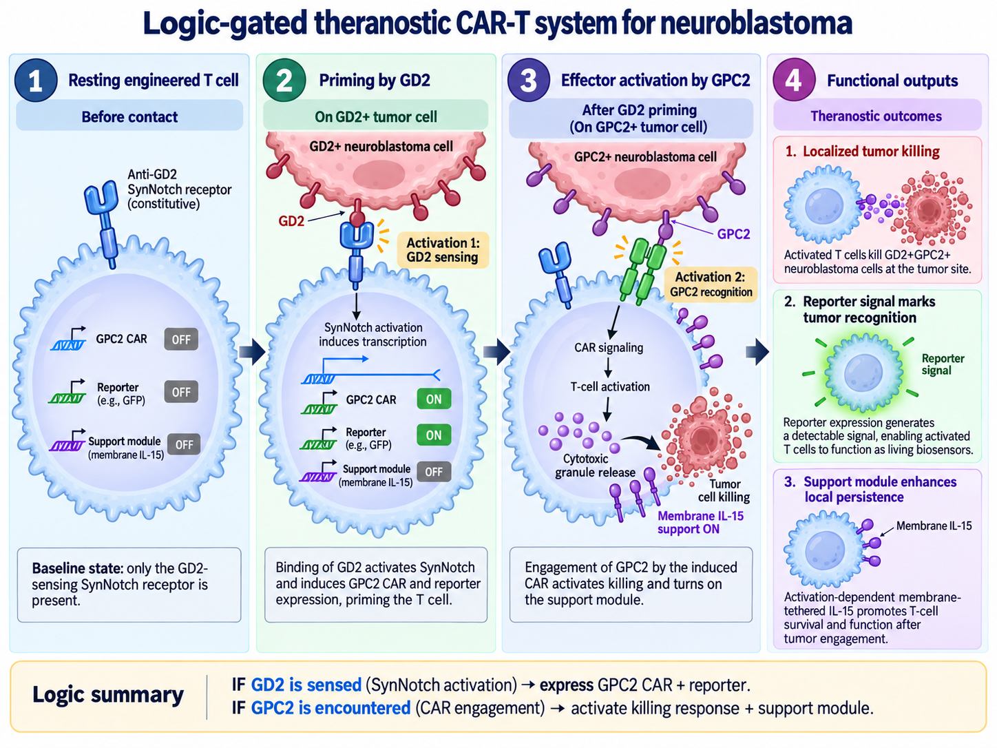
The engineered T cell would first recognize the neuroblastoma-associated antigen GD2 through a synthetic Notch (SynNotch) receptor.
GD2 recognition would act as a priming signal, releasing a synthetic transcription factor that induces expression of a CAR directed against a second neuroblastoma antigen, GPC2.
The resulting logic is:
IF GD2 is detected → THEN express GPC2 CAR
followed by:
IF GPC2 is encountered → THEN activate the T cell and kill the target cell
Additionally, an inducible fluorescent reporter (such as GFP) generate a detectable signal upon GD2-SynNotch binding and an activation-dependent, membrane-tethered IL-15 module is added to enhance T-cell survival and persistence after GPC2 recognition allowing the engineered T cells to function as both therapeutic effectors and living biosensors.
The concept is therefore not simply to manufacture a CAR-T cell, but to manufacture a living therapeutic system capable of sensing its environment, making a decision, and executing an antitumor response.
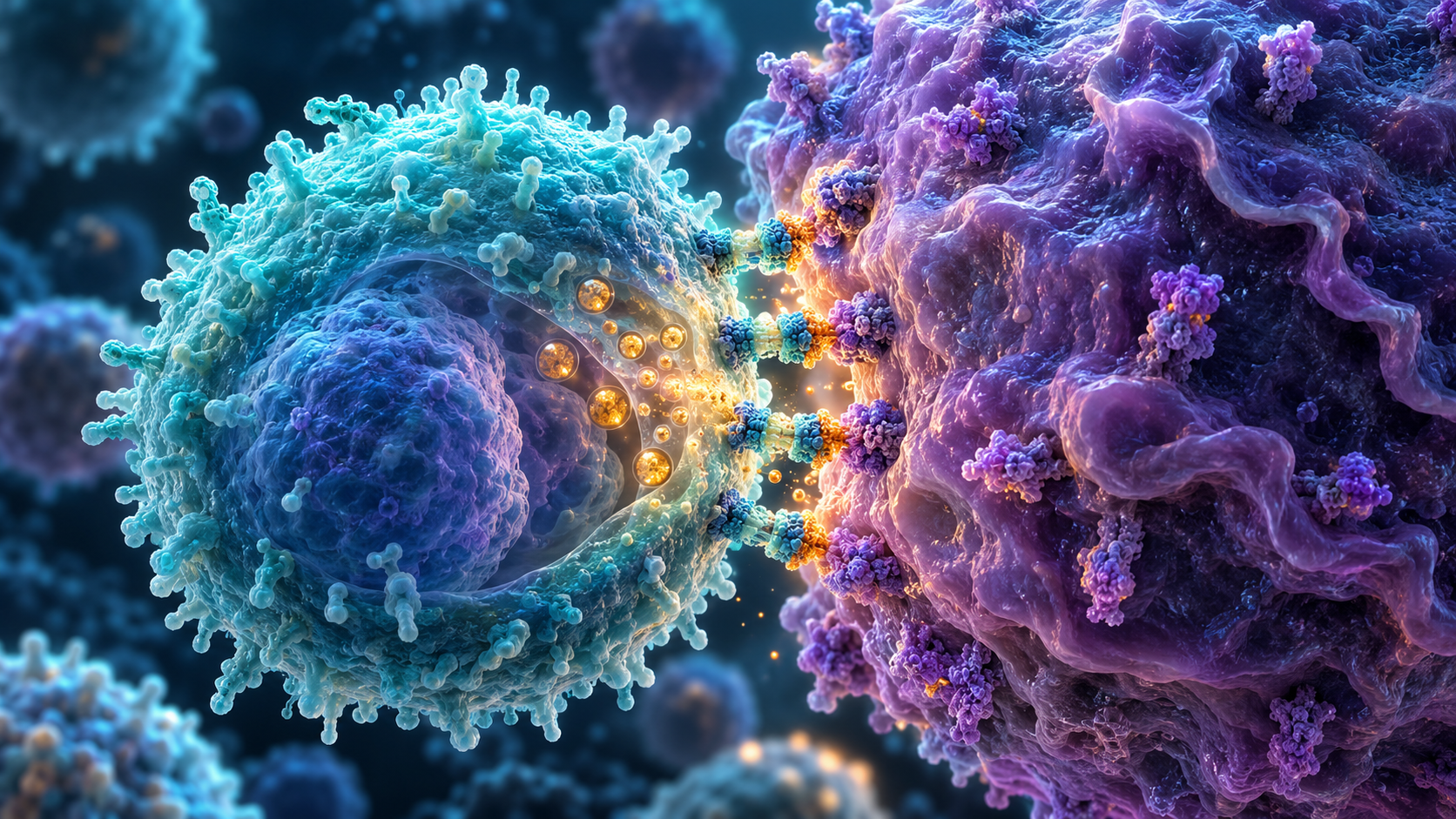
Mechanism of the GD2-SynNotch–GPC2 CAR-T Logic-Gated System (AI generated)
2.🦠 What living system would I use?
The manufacturing chassis would be primary human T lymphocytes obtained from the patient, producing an autologous engineered T-cell product.
The input into the manufacturing process would therefore be a patient-derived leukapheresis product containing T lymphocytes together with the genetic information encoding the synthetic recognition circuit.
At a conceptual level, the cells would be engineered to contain four functional modules:
GD2-sensing module:A GD2-specific SynNotch receptor constitutively present on the engineered T cell.
GPC2 effector module:Recognition of GD2 releases the SynNotch transcription factor, which activates transcription of a GPC2-specific CAR.
Reporter module:An inducible reporter module
Activation-support module:After CAR engagement, an inducible membrane-tethered cytokine signal, such as IL-15, would provide localized support to the activated T cell.
The inputs to the living system are therefore tumor-associated molecular signals, primarily GD2 and GPC2. The biological processes performed by the engineered T cell include antigen sensing, synthetic transcriptional regulation, CAR expression, signal transduction, cytotoxicity, cytokine-mediated support.
The final output is a population of engineered autologous T cells capable of conditional tumor recognition and regulated antitumor activity.
3.⚙️ How would I use automation?
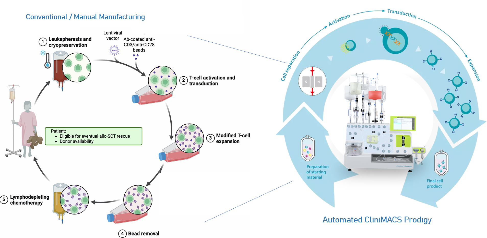
I would integrate the engineered T-cell system with a closed, automated cell-processing platform such as the CliniMACS Prodigy.
CAR-T-cell manufacturing involves multiple interconnected operations including T-cell enrichment, activation, genetic modification, expansion, sampling, washing, harvesting and final formulation.
Performing these operations manually increases operator workload and introduces opportunities for variability and contamination.
For this project, automation would be particularly useful for:
🩸 T-cell enrichment and preparation from the patient's starting material; 🧬 standardized cell activation and genetic modification; 🧫 controlled expansion and culture; 🤖 automated feeding and process monitoring; 🔬 in-process sampling; 🧪 final washing, harvesting and formulation.
Automation would also improve reproducibility and enable multiple patient-specific manufacturing batches to be processed in parallel.
During early-stage development, an Opentrons Flex platform could be used to automate high-throughput liquid handling, construct screening, and characterization of the engineered T-cell circuit.
Manual vs automated manufacturing of CAR-T Cells
Image sources:
Source 1 |
Source 2
4.a ⚖️ How much would I need to manufacture ?
The scale I define is manufacturing enough therapy for the approximate annual worldwide population of patients with high-risk neuroblastoma.
A global analysis estimated approximately 5,560 new neuroblastoma cases in children aged 0–14 years in 2021. Approximately half of neuroblastoma cases are classified as high risk.
Therefore:
5,560 cases/year × 0.50
2,780 potential high-risk patients/year
A recent phase 1/2 study of GD2-CART01 used a target dose of:
10 × 10⁶ CAR-positive T cells/kg
I will use 20 kg as an assumption.
For one 20-kg patient:
10 × 10⁶ CAR⁺ cells/kg × 20 kg
2 × 10⁸ CAR⁺ T cells
Therefore, the approximate annual number of infused CAR-positive cells for 2,780 patients would be:
556 billion therapeutic CAR-positive T cells per year
However, because the proposed treatment is autologous, approximately 2,780 separate patient-specific manufacturing batches per year would be required.
4.b ⏳ How long would it take?
Published automated CAR-T manufacturing processes using the CliniMACS Prodigy typically require approximately 7–13 days, depending on the product and manufacturing process.
I will assume a 12-day manufacturing cycle.
One instrument continuously occupied by 12-day manufacturing runs could theoretically complete:
365 days/year ÷ 12 days/run
≈ 30 runs per year
Since real facilities cannot operate at 100% utilization (time for scheduling, setup, failures, maintenance, etc.), assuming 80% effective utilization:
30 × 0.8
≈ 24 patient batches per instrument per year
For 2,780 patients:
2,780 ÷ 24
≈ 116 parallel manufacturing instruments
So, under these assumptions, treating every newly diagnosed high-risk neuroblastoma patient would require approximately 100–120 parallel automated manufacturing units.
The actual number could be higher because the proposed synthetic circuit is considerably more complex than a conventional single CAR.
SynNotch receptors, inducible CAR expression, cytokine modules and negative-feedback circuitry increase the genetic payload and may reduce genetic-modification efficiency or cell yield.
Thus, at a larger commercial scale, I would consider transferring the manufacturing process to a high-throughput closed platform such as the Cellares Cell Shuttle, which can handle up to 16 independent cartridges asynchronously, meaning different patient batches can run simultaneously.
For a 7-day process, the company reports about 800 batches/year per Cell Shuttle:
So theoretically, only 4 Cell Shuttles could provide the nominal manufacturing throughput required for 2,780 patient-specific batches per year.
Laboratory prototyping
At the prototyping stage, I would therefore initially manufacture and test the system at laboratory scale.
Process development & clinical scale
Only after determining genetic-modification efficiency, activation kinetics, cell fitness and functional performance, the manufacturing process could be transferred to a closed platform such as the CliniMACS Prodigy for process development and clinical-scale production.
Commercial scale
At larger commercial scale, manufacturing could be transferred to a high-throughput platform such as the Cellares Cell Shuttle to enable parallel production of multiple autologous patient batches
❓ whether the dual-antigen logic operates with appropriate kinetics and specificity in neuroblastoma
I would need to determine how much GD2 is required to activate the SynNotch receptor, how rapidly GPC2 CAR appears after priming, how long CAR expression persists after the priming signal disappears, and whether heterogeneous or antigen-low tumor populations can escape the circuit.
🧬 whether such a complex genetic program can be manufactured reproducibly without compromising T-cell fitness
SynNotch-CAR constructs are already substantially larger than conventional CAR constructs. Adding cytokine, reporter and feedback modules may decrease vector production and genetic-modification efficiency or generate heterogeneous cell populations.
The architecture may therefore eventually need to be simplified, divided between multiple genetic constructs, or implemented using an alternative genome-engineering strategy.
⚖️ The balance between T-cell persistence, antitumor activity, and safety.
Activation-dependent membrane-tethered IL-15 may enhance T-cell survival and persistence, but its expression level and duration would need to be optimized to avoid excessive stimulation, uncontrolled expansion, or toxicity.
Thus, the success of the project would ultimately depend not only on whether each individual module works, but also on whether the complete engineered cell behaves as a stable and predictable biological system.
👾 AI disclaimer
ChatGPT was used at three stages of the project. First, it was used during brainstorming to help generate ideas and assess their feasibility and applicability. Second, it was used to clarify key concepts. Third, it was used to refine the language and improve the clarity of the written proposal.
Some figures and visual elements in this project were generated or assisted by AI for illustration and conceptual visualization.
Prompts :
What forms of CAR-T cell manufacturing exist?
How can SynNotch receptor be used in biomanufacturing? What applications can it improve?
What potential conditions (malignancies or other diseases) could benefit from SynNotch?
How does the SynNotch system work exactly? Is it activated only through cell–cell interactions?
Explain the whole process of CAR-T cell manufacturing, from patient leukapheresis to therapy.
How does apheresis exactly work ?
What are the challenges in CAR-T therapy in Neuroblastoma?
How many CAR-T cells does a standard manufacturing protocol produce?
How many CAR-T cells are administered to patients?
What automation systems exist for CAR-T therapy?
How does these automation systems work?
What are the advantages and disadvantages of each system ?
Week 2 HW: DNA Design
Building the GD2-SynNotch–GPC2 CAR-T Genetic Circuit
This week, I translated the therapeutic concept proposed in Homework 1 into defined genetic parts and three buildable lentiviral constructs.
For my project, I will express two synthetic receptors (GD2-SynNotch and GPC2-CAR), one membrane-tethered cytokine (IL-15), and a fluorescent reporter (EGFP).
: GD2-SynNotch
Recognizes GD2 on the neuroblastoma cell surface and releases Gal4-VP64 to activate the downstream response cassette.
GPC2-CAR
Recognizes glypican-2 (GPC2) following SynNotch priming. Its activation triggers T-cell cytotoxicity.
Membrane-bound IL-15 (mbIL-15)
Provides localized cytokine support to improve T-cell survival and persistence. Its expression is activation-inducible through NFAT signaling.
EGFP
Serves as a fluorescent reporter of SynNotch-induced transcription and is expressed from the same inducible transcript as the GPC2-CAR through an IRES.
The current CAR response vector also retains constitutive tBFP expression as a marker of vector expression.
1.b Sequence sources
GD2-SynNotch. The design is based on Moghimi et al. (2021), using the E101K-mutated anti-GD2 scFv. The remaining domains use the established Notch1 regulatory core and Gal4-VP64 architecture.
Moghimi et al. (2021) | Addgene #79125
GPC2-CAR. I selected the humanized hCT3-1 anti-GPC2 scFv (VL–linker–VH), reported in patent US20250281538A1, SEQ ID NO: 12. It is incorporated into a second-generation CAR containing a CD8α hinge/transmembrane region, 4-1BB and CD3ζ.
GPC2 antibody patent | Addgene #79123
Membrane-tethered IL-15. The cytokine module is based on Hurton et al. (2016), using human IL-15 connected to IL-15Rα through a flexible linker. Reference proteins: IL-15 NP_000576.1 and IL-15Rα NP_002180.1.
Hurton et al. (2016)
EGFP. Enhanced green fluorescent protein derived from Aequorea victoria, NCBI GenBank U55763.1.
1.c Host / chassis
The chassis is primary human T lymphocytes, including CD4+ and CD8+ T cells. For the proposed autologous therapy, these cells would be obtained from the patient and genetically modified ex vivo.
1.d Where do the products go?
Membrane-associated proteins
GD2-SynNotch, GPC2-CAR and mbIL-15 require entry into the secretory pathway through a signal peptide and contain an appropriate transmembrane or membrane-anchoring region.
Cytoplasmic reporters
EGFP and tBFP remain intracellular and therefore do not require signal peptides.
🧩 Design the genetic parts
I organized the system into three functional constructs, each with a different regulatory role.
Construct 1 — Constitutive GD2 sensor
Expression: Constitutive
Architecture: PGK promoter → 5′ UTR → Kozak → CD8α signal peptide → GD2 scFv (14G2a-E101K) → Notch1 regulatory core → Gal4-VP64 → Stop
The constitutive PGK promoter maintains expression of the GD2 sensor. After GD2 recognition, the SynNotch regulatory core releases Gal4-VP64, which activates the downstream Gal4-responsive cassette.
Reference backbone: Addgene #79125, modified by replacing the original anti-CD19 scFv with the selected anti-GD2 scFv.
Construct 2 — SynNotch-inducible effector
Expression: Inducible by GD2-SynNotch activation
Architecture: Gal4 UAS → minimal CMV → GPC2-CAR → IRES → EGFP → poly(A)
CAR: CD8α signal peptide → GPC2 scFv → CD8α hinge → CD8 TM → 4-1BB → CD3ζ
Gal4-VP64 released by activated SynNotch binds the Gal4 UAS and induces GPC2-CAR expression. EGFP is translated from the same transcript through an IRES and reports activation of the response cassette. The current vector also contains a separate constitutive PGK → tBFP unit.
Reference backbone: Addgene #79123.
Construct 3 — Activation-dependent cytokine support
Expression: Activation-inducible through NFAT signaling
Architecture: 3× NFAT-responsive elements → minimal IL-2 promoter + Kozak → IgE signal peptide → human IL-15 → flexible linker → IL-15Rα membrane-anchoring component → Stop
Following T-cell activation, NFAT can activate transcription of the mbIL-15 cassette. Membrane tethering is intended to provide localized cytokine support rather than constitutive soluble cytokine secretion.
Reference backbone: pRRLSIN.cPPT.PGK-GFP.WPRE — Addgene #12252, with the original expression cassette replaced by the NFAT-inducible mbIL-15 cassette.
2.d Genetic-part summary
Part type Selected part Source / role
Constitutive promoter PGK Addgene #79125; constitutive SynNotch expression
Inducible regulatory element Gal4 UAS + minimal CMV Addgene #79123; activated by Gal4-VP64
Activation-responsive promoter 3× NFAT + minimal IL-2 promoter Activation-dependent mbIL-15 expression
Translation initiation 5′ UTR + Kozak Translation initiation in human cells
Antigen-binding domains GD2 scFv; GPC2 hCT3-1 scFv Tumor-antigen recognition
Signaling domains Notch core; 4-1BB; CD3ζ Synthetic transcriptional and CAR signaling
Targeting / membrane domains CD8α signal peptide, hinge/TM; IL-15Rα anchor Membrane localization
Translational control IRES EGFP expression from the inducible CAR transcript
Reporter EGFP; tBFP Inducible response reporter; constitutive vector marker
Lentiviral context WPRE / LTRs / backbone elements Retained from the selected vectors
🧬 Exact sequence record
The detailed imported/selected sequence parts used for the three constructs are kept here so that the main page remains readable.
Par type Sequence
SynNotch-GD2
Backbone used is
PGK promoter → 5′ UTR → Kozak → Signal peptide (CD8)→ GD2 scFv (14G2a) → Notch1 regulatory core → Gal4-VP64 → Stop codon → 3′ UTR → Poly(A) signal
HumanCD8 derived signal peptide
MALPVTALLLPLALLLHAARP
Atggctctgccagtcaccgccctcctgctgccgctcgcccttctgcttcatgctgctaggcca
GD2 scFv (14G2a) VL – linker – VH
3. 🧱 Assemble the constructs in Kernel
I assembled the three constructs in Kernel using the selected lentiviral backbones and imported genetic parts. The final maps were checked for part order, orientation and coding-region continuity.
Sequence checks:
✓ Intended 5′→3′ part order ✓ Coding regions in the intended reading frame ✓ Start and stop positions checked ✓ No unintended internal stop codons retained in the final CDSs
Construct 1 — GD2-SynNotch
Kernel schematic of the GD2-SynNotch construct.
Construct 2 — GPC2-CAR–IRES–EGFP
Kernel schematic of the Gal4-inducible GPC2-CAR–IRES–EGFP construct.
Construct 3 — NFAT-inducible mbIL-15
Kernel schematic of the NFAT-inducible membrane-bound IL-15 construct.
🔎 Optimize and quality-check
4.a Codon optimization
I codon-optimized the protein-coding regions of the three constructs for expression in Homo sapiens. Regulatory regions such as the PGK promoter, Gal4 UAS, NFAT-responsive elements, minimal promoters, IRES, WPRE and LTRs were left unchanged because their nucleotide sequences are functionally important.
The optimization changed synonymous codon usage while preserving the intended protein products. The optimized CDS versions were retained as the proposed sequences for synthesis.
Codon optimization of the coding regions for human-cell expression.
4.b Sequence verification by BLAST
I used BLASTp to verify the identity and architecture of the encoded proteins.
GD2 scFv: strong full-length matches to antibody variable-region sequences, with approximately 77–81% identity and E-values down to 2.3 × 10⁻¹²⁵.
Complete GPC2-CAR: highly significant matches to other engineered CAR proteins, with approximately 70–74% identity across most or all of the 497-aa sequence and E-values approaching zero.
GPC2 hCT3-1 scFv: direct comparison with the selected hCT3-1 reference confirmed 100% amino-acid identity.
mbIL-15 fusion: the strongest hit was human IL-15Rα, with 100% identity over residues 159–395 and an E-value of 9.15 × 10⁻¹⁶⁸, consistent with the engineered IL-15–linker–IL-15Rα fusion.
The complete CAR and mbIL-15 sequences are engineered fusion proteins, so they are not expected to be identical to one natural protein.
Representative BLASTp verification results.
4.c Biosecurity screening
I ran Kernel’s DNA biosecurity screen on each complete final construct. All three constructs passed without a reported biosecurity flag.
The screen evaluates submitted DNA sequences for matches associated with sequences of biosecurity concern. A clear result supports the required sequence-level security check, but it does not assess the biological or clinical safety of the engineered T cells themselves.
Kernel DNA biosecurity screening of the final constructs.
🧪 From design to build
5.a How would I build it?
I would reuse the three lentiviral backbones and synthesize only the new or modified DNA modules rather than complete plasmids. Each backbone would be PCR-linearized while excluding the sequence being replaced. The new fragment would be synthesized with homologous terminal overlaps and joined to the backbone by Gibson/HiFi assembly.
: GD2-SynNotch: replace the original anti-CD19 scFv with the GD2 scFv.
GPC2-CAR: replace the inducible payload with the GPC2-CAR–IRES–EGFP cassette.
mbIL-15: replace the original pRRLSIN expression cassette with the NFAT/minimal IL-2–mbIL-15 cassette.
Gibson assembly is suitable because it enables seamless joining of overlapping DNA fragments without depending on restriction sites (Gibson et al., 2009).
Proposed assembly strategy for the three lentiviral constructs.
5.b What does synthesis constrain?
The main constraints are sequence length, repeated regions and synthesis errors. My design contains repeated regulatory elements, particularly the NFAT sites and Gal4 UAS, as well as repetitive linker sequences. These regions may be harder to synthesize or assemble accurately.
If a complete cassette is difficult to synthesize, I would divide it into smaller fragments and assemble them together with the backbone. DNA synthesis is not inherently error-free, so the assembled plasmids would be sequence-verified before use. Error-correction approaches can reduce mutations in de novo synthesized DNA (Carr et al., 2004).
Main practical constraint
The repetitive NFAT-responsive region is the part most likely to complicate synthesis or assembly. If needed, I would order the regulatory region and mbIL-15 coding region as separate fragments rather than forcing the whole cassette into one synthesis fragment.
5.c Prototype vs. scale
Prototype
At the prototype stage, each construct would be assembled by PCR linearization and Gibson assembly, transformed into bacteria, sequence-verified and expanded to obtain plasmid DNA for small-scale lentiviral production and T-cell testing.
After the design is fixed
The constructs would not need to be resynthesized for every batch. Sequence-verified plasmid clones could be established as master bacterial stocks and expanded whenever additional plasmid DNA is required.
Larger / clinical scale
Scale-up would therefore focus mainly on controlled plasmid production, reproducible lentiviral-vector manufacturing and patient-specific T-cell processing rather than repeated de novo synthesis of the genetic constructs.
6. 🧠❓ What do I not know yet?
❓ Will the GD2-SynNotch gate be sufficiently specific and tightly controlled?
The main uncertainty is whether GD2 recognition will induce enough Gal4-VP64 activity to express the GPC2-CAR while maintaining low background expression in the absence of GD2.
I would compare EGFP/CAR expression after exposure to GD2-negative cells and cells expressing different GD2 levels. Synthetic transcriptional circuits can show behaviors that are difficult to predict from individual components alone, so this will need empirical tuning rather than sequence design alone (Elowitz & Leibler, 2000).
🧬 Will the three constructs function efficiently together in the same T cell?
Because the system uses separate lentiviral constructs, not every T cell may receive and express all three modules at appropriate levels.
I would measure expression of each module and compare different vector-delivery conditions, then test whether cells containing the complete circuit respond differently from cells missing one component.
⚖️ Will NFAT-inducible mbIL-15 remain sufficiently activation-dependent?
NFAT can also be activated through endogenous T-cell signaling, so mbIL-15 may not be exclusively triggered by the GPC2-CAR.
I would compare mbIL-15 expression in resting cells, after CAR stimulation and after independent TCR stimulation, together with proliferation and activation/exhaustion markers. More generally, maintaining functional separation between synthetic regulatory modules is an important consideration in engineered logic circuits (Stanton et al., 2014).
The main challenge is therefore not simply whether each individual genetic module works, but whether all three modules behave together as a predictable and controllable T-cell circuit.