Hi! I’m Zia. I studied biotechnology, where I got interested in how biology can move beyond the lab and be turned into useful, scalable products.
I’m especially interested in biomanufacturing, synthetic biology, and automation. I’m excited to learn how tools like microfluidics and cloud labs can make research more reproducible and accessible.
Outside of biology, I also like building things. Some of my past projects have included 3D-printed prosthetics and DIY self-driving cars. I’m excited to use HTBAA as an opportunity to combine biology with engineering, automation, and design.
Use the navigation bar on the left to follow along with my work throughout How To
Biomanufacture Almost Anything (Fall 2026)!
I’ll be documenting my homework, experiments, and semester-long project here!
Subsections of Zia Baig — HTBAA Fall 2026
Week 1 HW: Final Project Ideas
1. What would I like to biomanufacture and why?
I want to use bacteria to “grow” colour without dyes. Specifically, I want to use bacterial cellulose to create thin, flexible films that get their colour from their physical structure rather than added dyes.
The idea is to grow cellulose and process it into cellulose noncrystals. These nanocrystals can self-assemble into a helical structure. The spacing within the structure determines which wavelengths of light are reflected, creating colour (Droguet et al., 2022).
The material would likely have 2 layers:
A black bacterial-cellulose backing containing melanin to help the colour stand out.
A colour-producing layer that reflects the desired wavelength of light.
The material could be cut into sequins or flakes for glitter. It could also be used as sheets for textiles and packaging.
Why?
I want to explore an alternative to conventional decorative materials. Dyeing can create wastewater problems (Walker et al., 2025). Pigments can depend on mined materials and energy-intensive processing. Conventional sequins and glitter are often made from plastic films such as PET or PVC. These can shed microplastics and make clothes harder to recycle (Droguet et al., 2022; Green et al., 2021).
This proposed experiment could offer an alternative, but I would still need to evaluate the finished material’s environmental impact.
I’m also drawn to the idea of making colour through biology. Someone can appreciate the colour before knowing anything about biomanufacturing. It would be an interesting way to share what I’m learning throughout the course.
2. What living system would I use?
I would use Komagataeibacter rhaeticus. It is a cellulose-producing bacterium associated with kombucha.
It can grow in a sugar-containing medium and produce a cellulose sheet at the surface of the liquid. Researchers have also engineered this bacterium to produce melanin-darkened cellulose (Walker et al., 2025).
There is also a published genetic toolkit for working with it (Florea et al., 2016).
Input
The culture would use sugar (glucose or sucrose) for growth and cellulose production. It would also need a growth medium for nitrogen and other nutrients. It would be grown at temperatures of around 26 to 30 degrees Celsius, with access to oxygen at the liquid surface to allow a cellulose sheet to form (Florea et al., 2016).
For the pigmented layer, I would also add L-tyrosine and copper ions to support melanin production, since tyrosinase is a copper-dependent enzyme tyrosinase (Walker et al., 2025).
Process
The bacteria would use the sugar to make cellulose, which would grow into a thin sheet. Unlike cellulose from plants, bacterial cellulose does not have lignin or hemicellulose, so less material needs to be removed afterwards (Florea et al., 2016).
An engineered strain would also produce melanin, which would darken the cellulose (Walker et al., 2025).
Outputs
There would be two main outputs:
Unpigmented cellulose for the colour-producing layer
Melanin-darkened cellulose for the backing.
3. How would I use automation?
The main experimental challenge would be finding conditions that reliably produce the colour I want. Factors such as concentration, pH, temperature, and humidity could all affect the final result (Droguet et al., 2022).
Testing these combinations manually would be slow. Small differences in dispensing or drying could make the results difficult to compare.
I would use a robotic liquid handler to prepare and screen different colour formulations (Petzold & Mukhopadhyay, 2025).
The proposed workflow would be:
Prepare a set of formulations with the robot.
Dry them under controlled temperature and humidity.
Measure each film’s reflected colour.
Use the results to choose the next set of formulations.
The benefit would be consistency, making it easier to understand why the colour changes.
4. How much would I need to make, and how long would it take?
(a) How much?
For a semester prototype, I would aim to make one 10 x 10 cm sample and approximately 200 small sequins.
These are starting estimates that I would update after making the first samples:
I would aim to grow about 10 g of dry bacterial cellulose. Most of it would be used for testing different formulations and replacing failed samples.
The final prototype would require approximately 3 g of dry bacterial cellulose (assuming about 70% of the total is used for testing).
Cellulose needed for the colour layer: 1.2 g
At a 50% conversion yield, this requires:
1.2 g / 0.5 = 2.4 g of dry bacterial cellulose
Cellulose needed for the black backing, assuming it requires half as much: 0.6 g
Total dry bacterial cellulose needed:
2.4 g + 0.6 g = 3.0 g
(b) How long?
Using 5 dishes in parallel, I would estimate it would take around 20 days to grow 10 g of dry cellulose, assuming each dish produces approximately 1 g every 10 days:
1 g x 5 dishes x 20 days / 10 days = 10 g
Including strain preparation, cellulose extraction, and colour testing, I would allow 8 to 12 weeks for the prototype.
5. What don’t I know yet?
1. Will bacterial cellulose produce reliable visible colour?
Bacterial cellulose may behave differently from the plant cellulose used in other literature (Droguet et al., 2022). This could affect how the material assembles. The black backing may also interfere with film formation.
I would screen different formulations and repeat successful ones across new batches to see if the colour can be produced reliably.
2. Will the material survive use as a sequin or decorative film?
The film could crack when bent, separate from its backing, or change colour with moisture.
I would test bending, washing, and humidity exposure. I would also track changes in colour and physical condition. If a protective treatment is needed, I would also test how it affects the material’s intended behaviour.
3. Can I produce enough cellulose at the assumed yield?
The production estimate depends on growth rate and cellulose extraction or recovery. Melanin production could reduce cellulose output.
I would measure dry-cellulose production and usable CNC recovery. These results would let me replace the assumptions with better estimates where needed.
References
Droguet, B. E., Liang, H.-L., Frka-Petesic, B., Parker, R. M., De Volder, M. F. L., Baumberg, J. J., & Vignolini, S. (2022). Large-scale fabrication of structurally coloured cellulose nanocrystal films and effect pigments. Nature Materials, 21(3), 352–358. https://doi.org/10.1038/s41563-021-01135-8
Florea, M., Hagemann, H., Santosa, G., Abbott, J., Micklem, C. N., Spencer-Milnes, X., de Arroyo Garcia, L., Paschou, D., Lazenbatt, C., Kong, D., Chughtai, H., Jensen, K., Freemont, P. S., Kitney, R., Reeve, B., & Ellis, T. (2016). Engineering control of bacterial cellulose production using a genetic toolkit and a new cellulose-producing strain. Proceedings of the National Academy of Sciences, 113(24), E3431–E3440. https://doi.org/10.1073/pnas.1522985113
Green, D. S., Jefferson, M., Boots, B., & Stone, L. (2021). All that glitters is litter? Ecological impacts of conventional versus biodegradable glitter in a freshwater habitat. Journal of Hazardous Materials, 402, Article 124070. https://doi.org/10.1016/j.jhazmat.2020.124070
Petzold, C. J., & Mukhopadhyay, A. (2025). From bench to biofactory: High-throughput technologies and automated workflows to accelerate biomanufacturing. Current Opinion in Biotechnology, 94, Article 103320. https://doi.org/10.1016/j.copbio.2025.103320
Walker, K. T., Li, I. S., Keane, J., Goosens, V. J., Song, W., Lee, K.-Y., & Ellis, T. (2025). Self-pigmenting textiles grown from cellulose-producing bacteria with engineered tyrosinase expression. Nature Biotechnology, 43(3), 345–354. https://doi.org/10.1038/s41587-024-02194-3
Week 2 HW: DNA Design
This design advances my Week 1 proposal for structurally coloured cellulose films. I want a black, melanin-containing bacterial-cellulose backing to deepen the contrast of a separate colour-producing layer. For this assignment, I assembled a Tyr1 tyrosinase expression cassette in Kernel. The digital construct is a proposed component for a future prototype; I have not built or tested a pigmented film (Baig, 2026; Walker et al., 2025).
Kernel workspace
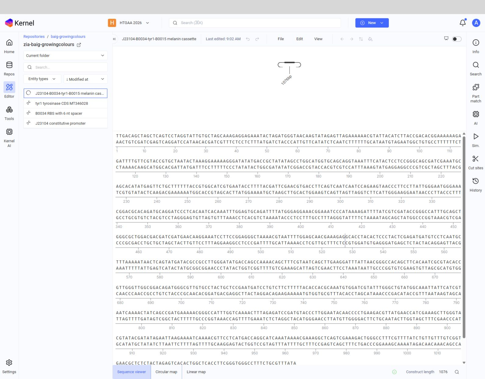
I used the HTGAA 2026 workspace and the baig-growingcolours repository. The saved construct is named J23104-B0034-tyr1-B0015 melanin cassette. Figure 1 shows it in Kernel (Asimov, 2026).
1. Expression target
The target is Tyr1, a 297-amino-acid tyrosinase derived from Bacillus megaterium. Tyr1 catalyses oxidation of L-tyrosine toward melanin formation. I would use the dark pigment as the backing to the cellulose-based colour layer (National Center for Biotechnology Information [NCBI], n.d.; Walker et al., 2025).
The proposed chassis is Komagataeibacter rhaeticus. This bacterial-cellulose producer was part of my Week 1 proposal and has been studied as a host for engineered cellulose and melanin production (Baig, 2026; Florea et al., 2016; Walker et al., 2025).
2. Parts and vector context
Layer and Kernel part
Source and length
Reason for choice
Promoter: J23104
iGEM BBa_J23104; 35 bp
Constitutive transcription. I would measure its strength with Tyr1 in K. rhaeticus (Florea et al., 2016; iGEM Registry of Standard Biological Parts, n.d.-c).
RBS: B0034 plus spacer
iGEM BBa_B0034, 12 bp; designed TACTAG spacer, 6 bp
Bacterial translation initiation (iGEM Registry of Standard Biological Parts, n.d.-b).
CDS: Tyr1
GenBank MT346028, positions 2277–3170; 894 bp
Tyrosinase associated with melanin production (NCBI, n.d.; Walker et al., 2025).
Terminator: B0015
iGEM BBa_B0015; 129 bp
Double terminator after the Tyr1 stop codon (iGEM Registry of Standard Biological Parts, n.d.-a).
Target backbone: pSEVA331Bb
Addgene plasmid #78269; approximately 2,893 bp
Proposed plasmid context for a K. rhaeticus prototype (Addgene, n.d.; Florea et al., 2016).
The 1,076 bp Kernel construct contains the expression cassette. The selected pSEVA331Bb backbone is a proposed build target and is not part of the saved Kernel cassette.
3. Assembled cassette and reading frame
Region
Kernel coordinates
Orientation and check
J23104 promoter
1–35
Forward
B0034 plus spacer
36–53
Forward; Tyr1 starts immediately after the spacer
Tyr1 CDS
54–947
Forward; ATG at 54–56 and TGA at 945–947
B0015 terminator
948–1076
Forward; follows the Tyr1 stop codon
The CDS has 297 sense codons followed by one stop codon. Kernel displayed a construct length of 1,076 bp. Figures 1 and 2 document the saved cassette; the schematic export does not label its individual parts, so the table above supplies their positions (Asimov, 2026).
Figure 1. Kernel editor showing the saved cassette in baig-growingcolours (Asimov, 2026).
Figure 2. Schematic exported by Kernel. The export lacks individual part labels; the table above supplies the coordinates (Asimov, 2026).
4. Optimization and sequence checks
Kernel codon optimizer
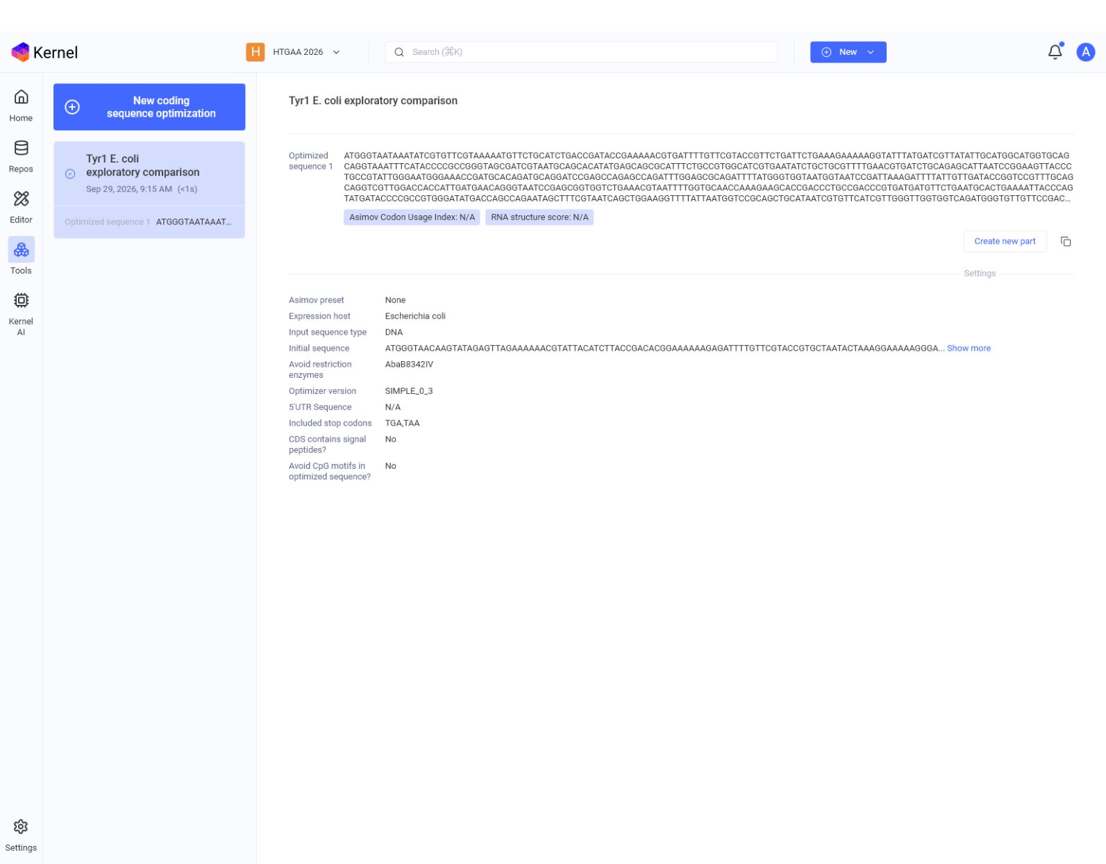
I submitted the native 894 bp Tyr1 CDS to Kernel SIMPLE_0_3 with Escherichia coli as the expression host because K. rhaeticus was absent from its four available hosts. The result is 897 bp, with 172 substitutions across the 891 sense-coding bases. GC content changed from 43.85% to 44.93%, and the output added a TAA after the native TGA. Local translation confirmed the same 297 amino acids. I used this E. coli variant only as a comparison; I did not substitute it into the K. rhaeticus construct (Asimov, 2026).
Figure 3. Kernel optimizer output and recorded run settings (Asimov, 2026).
Sequence identity
I compared the saved CDS base for base with the published MT346028 Tyr1 region used to create it: all 894 positions match, and the translated protein matches QLI61466.1 (NCBI, n.d.). This is a comparison with the selected reference sequence, not a broad similarity search. The revised document reports no biosecurity scan result.
5. From digital design to DNA
For one prototype, I would order an insert covering the 1,076 bp cassette and place it in the verified pSEVA331Bb multiple cloning site using overlap-based assembly. I would then sequence the full insert and both vector junctions in an isolated clone before testing pigment formation. Overlap-based assembly has been demonstrated for joining DNA fragments, and sequence verification is important because synthesized DNA can contain errors (Carr et al., 2004; Gibson et al., 2009; Kosuri et al., 2010).
I believe one verified clone is enough to begin promoter and pigment tests. The assembly, sequencing and pigment tests remain proposed work.
6. Main unknowns and next tests
Uncertainty
Test and decision
Tyr1 expression may be too weak or burdensome
Compare weaker promoter variants with the same Tyr1 CDS; measure transcript or protein level, growth and cellulose yield. Keep the lowest expression that gives adequate colour.
Active tyrosinase and pigment placement are unproven in this host
Measure Tyr1 activity, melanin accumulation, copper dependence and pigment distribution; test variants if the initial design underperforms.
A dark backing may not improve the final optical film enough
Test layers with and without pigmented cellulose; compare contrast and colour.
Carr, P. A., Park, J. S., Lee, Y.-J., Yu, T., Zhang, S., & Jacobson, J. M. (2004). Protein-mediated error correction for de novo DNA synthesis. Nucleic Acids Research, 32(20), e162. https://doi.org/10.1093/nar/gnh160
Florea, M., Hagemann, H., Santosa, G., Abbott, J., Micklem, C. N., Spencer-Milnes, X., de Arroyo Garcia, L., Paschou, D., Lazenbatt, C., Kong, D., Chughtai, H., Jensen, K., Freemont, P. S., Kitney, R., Reeve, B., & Ellis, T. (2016). Engineering control of bacterial cellulose production using a genetic toolkit and a new cellulose-producing strain. Proceedings of the National Academy of Sciences, 113(24), E3431–E3440. https://doi.org/10.1073/pnas.1522985113
Gibson, D. G., Young, L., Chuang, R.-Y., Venter, J. C., Hutchison, C. A., III, & Smith, H. O. (2009). Enzymatic assembly of DNA molecules up to several hundred kilobases. Nature Methods, 6, 343–345. https://doi.org/10.1038/nmeth.1318
Kosuri, S., Eroshenko, N., LeProust, E. M., Super, M., Way, J., Li, J. B., & Church, G. M. (2010). Scalable gene synthesis by selective amplification of DNA pools from high-fidelity microchips. Nature Biotechnology, 28, 1295–1299. https://doi.org/10.1038/nbt.1716
National Center for Biotechnology Information. (n.d.). Cloning vector pJV298-Tyr1, complete sequence (GenBank accession MT346028) [GenBank record]. https://www.ncbi.nlm.nih.gov/nuccore/MT346028
Walker, K. T., Li, I. S., Keane, J., Goosens, V. J., Song, W., Lee, K.-Y., & Ellis, T. (2025). Self-pigmenting textiles grown from cellulose-producing bacteria with engineered tyrosinase expression. Nature Biotechnology, 43(3), 345–354. https://doi.org/10.1038/s41587-024-02194-3
Week 3 HW: Cell-Free Reaction Design
Zia Baig | HTBAA 2026 | October 6, 2026
A. Comparing reaction components
Potassium glutamate helps maintain the ionic conditions needed for protein synthesis. Magnesium glutamate supports ribosomes and many enzymes, but nucleotides and phosphate bind magnesium and change how much remains available. Magnesium is therefore the more sensitive variable: small changes can sharply reduce expression, while potassium usually has a broader useful range (Olsen et al., 2026).
A PEP/NTP formulation supplies nucleotide triphosphates for transcription and uses PEP to regenerate ATP. It is well established, but its inputs are costly and phosphate can accumulate. A ribose/NMP formulation relies on the lysate to generate ATP and convert monophosphates into triphosphates. This can lower reagent costs, but performance depends more on lysate metabolism and oxygen transfer (Olsen et al., 2026).
Lower reagent cost, but greater dependence on lysate metabolism and oxygen
B. Proposed 20 µL reactions
The table shows one reaction for each target. All volumes are in nL, in the designer’s 25 nL increments, and each reaction totals 20,000 nL. Each uses the matching DNA template. The fixed buffer contributes 200 mM potassium glutamate, 2.6 mM magnesium glutamate, 30 mM HEPES, and 1 mM each of the amino acid mix, tyrosine, and cysteine. All listed reagents were in stock on October 6, 2026 (Ginkgo Bioworks, 2026; HTBAA, 2026).
Reagent (nL)
sfGFP
PETase
Reteplase
Base buffer
2,000
2,000
2,000
Lysate
5,000
5,000
5,000
Target DNA template
2,000
2,000
2,000
17 amino acid mix
1,600
400
400
Ammonium glutamate
—
200
200
AMP
600
—
—
CMP / GMP / UMP
425 each
—
—
ATP
—
250
250
CTP / GTP / UTP
—
175 each
175 each
Coenzyme A
—
100
100
DsbC
—
500
800
Folinic acid
—
50
50
HEPES KOH pH 7.5
900
550
750
Magnesium glutamate
225
300
225
NAD
—
75
75
Nicotinamide
800
—
—
Oxidized glutathione GSSG
—
—
475
Reduced glutathione GSH
—
—
200
PEP
—
6,000
6,000
Potassium glutamate
3,325
—
—
Potassium oxalate
—
150
150
Potassium phosphate mix
600
—
—
Putrescine
—
50
50
Ribose
1,500
—
—
Spermidine
50
125
125
Nuclease free water
125
1,725
625
Total
20,000
20,000
20,000
sfGFP. I would add 0.625 mM spermidine to the site’s ribose/NMP reference recipe and reduce the water by 50 nL. This changes one ingredient, so I could compare sfGFP fluorescence and cost per gram with the original recipe (Ginkgo Bioworks, 2026; Olsen et al., 2026).
PETase. I would use standard lysate and reduce DsbC from 5 to 2.5 µM, replacing the removed 500 nL with water. At the listed stock price, this saves $2.816 per reaction. I would check whether PETase activity remains high enough to improve activity per dollar (Ginkgo Bioworks, 2026; HTBAA, 2026).
Reteplase. I would keep the oxidizing DSB lysate and GSSG/GSH mixture, raise HEPES from 57.5 to 67.5 mM, and reduce DsbC from 5 to 4 µM. The lower DsbC amount saves about $1.126 per reaction. I would use IPR-AMC cleavage to test whether the reaction maintains activity (Ginkgo Bioworks, 2026; HTBAA, 2026).
I would compare each design with its unchanged reference and a no-DNA control, then repeat any promising result. The PETase and Reteplase assays report activity, not protein mass, so they cannot establish cost per gram on their own (HTBAA, 2026).
Community reagent: FkpA
FkpA is my choice because it may help fold disulfide-rich Reteplase. In an E. coli cell-free system, FkpA and DsbC improved antibody folding together (Groff et al., 2014). Its effect on Reteplase activity and cost remains to be measured.
Groff, D., Armstrong, S., Rivers, P. J., Zhang, J., Yang, J., Green, E., Rozzelle, J., Liang, S., Kittle, J. D., Steiner, A. R., Baliga, R., Thanos, C. D., Hallam, T. J., Sato, A. K., & Yam, A. Y. (2014). Engineering toward a bacterial ‘endoplasmic reticulum’ for the rapid expression of immunoglobulin proteins. mAbs, 6(3), 671–678. https://doi.org/10.4161/mabs.28172
Jewett, M. C., & Swartz, J. R. (2004). Mimicking the Escherichia coli cytoplasmic environment activates long-lived and efficient cell-free protein synthesis. Biotechnology and Bioengineering, 86(1), 19–26. https://doi.org/10.1002/bit.20026
Olsen, M. L., Copeland, C. E., Sundberg, C. A., Aw, R., Shaver, Z. M., Rao, G., Swartz, J. R., Karim, A. S., & Jewett, M. C. (2026). Design-driven optimization of low-cost reagent formulations for reproducible and high-yielding cell-free gene expression. Nature Communications, 17, Article 3478. https://doi.org/10.1038/s41467-026-69605-8