I’m Zubair Talib. I studied EE/CS at MIT in the 90’s. My master’s work was at MTL, building an image processing system for driverless vehicles. After presenting our work at an Intelligent Vehicle Control conference in the mid-90’s, I was certain we were late to market. :)
Since then I’ve spent my career in AI and software: enterprise search and information retrieval before Google, conversational AI for hiring, and LLM-based voice agents before ChatGPT. Today I run Celara, an AI and technology services company. I also do advisory work and special projects through Unigram Labs.
I’m completely new to biology. I’ve become fascinated by the combination of AI protein design models and automated wet labs, and I’m excited to learn about all aspects of synthetic biology.
My current interest is quite specific. My wife and two daughters all have celiac disease, an autoimmune condition that makes them react to gluten: wheat, bread, pasta, soy sauce. We love Asian food, and eating out is a constant challenge.
I’m interested in anything that improves the condition and ultimately solves celiac. For now my ambitions as a non-biologist are more modest: a reliable gluten detector so we can eat out safely.
See the nav bar on the left to look through my work for How To
Biomanufacture Almost Anything (Fall 2026)!
Subsections of Zubair Talib — HTBAA Fall 2026
Week 1 HW: Final Project Ideas
Idea 1 — Glumen: a de novo gluten-binding protein
Framing questions
What do you want to make? →
What living system could make it? →
How could automation help you make it? →
What would it take to make it at a meaningful scale?
(1) What you would like to biomanufacture and why?
Describe the target product or output you would like to make.
Why are you interested in making it? Your motivation might be utilitarian—for example, addressing a problem in health, sustainability, food, energy, or manufacturing—but it does not need to be. Your project might instead have a social, cultural, artistic, or personal motivation.
Clearly articulate what you want to make, who or what it is for, and why you think it is interesting or important.
I’m interested in producing a gluten binding protein to be a key component in a gluten detector - Glumen.
The goal is to make a very reliable gluten detector for individuals with Celiac Disease - something that can be used remotely at resturants, that is compact, fast, and highly reliable for a broad range of foods.
My wife and 2 daughters have celiac disease and eating out is quite challenging. Even food that is nominally gluten free - free of wheat or obvious gluten containing ingredients is not always safe. Hidden ingredient in sauces, cross contamination, lack of knowledge within the kitchen, all contribute to such challenging circumstances.
The human digestive/immune system obviously reacts to even these small amounts of gluten - and we’d like to detect small amount of gluten directly in a non-lab/compact consumer device.
I think the total system for gluten detection will be complicated including some method (e.g. ethanol, heat, and mechnical agitation) to release the gluten from food and to detect and calibrate thea mount of gluten present. But the key piece would be this de novo protein binder that can be used to bind to gluten and provide opportunity for identification. Existing gluten detection systems have limitations across food types particularly for fermenetd and hydrolyzed foods (e.g soy sauce, fried rice, pho, etc).
(2) What living system would you use?
Propose at least one living-system manufacturing chassis that could produce your target—for example, a microbial, mammalian, or cell-free system.
At a high level, describe how the system would make your target product. What goes into the system? What biological process or processes occur? What comes out?
You do not need to design the complete biological system yet. Make reasonable assumptions where necessary and identify them.
The living system I unerstand could be an E. coli expression system.
We would introduce the DNA that encodes our protein binder into the E. coli cells. Grow those cells and then extract and purify out our protein.
(3) How would you use automation?
Identify at least one automation technology that you would combine with your living system.
This could include a large-scale Cloud Lab, a desktop liquid-handling robot such as an Opentrons, a programmable microfluidics platform such as Nuclera’s, or another automation technology.
Describe what part of the biomanufacturing workflow you would automate and why. For example, you might automate liquid handling, culturing, assembly, screening, sampling, measurement, optimization, or another step.
While we can design the protein in-silico - using RF Diffusion, Protein MPNN, and predict the structure with Boltz/Protenix or AlphaFold3 - we will not actually know if the protein folds properly, if it will bind to the gluten fragment as expected, and if it has the selectivity needed to not identify other confoudners. I believe an automated wet lab can help experimentally determine effectiveness.
I had been looking at Adaptyv as a potential vendor/CRO lab that could both express the protein and then test the protein folding, binding strength, binding time, selectivity, etc.
(4) How much would you need to make, and how long would it take?
Perform a back-of-the-envelope calculation estimating what it would take to manufacture your target at a meaningful scale.
(a) How much?
Define a scale relevant to your project and calculate approximately how much of your target product you would need to manufacture.
For example, if you are producing a medicine, you might estimate the amount required to treat 1,000 people, everyone in Massachusetts, or everyone in the world. If you are producing a building material, you might calculate the amount required for one structure or an entire neighborhood. If you are producing an art or design artifact, define an appropriate scale yourself.
Clearly show your calculation, including units, assumptions, and sources for important numbers.
While we dont know the exact amount of binder we’d need, for planning we can estiamte 250ug of binder per test. Lets say we are looking to equip a device with 20 tests to begin with.
And we want to get 500 trial devices x 20 ==> 10000 test x 250 ug = 2.5 g of binder
Lets assume some loss (20%) - round up to 3.0g.
This is for production.
For the automation run - I think we need small amount of samples to test binding affinity, binding time etc.
Assay volumes
Food
Extraction Liquid
Binder Liquid
Amount
1/2 tsp (2.5g)
4.5 ml 3.0 ml ethanol
2.5 ml 250 ug binder
Water
1.5 ml
1.5 ml
2.5 ml
Total Liquid in Cup
1.5 ml
6.0 ml
8.5 ml
Ethanol concentration
—
50%
35%
Gluten Concentration
Concentration
Gluten in sample
10 ppm
25 ug gluten
20 ppm
50 ug gluten
100 ppm
250 ug gluten
(b) How long?
Using your proposed living system and automation technology, estimate how long it would take to manufacture that amount.
Consider quantities such as production rate, yield, volume, number of parallel experiments or reactors, automation throughput, or whatever parameters are most appropriate for your system.
You may discover that the automation technology you would use to prototype your system is very different from what you would use to manufacture at scale. If so, describe both.
Again, we are looking for a reasonable engineering estimate. Show your assumptions and calculations.
According to Adaptyv Bio - they can express the protein in about a week, run the measurements by week 2, and have the full data by week 3. 16-21 days in total.
I believe the in-silico design can be done in a few days. So maybe the full loop is 3-4 weeks in total - from start of a design to getting results from wet-lab.
(5) What don’t you know yet?
After completing your analysis, identify the 2–3 biggest technical unknowns, bottlenecks, or assumptions in your proposed biomanufacturing system.
What would you need to learn, measure, or experimentally determine to know whether your idea could actually work?
LOTS. There are a few classes of problem in general with this device.
can we separate enough signal of the gluten from the food - this is pure extraction or “gluten release”
can we get a binder that can bind to enough of the types of gluten but be selective enough to not bind to the other type of confounders (e.g. maize etc)
will the binder survive the diltuion liquid (e.g ethanol)
Will the binder provide enough signal - can we attaach a flouresence or something else - so that we can detect the amount of gluten that binds.
I think for this project - we should focus on step 2 - as that is the biomanufacturing part of making a de novo protein
The first test would be to see if we can make a binder that binds strong enough to these small fragments. And test folding, binding affinity etc.
I think the concern will then be is the binder will survive the gluten release step (ethanol, heat, etc.) and if its selective enough amongst the confoudners like maize and those that have similar amino acid seqeunces.
AI use
I’ve been using AI substantially over the past several weeks and months to
learn the basics of AI protein design — RFdiffusion, ProteinMPNN, and
structure prediction with Boltz/Protenix and AlphaFold3.
For this write-up specifically:
The idea, motivation, chassis choice, and open questions are mine.
AI assisted with the estimates in section 4(a) — the per-test binder
quantity, the assay volumes, and the resulting ethanol concentrations.
Claude Code handled formatting and
publishing: it converted my plain-text draft into Markdown, built the two
tables from my values, and committed and pushed to this repository. It was
instructed not to change my wording, and did not. Although it did draft this note :)
Prompt to Claude Code: “can you put Idea 1.txt in very nice attractive
markdown. Update the table. DO NOT change the text from my language. Just
format nicely.”
Week 2 HW: DNA Design
Project scope and AI assistance
My HW1 project, Glumen, aims to produce a gluten-binding protein for a gluten detector. For HW2, I used an existing computational protein candidate to explore how to move from a protein sequence to a DNA expression design while the gluten-binder work continues.
Much of this write-up was written by OpenAI Codex as an AI-assisted exploration of the topic. Codex also did substantial technical work: retrieving the candidate, generating and checking sequence files, helping inspect the design in VectorBee, and capturing screenshots. This is a learning exercise, not a claim that I independently designed or validated every element. I am still developing my understanding of the terminology and design choices.
We did not have access to Kernel. We used VectorBee to inspect the constructs, with Biopython and DNA Chisel for local sequence generation, optimization, and checks. No Kernel repository was created and no Kernel tools were run. Database similarity searching and biosecurity screening remain incomplete, so this draft does not claim that every assignment requirement has been fulfilled.
The practice candidate was designed to bind human transglutaminase 2 (TG2). It is not a gluten binder, and its binding has not been experimentally validated. Using it gave us a concrete sequence for exploring the DNA-design workflow.
Part 0 — Workspace and tools
The local workspace is HTBAA/HW2/practice-v1. It contains source sequences, an annotated construct, a saved VectorBee workspace, screenshots, and a detailed step-by-step walkthrough. (The data files linked below are published with this page; the full walkthrough is kept in that local workspace.)
The original backbone and modified construct were opened in VectorBee to inspect their maps and sequence annotations. The local folder serves as the homework workspace; it is not a substitute for a findable Kernel repository.
Part 1 — Define the expression target
The intended product is a small protein binder. For this exercise, we used the 90-amino-acid candidate rf_9_dldesign_0, generated in my earlier RFdiffusion and ProteinMPNN work against TG2.
The proposed expression host is E. coli BL21(DE3). The design uses a T7 promoter, so the host must supply T7 RNA polymerase. The protein would remain inside the cell, in the cytoplasm, followed by extraction and purification.
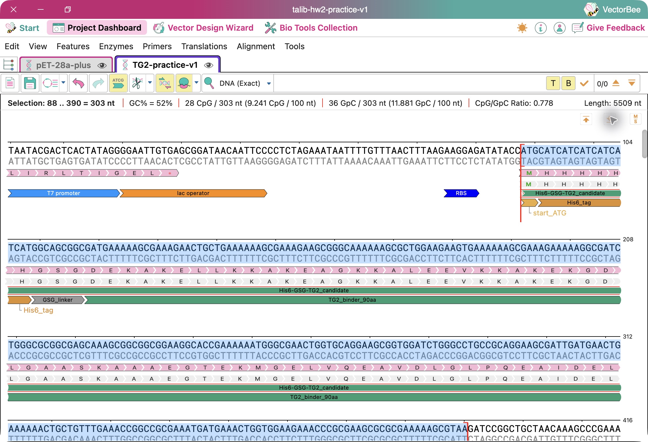
The six histidines, called a His6 tag, provide a handle for affinity purification. The GSG spacer is three amino acids—glycine, serine, glycine—connecting that handle to the candidate. Together with the starting methionine, these additions give a 100-amino-acid protein. The candidate itself is unchanged.
No secretion signal or dedicated tag-cleavage site is included. The proposed purified protein would retain its tag; whether the tag affects binding remains an experimental question.
Part 2 — Start with an existing plasmid and replace one section
I used the pET-28a(+) backbone based on the sequence from SnapGene’s sequence resource, and replaced the original tag and cloning region with the candidate coding sequence.
A plasmid is a circular piece of DNA. Its backbone is the reusable supporting DNA: it lets bacteria maintain the plasmid and provides controls for reading the protein instructions.
The before-and-after comparison is:
Section
Original plasmid
Our design
Controls for reading the protein instructions
T7 promoter, lac operator, and ribosome binding site
Kept unchanged
Protein-coding and insertion region
Original tags and cloning region: 163 DNA bases
Replaced with 303 bases encoding our tagged candidate and its stop codon
Signal to stop copying the instructions into RNA
T7 terminator
Kept unchanged
Supporting DNA elsewhere around the circle
Replication, selection, and regulation features
Kept unchanged
We removed the original tag-and-cloning section and supplied our own defined version. We did not add a second set of tags on top of the existing ones. The replacement corresponds to bases 88–250 in the locally oriented source record; those coordinates refer to that record, not necessarily other displays of the same circular plasmid.
Part
Source
Plain-English role and reason for including it
T7 promoter and lac operator
Existing backbone
Control when the protein instructions are copied into an RNA message.
Ribosome binding site (RBS)
Existing backbone
Provide a landing site on that message for the protein-making machinery; retain its original spacing to the start codon.
Tagged candidate coding sequence (CDS)
Earlier protein design; DNA generated locally
Encode the complete proposed protein, including its purification handle and spacer, with start and stop instructions.
His6 tag and GSG spacer
Added in this design
Provide a purification handle and a short flexible connection to the binder. These are already included within the CDS, not separate proteins.
T7 terminator
Existing backbone
Signal the end of copying DNA into the RNA message.
Origin of replication
Existing backbone
Allow bacteria to copy and maintain the plasmid.
Kanamycin-resistance marker
Existing backbone
Allow selection of bacteria carrying the plasmid.
lacI and its promoter
Existing backbone
Provide the repressor that helps control expression.
The exact sequences, coordinates, and sources are recorded in the parts table.
Part 3 — Assemble and inspect the construct
The expression region follows this order:
Reading controls → ribosome landing site → instructions for our tagged protein → stop-copying signal
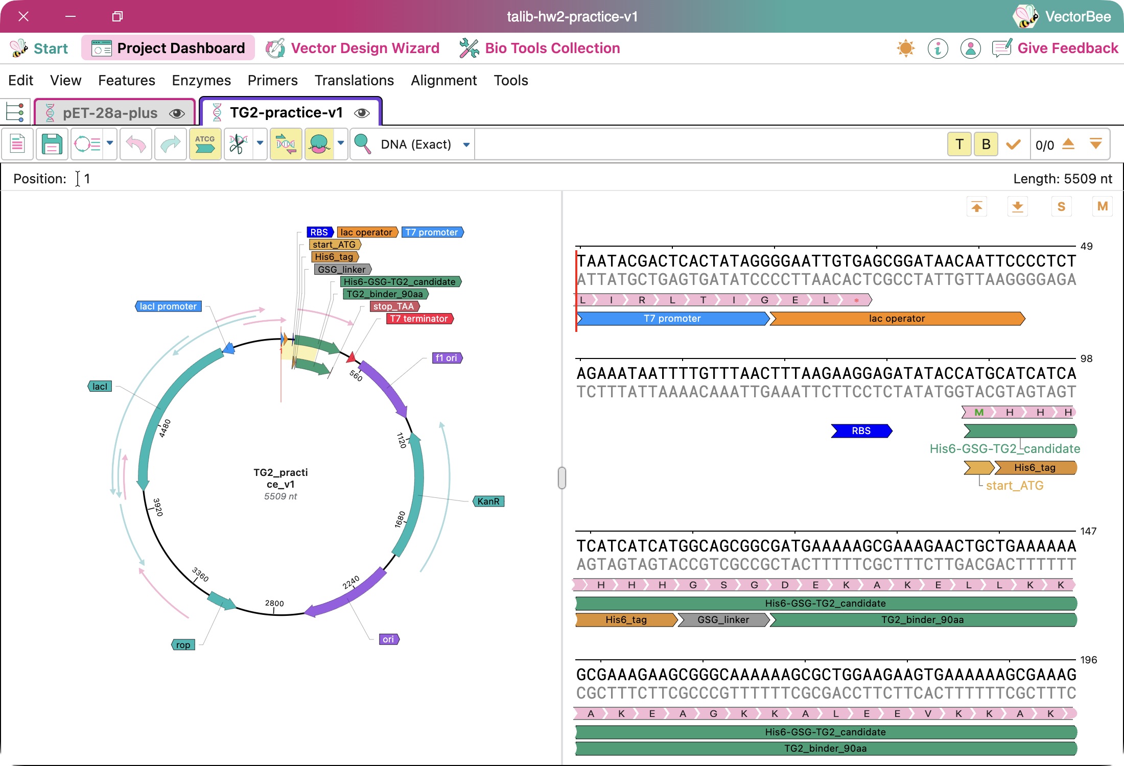
The final circular plasmid is 5,509 base pairs: 5,369 original bases − 163 removed bases + 303 inserted bases. The 303-base CDS contains 300 bases encoding the 100-amino-acid protein and three bases forming the stop codon.
The annotated sequence was generated locally and inspected in VectorBee. Saved computational checks confirm that:
The CDS translates into the intended tagged protein.
There are no internal stop codons.
Backbone DNA outside the intended replacement is unchanged.
The proposed assembly fragments reconstruct the final circular sequence.
These checks establish consistency of the sequence design. They do not establish expression, folding, or binding.
Figure 1. The final plasmid in VectorBee, showing the candidate CDS and retained backbone features.
Figure 2. The selected 303-base CDS, with the start codon, His6 tag, GSG spacer, and candidate translation. Overlapping annotations and translation rows describe the same sequence; they are not additional protein copies.
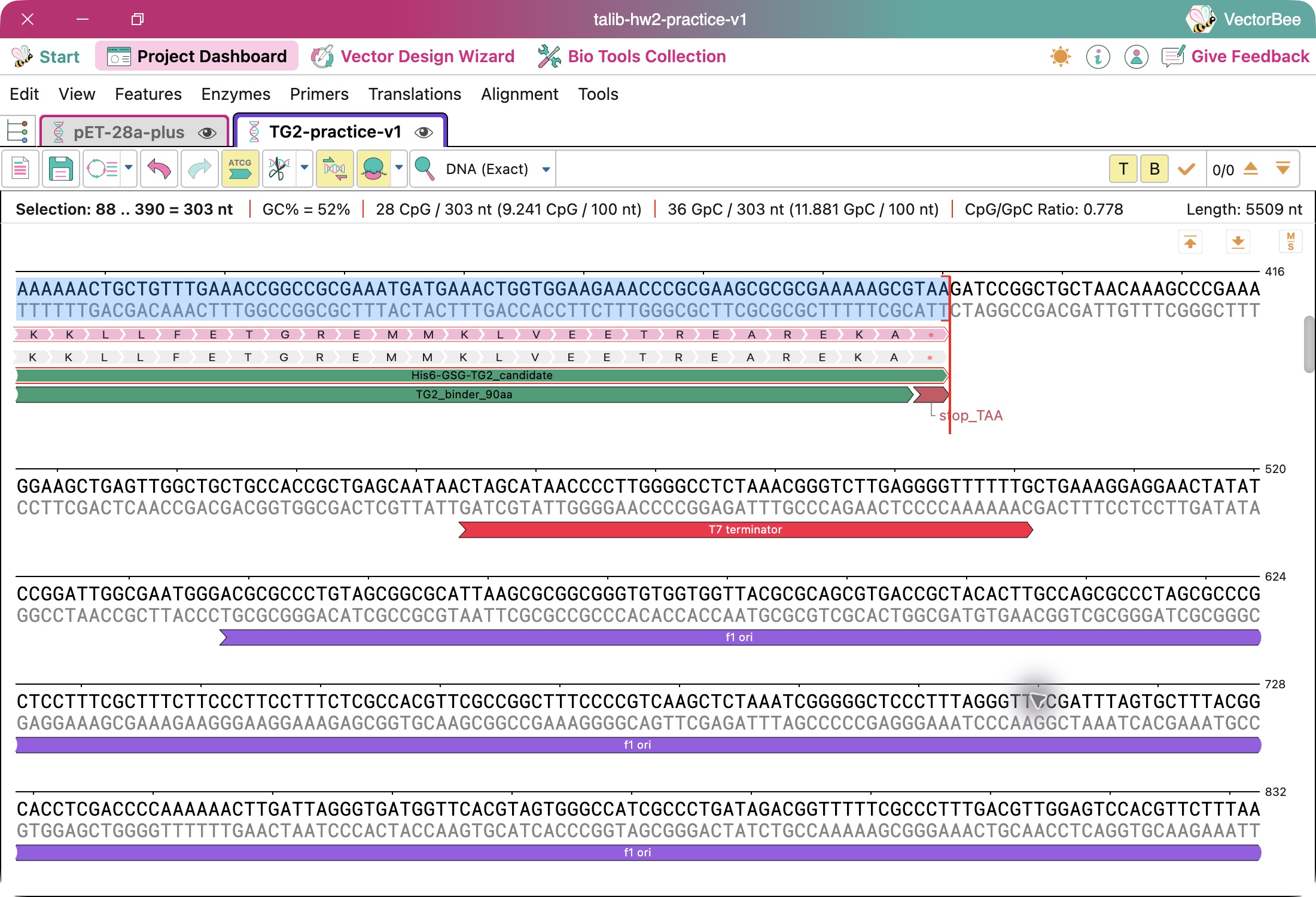
Figure 3. The TAA stop codon tells the cell to stop making this protein. The separate downstream T7 terminator signals the end of copying DNA into RNA.
Because this is a de novo protein candidate, there was no natural gene sequence to use as the starting DNA. We generated a synthetic baseline and used the open-source tool DNA Chisel to optimize codon usage for E. coli while preserving the protein sequence.
A codon is a group of three DNA letters that specifies an amino acid or a stop. Different codons can specify the same amino acid, allowing the DNA to change without changing the protein.
Metric
Before
After
GC content, excluding the stop codon
50.33%
52.33%
Codon adaptation index against the saved usage table
0.5828
1.0000
Intended protein sequence
100 amino acids
Unchanged
GC content is the percentage of DNA letters that are G or C. Roughly half the letters are G or C in both versions. We tracked it as a sequence-design constraint; higher is not automatically better.
Of the 100 amino-acid-encoding codons, 62 changed, involving 67 nucleotide substitutions. Constraints included limits on overall and local GC content, avoiding long single-base repeats, and excluding selected restriction sites from the coding region.
A codon adaptation index of 1.0 means the selected codons maximize the score against this particular usage table. It does not mean perfect expression. RNA structure, protein folding, and other biological factors remain relevant. The before-and-after codon comparison records the changes.
Sequence similarity search — incomplete
We verified that the designed DNA encodes the intended candidate, but have not searched it against a database of known sequences. I therefore cannot report a closest natural match or conclude that the sequence is novel.
A later step would be to search both the untagged protein and designed CDS, recording the database, settings, closest matches, identity, coverage, and E-values.
Safety screening — incomplete
We did not have access to Kernel’s screening tool and have not run an equivalent biosecurity screen. The local checks address sequence integrity and selected design constraints; they do not substitute for screening against defined biological hazards.
No screening pass or absence of flags is claimed.
Part 5 — From design to physical DNA
For a first prototype, my proposed approach is to obtain a synthetic insert and assemble it with the retained backbone. The saved design specifies a 363-base insert including overlap regions and a 5,206-base retained backbone. The overlapping ends allow the two pieces to join; those shared bases are not counted twice in the final plasmid.
This approach reuses most of the existing vector and introduces only the new coding region. Gibson et al. describe joining DNA fragments through overlapping ends, supporting this proposed assembly strategy. Fragment joining has been checked computationally; no physical assembly has been performed. Gibson et al., 2009
The coding sequence is short and has moderate GC content, but repeated codons and other local features could still affect synthesis. A supplier has not assessed or accepted the design. Primer design and laboratory conditions also remain outside the completed work.
Carr et al. describe error correction for synthetic DNA. The practical lesson for this construct is that synthesis can introduce errors. I would verify the resulting sequence and assembly junctions before interpreting protein experiments; error correction during synthesis would not replace final verification. Carr et al., 2004
For repeated production of the same protein, the goal would be to retain a verified production clone and scale expression and purification. The gene would not need to be chemically resynthesized for every batch. Producing many different binder candidates would require a separate strategy for DNA synthesis, assembly, and identification.
Part 6 — What remains uncertain?
Unknown
How I would investigate it
Will the construct produce enough protein without excessive burden on the cells?
Compare induced and uninduced expression, cell growth, and protein abundance.
Will the protein be soluble and suitable for further testing?
Compare soluble and insoluble fractions, then assess recovered yield, purity, and aggregation.
Will the candidate bind TG2 specifically, and does the tag affect binding?
Measure concentration-dependent binding with unrelated-protein controls; compare a tag variant if needed.
Even a positive result against TG2 would not establish gluten binding. Moving back to Glumen would require an actual gluten-binder candidate, an updated construct, and appropriate binding and selectivity tests.
Open-source follow-up to explore later
We have already used open-source tools for sequence generation, optimization, and checking. If time permits, the next priorities are:
Follow-up
Possible tool
Intended result
Search for related known sequences
NCBI BLAST+, using downloaded databases
A documented similarity-search result without submitting the candidate to a public search service.
Investigate an alternative biosecurity screen
SecureDNA’s open-source tools and screening infrastructure
Establish access requirements and, if available, obtain an actual screening report.
These are proposed follow-ups, not completed results.
AI use and prompts
OpenAI Codex wrote much of this document and performed substantial portions of the computational workflow. My role included providing the project context, requesting the exploration, reviewing the explanations, and asking for clarification of concepts such as the tag, spacer, coding sequence, and GC content. The document is an account of AI-assisted exploration rather than independent experimental work. No DNA was ordered and no expression or binding experiment was performed as part of this exercise.
The original design-work prompt, as recorded in the walkthrough, was:
“ok if you were going to go ahead and help with this assignemtn can you get th binder sequence or a sample one? maybe we can start with one we have and then we can improve it . make it later? and contuie with the rest of the steps . make sure each step is super clear what you do what happend and that I can follow - take screenshots if need be. I want to follow step by step and see the full restults”
For this write-up, I asked:
“ok can you suggest good answers here - that I can review - say we didnt have Kernel but show the parts we did do and what we could do with open source if we have time (which we shoudl look at later)”
I also requested VectorBee screenshots and a plain-English explanation of the original plasmid and the section replaced. For finalization, I asked:
“lets go ahead and finalize this draft Md - and acknwoledge much of this was written by Ai as an exploration of this topci and didnt have access to Kernel”
Week 3 HW: Cell-Free Reaction Design
Part A — Compare cell-free reaction compositions
Each question below shows my own working answer — arrived at with substantial
AI research and help — followed by an AI cleaned-up version. See the
note on AI use at the bottom.
Part B — publishing 20 µL reaction compositions for sfGFP, PETase, and
Reteplase on coopetition.htgaa.org, plus the Community Reagent contribution,
is not included in this write-up yet.
Q1 — Why does the concentration of potassium glutamate and magnesium glutamate differ between compositions? Which is more sensitive?
My answer
Short answer
a/ Why they differ. The concentrations differ because (a) they serve and
help catalyze different reactions, and (b) different recipes bind differently
and require different amounts of K⁺ or Mg²⁺.
b/ Which is more sensitive. Magnesium is more sensitive, as we can see in
Table II — it shows that if you overdo the concentration the yield falls off
substantially as compared to potassium for the same increase %.
Longer answer
CFPS requires various reagents and buffers to enable the protein generation
process to take place, and potassium glutamate and magnesium glutamate both
offer various chemical behaviors (providing charged particles) that facilitate
that process.
Potassium has one positive charge and magnesium has two — and therefore they
have different binding behaviors and serve different purposes.
Imagine trying to fold a long ribbon whose different sections keep pushing
one another away.
RNA has a version of that problem: it has many negatively charged parts, which
repel one another. Positive particles help soften those electrical pushes. This
allows RNA to adopt useful shapes and interact with other molecules. That
matters because a ribosome — the machine that assembles protein — is itself
made largely of folded RNA and proteins. Potassium helps establish suitable
electrical surroundings for these interactions.
Magnesium:
Keeping the machinery’s shape — it helps stabilize folded RNA and the ribosome.
Helping chemical reactions happen — many enzymes need magnesium together with ATP.
Supporting the right molecular connections — the ribosome must attach to
molecules, move, and release them as it builds a protein.
When you add magnesium:
Some associates with RNA, ATP, and other molecules.
Some remains free in the liquid.
Changing the ingredients changes that distribution.
For example, PEP binds magnesium more strongly than pyruvate. When the
researchers changed the energy ingredient, they therefore retested the
magnesium concentration.
For potassium, the authors also tested its concentration within each complete
mixture. Its preferred amount depends on the surrounding salts and other
ingredients.
So in short, the concentration differs between compositions because of what
each of potassium and magnesium binds to — the contents of the solution.
AI cleaned-up answer
1. Why do the concentrations differ between compositions?
Different recipes contain different ingredients and different starting salt
levels. Potassium maintains the ionic environment the protein-making machinery
needs. Magnesium supports RNA folding, ribosome structure, and every reaction
that runs on ATP — but it also binds other components in the mixture, so only
part of what you add stays available. Change the ingredients and you change
that distribution. For example, PEP binds magnesium more strongly than
pyruvate, so switching the energy substrate requires re-tuning magnesium.
2. Which is more sensitive?
Magnesium, clearly, in the formulations tested in Table II:
Salt
Change
Relative yield
Magnesium glutamate (Cytomim)
8 → 12 mM (+50%)
100% → 67%
Potassium glutamate (Cytomim)
130 → 200 mM (+54%)
100% → 95%
A comparable percentage increase costs about a third of the yield for
magnesium, versus a few percent for potassium. The practical implication: in a
screen, magnesium deserves a narrow, finely-spaced range around the optimum,
while potassium can be scanned coarsely.
Source: Jewett & Swartz (2004), Table II and accompanying discussion.
Q2 — Explain how a PEP+NTP reaction formulation is different from a Ribose+NMP reaction formulation.
My answer
PEP — helps replenish ATP so energy-dependent tasks can continue.
NTPs — supply the nucleotide form needed to make RNA; ATP and GTP also
support energy-consuming tasks.
PEP + NTP: the NTPs supply the building blocks for making RNA from DNA.
ATP also powers energy-consuming processes. As ATP is used and ADP is
produced, PEP uses that ADP and helps regenerate ATP so the reaction can keep
working.
Ribose — a sugar that enzymes in the lysate process to help regenerate ATP
from ADP.
NMPs — nucleotides with one phosphate group. Enzymes transfer additional
phosphates from ATP onto NMPs, turning them into NTPs with three phosphate
groups. RNA polymerase then uses these NTPs to make RNA by following the DNA
template.
Ribose + NMP: ribose helps regenerate ATP from ADP. Enzymes use ATP to
add phosphates to NMPs, converting them into NTPs. RNA polymerase then uses
those NTPs to make RNA from DNA.
Deeper dive
When ATP powers a task, it is chemically changed. Two common outcomes are:
ATP → ADP — ATP loses one phosphate group.
ATP → AMP — ATP loses two phosphates together. Attaching an amino acid to
tRNA uses this route.
So ADP comes from ATP being used by reactions in the mixture. Some ATP, ADP,
and AMP can also arrive with the lysate.
If we keep consuming ATP without replenishing it, energy-dependent steps slow
down or stop — even if amino acids and ribosomes are still available. PEP helps
turn used ADP back into usable ATP.
AI cleaned-up answer
The two formulations differ in how far upstream they start, and that single
choice drives both cost and reaction behavior.
PEP + NTP
Ribose + NMP
RNA building blocks supplied as
NTPs (triphosphates) — ready to use
NMPs (monophosphates) — must be phosphorylated first
Energy substrate
PEP, a high-energy phosphate donor
Ribose, a sugar fed into central metabolism
ATP regeneration
Direct: PEP + ADP → pyruvate + ATP, one enzyme step
Indirect: lysate glycolytic enzymes catabolize ribose over many steps
Reliance on lysate metabolism
Low
High
Cost
High — PEP and NTPs are the expensive reagents
Low — ribose and NMPs are commodity-cheap
PEP + NTP is the direct route. The nucleotides arrive already in the
triphosphate form RNA polymerase needs, and PEP regenerates ATP from ADP in a
single enzymatic step. It is fast and predictable, but PEP is consumed
stoichiometrically, releases inorganic phosphate that accumulates and
inhibits the reaction, and both PEP and NTPs are costly.
Ribose + NMP pushes work onto the lysate’s own metabolism. Ribose is
catabolized through the enzymes already present in the extract to regenerate
ATP, and that ATP is then used by kinases in the lysate to phosphorylate NMPs
up to NTPs before transcription can proceed. More steps have to work, so the
formulation depends more heavily on extract quality — but the inputs are
dramatically cheaper, and the slower, metabolically-buffered energy release
avoids the phosphate accumulation problem.
The trade-off: PEP + NTP buys speed and simplicity with reagent cost; Ribose +
NMP buys cost and sustained energy supply by trusting the lysate to do more of
the chemistry. This is exactly the trade-off Q3 quantifies.
Q3 — Choose two cell-free reaction compositions from the literature; compare and contrast the choice of reagents and the concentration of reagents.
My answer
Let’s use Olsen (2026) — we can compare PANOx-SP and RF₀ₚₜ.
All concentrations in mM unless otherwise noted.
Component
PANOx-SP
RF₀ₚₜ
Energy ingredients
PEP: 30
Ribose: 50; glucose: 10
Nucleotides
NTPs: ATP 1.2; CTP/GTP/UTP 0.85 each
NMPs: AMP 3; CMP/GMP/UMP 2.15 each
Amino acids
2 each
5 each
HEPES buffer
57
75
Potassium glutamate
130
362
Magnesium glutamate
8
8
Ammonium glutamate
10
Not added
Polyamines
Spermidine 1.5; putrescine 1
Not added
Other metabolic additives
NAD 0.4; CoA 0.27; oxalic acid 4
Phosphate 15; nicotinamide 4
Additional translation components
tRNA 0.17 mg/mL; folinic acid 0.03 mg/mL
Not added
Like question 2, we have PEP + NTP vs. Ribose + NMP.
We have:
2 vs. 5 mM amino acids
57 vs. 75 mM HEPES buffer
130 vs. 362 mM potassium glutamate
In RF₀ₚₜ we did not add ammonium glutamate. We also did not add spermidine and
putrescine. And there are some different metabolic additives between the two.
The RF₀ₚₜ is double the yield at 1% the cost.
We have more protein building material — with the more amino acids. It seems
that the engine Ribose + NMP outperformed the PEP + NTP.
AI cleaned-up answer
I compared PANOx-SP (Jewett & Swartz 2004) and RF₀ₚₜ, the cost-optimized
formulation, as evaluated by Olsen et al. (2026).
The energy and nucleotide system is the headline difference. As discussed
in Q2, PANOx-SP runs on PEP and NTPs; RF₀ₚₜ runs on ribose, glucose, and NMPs.
RF₀ₚₜ also raises several concentrations:
Amino acids: 2 → 5 mM each — more protein-building material, and a larger
buffer against depletion of whichever residue is limiting.
HEPES: 57 → 75 mM — greater capacity to resist the pH drift that comes
with sustained metabolic activity.
Potassium glutamate: 130 → 362 mM — a substantially different ionic and
metabolic environment.
Magnesium glutamate: unchanged at 8 mM — consistent with Q1: magnesium is
the sensitive parameter and sits near its optimum in both.
RF₀ₚₜ also removes components: ammonium glutamate, spermidine, putrescine,
tRNA, folinic acid, NAD, CoA, and oxalic acid are all dropped. In their place it
adds phosphate (15 mM) and nicotinamide (4 mM). Several of the removed
supplements are among the most expensive per-reaction line items, so removal is
doing real work on the cost side as well as the chemistry side.
Result:
PANOx-SP
RF₀ₚₜ
sfGFP yield
1.11 g/L
2.39 g/L (≈2.2×)
Reagent cost per gram protein
$4,083
$60 (≈1.5%)
The complete RF₀ₚₜ formulation therefore produced roughly 2.2× more protein at
about 1.5% of the reagent cost — a ~150-fold improvement in cost per gram.
Important caveat: because many ingredients changed simultaneously, this
comparison does not establish which individual changes caused the improvement.
The formulations were co-optimized as whole systems, and the components likely
interact (the elevated potassium, for instance, may only be beneficial in the
context of the ribose-driven energy system). Attributing credit would require a
component-by-component ablation.
AI use
Consistent with Week 1, here is how AI was used
on this assignment.
My answers are substantially supported by AI help. I’m new to biology, and
I used AI heavily to get through these papers — to explain the chemistry, to
check my understanding, and to help me pull the numbers out of the published
formulations. The “My answer” sections are my own working answers in my own
words, written after that back-and-forth; they appear here essentially as I
drafted them, with spelling corrected and Markdown formatting applied, but the
wording and the reasoning are unchanged. The understanding behind them was
built with AI, not independently of it.
The “AI cleaned-up answer” sections are exactly what the label says: Claude
restructured my answers into tighter prose, pulled the specific numbers out of
my working notes into tables, and in a few places added framing I had not
written (the screening implication in Q1, the comparison table and
phosphate-accumulation point in Q2, the ablation caveat in Q3). I reviewed each
one against the sources.
Claude Code handled formatting and publishing — converting my plain-text
notes into this Markdown page and committing it to this repository.
References
Jewett, M. C. & Swartz, J. R. (2004). Mimicking the Escherichia coli
cytoplasmic environment activates long-lived and efficient cell-free protein
synthesis. Biotechnology and Bioengineering.
Olsen et al. (2026). Cost-optimized cell-free formulation (RF₀ₚₜ).
Smith et al. (2026). AI-designed cell-free formulations.